Acute Rheumatism
Historical document, translated for reference. It reflects medical knowledge of the 1920s–30s and is not medical advice.
Summary
This article from the 1928–1936 Soviet Great Medical Encyclopedia defines acute rheumatism as a systemic infectious disease of the mesenchyme, primarily affecting the cardiovascular system and joints. It discusses the historical shift in understanding the disease from a purely articular condition to a complex systemic pathology, explores theories of its allergic-infectious etiology, and notes its significant social impact as a cause of heart disease.
Encyclopedia article (1928–1936)
ACUTE RHEUMATISM. Contents: Geographical distribution and statistics.
Acute rheumatism [synonyms: “rheumatic fever” (Anglo-American authors), “rheumatic infection” (German and partially Russian authors), “morbus Bouillaud” (French authors), “rheumatic granulomatosis” (Fahr), “infectious specific rheumatism” (Graff), “true rheumatism” (Buchstab), “rheumatic state” (Coburn), “febrile rheumatism” (Klinge); former names: “acute articular rheumatism” or “acute rheumatic polyarthritis”], is a general infectious disease with a sharply pronounced tendency toward relapses, characterized by a peculiar systemic inflammatory lesion of the body's mesenchyme, with the primary localization of this lesion in the cardiovascular system and with the involvement—often multiple—of synovial and serous membranes in the inflammatory process. The first clear descriptions of the disease, accompanied by swelling of the joints, can be found in Prosper Alpinus (1611) and Cullen (1789). Bouillaud was the first to clearly establish the frequency of involvement of internal organs, and in particular the heart, alongside the joints in acute rheumatism, having established a close internal connection between these lesions; he also pointed to a peculiar “kind of general inflammatory diathesis” as the cause of acute rheumatism, anticipating in essence the modern theory of the allergic origin of acute rheumatism. Despite the work of Bouillaud, until recently the prevailing view in medicine was that the primary localization of acute rheumatism is the involvement of the joints (hence the former name of the disease, “acute articular rheumatism”), whereas the involvement of the heart and other internal organs is merely a complication of this disease. From the beginning of the 20th century, when in 1904 Aschoff described the granulomas in the myocardium characteristic of acute articular rheumatism (Aschoff bodies), the development of the pathological anatomy of this disease began (Geipel, Fraenkel, Huzella, Fahr, and especially Talalaev, Graeff, and Klinge), which leads to the concept of the presence in this disease of special histological changes of the mesenchyme characteristic of it. On the basis of these pathological-anatomical works, on the one hand, and the expansion and deepening of clinical observations, on the other, the above-mentioned concept of Bouillaud gradually develops and strengthens, and the position is accepted that the involvement of the joints is only one and far from an obligatory localization of this disease, that in any case, involvement of the myocardium and endocardium is very often observed, and that, furthermore, other internal organs are also frequently affected. This has led to the fact that now almost all authors are abandoning the term “acute articular rheumatism,” replacing it with other, above-mentioned names, of which, however, not one can yet be recognized as entirely satisfactory and generally accepted. (In particular, the term “acute rheumatism” adopted here is already unsatisfactory because clinically we very often deal with a protracted course of the disease; the pathological-anatomical data correspond fully to this.) At the present time, the question of the etiology and pathogenesis of acute rheumatism is on the agenda. There is no doubt that acute rheumatism is an infectious disease, but until now all attempts to find its specific pathogen, if one does not consider the streptococcus to be it, have not been successful. Recently, the view is becoming increasingly established that acute rheumatism is a manifestation of an “allergic” or hyperergic reaction of the organism, specifically its mesenchyme, to repeated infection of the organism with streptococci. If one adopts this point of view, then the task arises of distinguishing acute rheumatism from other streptococcal diseases—streptococcal sepsis, scarlet fever, local streptococcal suppurative infections, erysipelas, etc. Thus, the problem of rheumatism seems to be turning at the present time into a part of the large immunobiological problem of streptococcal infection, i.e., the problem of the interrelationships between the streptococcus (or streptococci) and the human organism—a problem, by its nature and significance, analogous, for example, to the immunobiological problem of tuberculosis. As an important—primarily purely practical—point in the history of acute rheumatism, it is necessary to point out the introduction into medical practice of salicylic acid preparations (Maclagan in 1874 and Buss and Riess in 1875). In recent years, starting from the World War, a huge number of studies have been devoted to acute rheumatism, as its role as a social disease, entailing huge material damage to society—mainly by causing heart disease that ends in disability or death at a young and middle age—is becoming clearer and clearer every year.
Geographical distribution and statistics. Geographical distribution of acute rheumatism. Cases of acute rheumatism are known in all countries with the most diverse climatic conditions. The maximum number of cases still falls on countries with a temperate climate, the minimum—on tropical and especially polar regions. In Western Europe, it is encountered especially often in England, Denmark, Scandinavian countries, and in certain parts of Germany, and less often in the Mediterranean basin. In the USSR, according to the tentative data of Danishevsky, relating to “rheumatic” diseases in general and covering only the draft age, the maximum number of cases is observed in the north-eastern part of the country (see Rheumatism). In the USA, acute rheumatism is a frequent disease, being especially widespread in certain parts of the country (e.g., in New York). Of the Asian countries, cases are observed most often in India, rarely in China, and very rarely in Japan. Certain places—the islands of Samoa, Wight, Guernsey, Madagascar, Zanzibar, the Antilles—hardly know this disease; in Europe, such places include the county of Cornwall in England and the district of Beauraing in Belgium. Statistics of acute rheumatism. There are still no exhaustive and reliable statistical data on acute rheumatism in most countries. The reasons for this are the still insufficient clarity in distinguishing acute rheumatism from other similar forms of diseases, the complexity of its clinical and often pathological-anatomical diagnosis, and as a consequence of this—the lack of established and, most importantly, uniform recording of it. Moreover, even in those countries (England, America) where the recording of morbidity and mortality from acute rheumatism (including its non-articular forms) is more or less established (insofar as this is possible due to the reasons just indicated), the comparison of data for different periods of time encounters difficulties, because the scope of the concept itself has undergone changes over time in accordance with the fact that clinical ideas about it have also changed: in earlier data, the discussion is only about acute articular rheumatism, in later ones—about articular and non-articular forms of acute rheumatism. In the overwhelming majority of countries, the bulk of the available statistical data on acute rheumatism relates either only to the articular form of acute rheumatism or more often concerns “rheumatic” diseases in general; in addition, a very large group of acute rheumatism cases, which proceeds with involvement of the cardiovascular system but without articular phenomena or ends in the development of valvular defects, falls into the category of cardiovascular diseases. Therefore, to form an idea at the present moment about the true prevalence of acute rheumatism in most countries is possible only by way of...
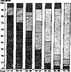
| syphilitic. Figure 1. Distribution of morbidity of organic heart diseases by age and type of disease. (According to Wyckoff-Lingg.) The isolation of cases of acute rheumatism from the number of: 1) all so-called "rheumatic" and 2) all cardiovascular diseases, which can be done only roughly and tentatively. In particular, regarding the question of what part of all "rheumatic" diseases is constituted by acute rheumatism, there are no sufficiently clear data, which is quite understandable in view of the lack of clarity of the boundaries between acute rheumatism and so-called chronic rheumatism. In this direction, perhaps, some idea is provided by Gelman's figures for Moscow for 1927-29, according to which, out of approximately 7,000 hospitalized rheumatic patients, about 4,500 were patients with acute articular rheumatism. As for cardiovascular diseases, an idea of the large place occupied by diseases caused by acute rheumatism in this group can be obtained to a certain extent from Fig. 1, which indicates the role of individual diseases in the etiology of organic heart diseases. Even the just-cited completely fragmentary data, which have only an indicative value, point with complete certainty to the significant prevalence of acute rheumatism. When evaluating statistical data on acute rheumatism, it is also necessary to take into account the significant fluctuations in the frequency of acute rheumatism depending on a number of geographical, socio-living, and other conditions, as well as its significant fluctuations by individual years, and finally, the completeness of the data, which is in close connection with the methods of accounting themselves. If, with the reservations just mentioned, one turns to the statistical material on acute rheumatism currently available (including, if there are no special reservations, its non-articular forms, insofar as they are subject to accounting), then, based mainly on the most complete and accurate Anglo-American data, one can obtain the following idea about this disease. Lethality of acute rheumatism. Mortality during an acute attack of the disease in relation to the total number of patients with acute rheumatism, according to the summary table of Atwater, based on 21,608 cases, is 1.74%, and according to Southey - 1.2%. It is highest in childhood, especially under 15 years of age (Table 1); this is undoubtedly in direct connection with the very frequent severe visceral lesions at this particular age, in particular with heart lesions. With increasing age, lethality falls, especially decreasing after 30 years. Mortality from acute rheumatism. If in general "mortality statistics without anatomical control give a completely false picture" (Lubarsch), then this should be said in particular about mortality statistics from acute rheumatism, since a significant part of the corresponding patients are treated during their lifetime under the diagnosis of cardiovascular diseases and thus fall into this same category of mortality. The extremely small percentage of autopsies in the vast majority of countries (e.g., in the USA, according to Karsner, it is equal to only 0.7% of all deaths; in Germany for 1920-21, according to Lubarsch, it does not exceed 5%) does not yet allow for complete clarity on this issue. The most complete sectional data of Davydovsky (38.8% of sections of all deaths in Moscow for 1923-27), based on 53,959 cases, give for acute rheumatism (obviously for its articular forms) only 18 cases of death (0.03% of all sections). If, however, one includes here cases of chronic verrucous endocarditis, then the total number of deaths immediately rises to an impressive figure of 797 cases (1.48% of all sections); the addition here of the group of endocarditis lenta gives an increase in the figure to 1.83% of all deaths, i.e., a figure almost 2 times larger than the figure of total mortality for these years from such common and severe infections as typhoid fever and diphtheria, and reaching 35.8% of mortality from cancer of all organs. In addition, valvular defects, which obviously developed on the basis of acute rheumatism, served as the cause of death in 405 cases (0.75% of all sections) according to the same data. Atwater, based on data on mortality from acute rheumatic fever, acute endocarditis, and pericarditis, determines the mortality figure for only acute forms of the disease as equal to 1% of deaths from all diseases in the USA for 1923. Some idea of the mortality from articular forms of acute rheumatism in different countries and cities can be provided by Table 2. These data, however, are 30-40 years old. More recent data for Denmark show mortality from acute rheumatism equal to 0.87%. Age has a great influence on the level of mortality from acute rheumatism (Table 3). Under 15 years of age, the percentage of mortality from acute rheumatism is the highest, and, for example, for England and Wales, the number of persons under 15 years of age dying from acute rheumatism exceeds 1/3 of the total mortality from this disease for all ages, while at the age of under 25, according to Anglo-American data, it constitutes about half of it. According to Askins, rheumatism accounts for 1/24 of all deaths in childhood. Anglo-American statistics note a somewhat higher mortality among women; only during the years of the World War did male mortality in England and Wales clearly predominate over female. The indications in the literature of higher mortality of the colored population compared to whites (see, for example, Table 4) are more correctly considered, it seems, as an indicator of the influence, first of all, of the incomparably worse socio-living conditions of their life in the corresponding capitalist countries. Data on the fluctuation of mortality from acute rheumatism by individual years show a clearly expressed downward trend for England and Wales and for the USA over the period 1900-24 (Figs. 2 and 3), which is linked by many authors to an increase in the cultural level of the population and an improvement in its socio-living conditions; however, one should approach the evaluation of these data with great caution, since - at least in the USA - the scope of the concept of acute rheumatism was different in different periods of time. Corresponding data for the continent of Europe are not available. Within individual population groups, American statistics indicate a somewhat higher mortality of the urban population (the average mortality rate per 100,000 population for 1910-23 is 4.85) compared to rural residents (the corresponding rate is 4.23). We have similar indications in the works of English authors (Coombs and others) and in old works (Newsholme) (Table 5). Morbidity of acute rheumatism.
The high prevalence of acute rheumatism is indicated by the data presented in Tables 6 and 7 regarding the incidence of acute articular rheumatism in certain Western European countries and the percentage of cases of the articular form of acute rheumatism alone relative to the total number of hospital admissions in a number of Western European cities. According to American data, there are 14.9 rheumatic heart disease patients per 1,000 population, and 4.5 per 1,000 schoolchildren. For the USSR, based on insurance fund materials for 1926 (1,832,165 cases of temporary disability), acute articular rheumatism accounts for, according to Gelman, 0.43% of all diseases resulting in loss of ability to work (0.5% for men and 0.35% for women).
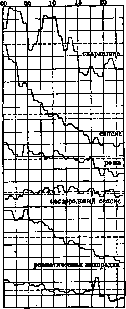
Figure 2.
Figure 3. Figure 2. Comparative curves of mortality rates from scarlet fever, septicemia and pyemia, erysipelas, puerperal sepsis, and rheumatic fever for 1905-1924 in England and Wales. The rates for each year and for each disease are shown as percentages of the average rate for the entire period; each division of the vertical scale corresponds to 5% above or below the average rate. Figure 3. Similar data to Fig. 2 for the USA (for regions where registration of the corresponding diseases has been introduced) for 1900-1923. The rates are the same as for Fig. 2. If one takes the number of cases of acute articular rheumatism that passed through Moscow outpatient clinics in 1926, converted per 100 people of the population, i.e., including children and adolescents and, to a certain extent, uninsured population groups, it is observed extremely rarely. Rauchfuss saw acute articular rheumatism in only 2 cases out of 15,000 infants; Widerhofer, 1 case out of 7,000. After 5 years, the morbidity curve for acute rheumatism begins to rise quite steeply, showing, according to Rolly, Danishevsky, and others, a particularly sharp rise between 15-20 years of age; after 30 years of age, morbidity falls rapidly, decreasing especially sharply after 40 years of age (cf. Table 1). According to Church, 96.7%, and according to Rolly, 90.3% of acute rheumatics are persons under 40 years of age; according to Southey, 90.4% are under 44 years of age, with 80.5% of all cases falling in the 15-44 age range; according to Wilk and Rabinovich (Moscow), 76.7% of patients with acute articular rheumatism are between 20 and 40 years old. Data on the influence of sex on the morbidity of acute rheumatism are very contradictory. Nevertheless, data based on vast statistical material for England and Wales and for the USA indicate a somewhat higher morbidity among women. Data processed by Danishevsky for Moscow, Tver, and the Moscow Governorate (see art. 454, Table 1) also point to a higher incidence among women, especially in villages. The higher morbidity of the colored population noted by Atwater for the USA and by English military statistics for the West Indies and Western America should, one must think, be attributed mainly to the same factors as its higher mortality compared to whites. The frequency of acute rheumatism cases in individual years can sometimes fluctuate very sharply. The fact of the almost complete disappearance of acute articular rheumatism during the years of the revolution and civil war in Petrograd is extremely interesting; according to materials from the Obukhov Hospital (A. A. Nechaev Jr.), the percentage of admissions of patients with acute articular rheumatism for 1910, 1918, and 1923 fluctuated as follows: in 1910 - 1.4% of all patients, in 1918 - 0.14%, and in 1923 - 0.6%. Observations from the Therapeutic Clinic of the 1st Leningrad Medical Institute from 1922 to 1930 also allow us to note a rapid, systematic increase in cases of acute articular rheumatism after the years of famine and devastation. Fluctuations in morbidity by months of the year for different localities of the same state have a different character, which is possibly connected with their different climatic conditions. However, even if one can note for many localities an increase in morbidity in the cold and damp months of the year (for the temperate zone of the Northern Hemisphere, mainly late autumn, winter, and the first spring months), these fluctuations do not always and not in all population groups have the same regular character. Observations on the morbidity of acute articular rheumatism in the Red Army, for example, allow us to note a higher morbidity among Red Army soldiers in the months they go to camps (May for the Leningrad garrison, according to L. S. Girshberg). In general, the influence of meteorological factors on the morbidity of acute rheumatism within individual population groups is, one must think, closely connected with their profession: the more a given category of persons is exposed to the influence of unfavorable meteorological factors due to their working conditions (work in the open air, in conditions of sharp changes in temperature, in damp and cold rooms, etc.), the more natural it is to expect a greater increase in the morbidity of acute rheumatism in them during periods of worsening weather compared to another group of the population working in completely different conditions (e.g., in closed, warm rooms). The clinical impressions of old authors regarding the highest general morbidity of acute rheumatism among representatives of those professions that require work in damp and cold rooms, a lot of walking, long periods of standing, and are accompanied by frequent and sharp changes in temperature (grooms, coachmen, laundresses, waiters, cooks, boys in wine and grocery stores, blacksmiths, railway employees, and to a significant extent servants and housewives, etc.), find full confirmation in the latest research based on the vast and, most importantly, comparatively very complete and uniformly collected material of the social insurance funds of the USSR (Table 8). The influence of the conditions and nature of work on the morbidity of acute rheumatism is revealed with particular clarity when comparing materials relating to persons who work in the same branch of the national economy but under completely different conditions. The corresponding data of Danishevsky and Gelman regarding railway workers not only clearly show a higher morbidity of acute articular rheumatism among locomotive personnel and workers in hot and cold workshops compared to office and senior administrative personnel (Table 9), but also indicate a longer duration of disability in the first three categories. Similar data from military statistics, relating to material that is homogeneous in terms of age and physical development, note, for example, a higher morbidity of acute articular rheumatism in mountain troops, whose work takes place in more unfavorable meteorological conditions and requires greater physical exertion (Andrieu). The influence of professional factors on the morbidity of acute rheumatism is closely intertwined with the influence of social and living factors in general, as can be seen with extreme clarity in the example already mentioned above of the much higher morbidity of peasant women in the Moscow Governorate compared to female residents of Moscow (see art. 455, Table 2). Although acute rheumatism affects representatives of all classes of society, a higher morbidity is observed mainly among the least well-off and, to a certain extent, among the least cultured strata of the population, who are in worse social and living conditions. Hence, one must think, comes the higher morbidity of the urban population compared to the rural in Western Europe and America, and the higher morbidity, resp. mortality, of the colored population in the USA, and the fall in mortality from acute rheumatism in England and the USA during 1900-24. In application to the USSR, the influence of social affiliation on the morbidity of acute rheumatism finds its manifestation in a significantly higher (2.2 times) morbidity of the rural population compared to the urban (at least for 1926 for Moscow and the Moscow Governorate) and to a certain extent compared to the population of factory-industrial districts (Danishevsky). Thus, here in 1926, we observed—at least in certain regions of the country—the exact opposite situation compared to the USA and Western Europe. The absence of more recent data makes it impossible for now to reveal the influence of the colossal shifts in the methods of agricultural management over a number of recent years in the USSR on the morbidity of acute rheumatism among the rural population. The average duration of a patient's stay in a hospital for the articular form of acute rheumatism is approximately 30-35 days. It is extremely important to note here that the later an acute rheumatic patient enters the hospital from the moment of the onset of the disease, the longer they lie in it on average, and the worse the final results of their treatment. The average duration of the entire disease, resp. the average duration of absence from work, is, however, significantly longer than the time the patient spends in the hospital. If the pathological-anatomical cycle of the development of the rheumatic inflammatory process is defined by Talalaev as 6 months, then its clinical manifestations disappear relatively quickly, and as a rule, patients begin work significantly earlier than the term indicated by Talalaev. The corresponding statistical data on this issue are still extremely insufficient. The total number of days of disability from acute articular rheumatism in the USSR for 1925, according to social insurance fund data, was 178,916 (Gelman). There are no satisfactory data on the magnitude of the disability caused by acute rheumatism. It is undoubtedly the case that the main mass of rheumatic invalids should be sought among the cardiovascular group of invalids (Fig. 1), and at an age mainly under 40 years.
There are far fewer invalids among those with a prolonged articular process (according to Kizilov's data for 1929, they constitute slightly more than 0.5% of all invalids who passed through the Moscow Bureau of Medical Expertise that year); moreover, with very rare exceptions, these individuals are found to be healthy in regard to their joints after a relatively short period (according to Kizilov, at most after 1 1/2 years). It is not yet possible to form an accurate idea of the national economic damage caused by acute rheumatism due to the lack of reliable data. There is no doubt that it is very great, both in view of the significant prevalence of acute rheumatism and in view of the fact that it affects mainly persons aged 20-40, i.e., in the most active period of life, and even more so because it very often (on average, according to modern concepts, in approximately 50% of cases) leaves behind valvular defects, or partially or completely turns the corresponding individuals into actual or potential invalids. If, along with these factors, one takes into account: 1) the predominant affection and more severe course of the disease in those classes of society that are in the worst social, living, and general cultural conditions, 2) the influence of professional factors on the level of morbidity in individual population groups, then we have every reason to classify acute rheumatism among the most important social diseases, the fight against which must be waged by the means and forces not only of state bodies but also of public organizations. Epidemics of acute rheumatism. The overwhelming majority considers acute rheumatism not to be a contagious disease. True, individual authors describe small "epidemics" of this disease, which, however, are usually limited in cities to individual streets, houses, or families, and in hospitals to individual wards. But the precise study of several "epidemics" of acute rheumatism in recent years (Sheldon, Collis, Coburn) has shown that they occur 1-3 weeks after outbreaks of other infectious diseases (in all investigated "epidemics," after infections of the upper respiratory tract caused by hemolytic streptococcus). This gives reason to think that it is not acute rheumatism itself that is contagious, but the disease that apparently usually precedes it in such "epidemics," with acute rheumatism developing after this disease only in persons predisposed to acute rheumatism. The latter is supported by observations of greater susceptibility in families where someone has previously had acute rheumatism (Botkin), or in persons with a history of acute rheumatism (Coburn). Etiology and pathogenesis. To date, the questions of the etiology and pathogenesis of acute rheumatism cannot yet be considered finally resolved. Since most of the numerous assumptions expressed in their time about the causes that trigger the development of acute rheumatism are currently of only historical interest, it is sufficient to limit ourselves to a very brief list of them. According to the oldest concept, "rheuma" is the result of the outflow of a caustic fluid from the brain, spreading throughout the body (hence the very Greek name "rheuma" - flow, stream). This purely humoral concept was subsequently, as the group of acute rheumatism was isolated, replaced by the concept of it as a cold-related disease (P. Frank, Cullen, 1784). Then, hypotheses were put forward about the nervous origin of acute rheumatism (Mitchell, Heymann, Friedlander, and others), about its origin due to an excessive accumulation of acids in the body, especially lactic acid (Prout, Fuller, Senator, and others), or due to the absorption of toxic substances from the intestines (Bell). With the development of bacteriology, a countless number of works appeared attempting to link acute rheumatism with the penetration of one or another pathogen into the body. For the first time, the idea of the importance of infection in the development of acute rheumatism was put forward in 1876 by Hueter, who suggested that acute articular rheumatism arises as a result of the introduction of emboli into the joints from an endocardium affected by infection. Indeed, a number of clinical features characteristic of acute rheumatism (initial angina, usually acute course at the beginning of the disease, high temperature, the general condition of the patient, the entire general clinical course of the process) bring it very close to other infectious diseases. However, all attempts to isolate a specific pathogen of acute rheumatism, if one does not recognize streptococcus as such, have not yet been crowned with success, although the most varied methods and the most diverse material have been used to detect the pathogen (articular effusion, blood, deposits on the endocardium, tissue and smears from the surface of the tonsils and the upper respiratory tract in general, pleural effusion, such lesions as Aschoff bodies in the myocardium and noduli rheumatici, and even urine). A fairly numerous part of researchers systematically obtained negative results in this; others found the most diverse microbes (streptococci and staphylococci, E. coli, Bacillus perfringens, and others; recently, Reiter and Lowenstein found the tubercle bacillus). Others—and their number has been growing recently—isolate streptococcus (especially in a large percentage of cases of rheumatic endocarditis), and authors working with blood most often find the so-called Streptococcus viridans, while authors examining the flora of the upper respiratory tract and, in particular, the tonsils in rheumatics, most often detect hemolytic streptococcus. The percentage of streptococcus detection in the blood fluctuates among different authors between 58-92%, with a tendency to increase in recent years, which is obviously explained by the improvement of bacteriological technique. Finally, there are isolated attempts to link acute rheumatism with an as yet unknown invisible filterable virus (Carrieu and some others); however, no weighty evidence in favor of this point of view has been presented. Many authors have tried to reproduce acute rheumatism, polyarthritis, and endocarditis experimentally by injecting animals with microbes, mainly streptococci, sometimes isolated from acute rheumatics. In a number of cases, they managed to obtain either changes in the endocardium (first by Vysokovic in 1882), or changes in the joints (J. Koch, Wassermann, and others), or both simultaneously (Fr. Meyer, Poynton and Paine, Menzer, and others; recently, Klinge and Vaubel). Summarizing the results of these studies, it can be said that at the present time, the role of an infectious agent in the development of acute rheumatism is disputed by almost no one. Disagreements between authors begin when deciding the question of the nature of the infectious agent causing acute rheumatism: while a fairly significant number of authors consider its nature not established, the majority of researchers at the present time lean toward recognizing the etiological role of streptococcus in acute rheumatism. In favor of the latter opinion, in addition to the bacteriological and partly experimental data mentioned above, one can also cite: 1) the fact of the frequency of anginas preceding acute rheumatism, the pathogen of which is almost always streptococcus in its various varieties; 2) the high percentage of positive skin tests in rheumatics upon the introduction of streptococcal cultures, as well as their extracts or filtrates. To a certain extent, the data of Anglo-American statistics, which note an almost complete parallelism in the annual fluctuations of the frequency of streptococcal infections and acute rheumatism (Figures 2 and 3), also speak for streptococci as the pathogen of acute rheumatism. Some consider streptococcus to be the specific pathogen of acute rheumatism and partially try to prove that certain varieties of streptococcus, characterized by peculiar cultural and biochemical features—Streptococcus viridans, Micrococcus rheumaticus, Streptococcus cardioarthritidis, Streptococcus haemolyticus, etc.—are responsible for the development of this disease; others speak only of the predominant role of streptococcus in the development of acute rheumatism, regardless of its type, but along with this, they admit, although in significantly rarer cases, the participation of other, most diverse microbes as an etiological factor. If in the question of the nature of the infection in acute rheumatism, a greater or lesser unanimity is already emerging at the present time in the sense of recognizing a very great, if not exclusive, importance for streptococcus, then in the sense of understanding the pathogenesis of acute rheumatism, such unanimity is still far away. Some authors, considering acute rheumatism to be a peculiar infectious disease, believe that this disease always develops following the penetration—usually from some local focus of infection—into the body, or into the bloodstream, of the corresponding pathogens or their toxins; some authors from this group speak of the entry of an infectious agent with each attack of acute rheumatism from a specific focus, usually located in the upper respiratory tract, while others (for example, Schottmuller) consider this mandatory only for the first attack of acute rheumatism, and subsequently, in their opinion, relapses of the disease can be caused by the entry into the blood of microbes or their toxins from foci of infection that formed in the body already during the first attack (in particular, from the heart valves). Finally, individual authors view acute rheumatism as one of the forms of a septic process.
However, the presence in acute rheumatism of a whole series of peculiarities in clinical manifestations and in its course indicates that infection plays only a subordinate role in the origin and development of the disease, whereas an exceptionally great significance in this regard belongs to the special reactive capacity of the macroorganism. This circumstance made it impossible to explain the entire rheumatic process from the point of view of ordinary infections and therefore led to the emergence 20 years ago of the so-called "allergic theory" of the origin of acute rheumatism, which has recently been gaining an ever-increasing number of supporters (Weintraud, Fr. Muller, Stettner, Leichtentritt, the Hueck school led by Klinge in Germany, Swift, Zinsser, and Small in America, Talalaev, Strazhesko, Diterikhs in the USSR, and others). Expressed initially in a rudimentary form by Chwostek (1895), Menzer (1902), and Friedberger (1913), it first received a coherent exposition by Weintraud (1913). At the present time, the allergic theory can be formulated in general terms as follows: from a specific, usually infectious focus, localized in various parts of the body, but most often in the upper respiratory tract—in particular in the lymphatic apparatus of the pharynx—corresponding proteins, or allergens and antigens (according to American authors, these are nucleoproteins), usually of bacterial (according to the majority of authors, streptococcal) origin, repeatedly enter the organism in small quantities. According to some authors, these are the bacteria themselves, which at times penetrate from the focus of infection into the bloodstream; according to others, they are the products of their vital activity, in particular their toxins; still others, along with both of these possibilities, allow for the toxic effect of proteins absorbed from the infectious focus, products of tissue decay of the focus, and inflammatory exudate. Upon parenteral entry into the organism, these protein substances, or bacteria, cause over time the development in the organism, and in particular in its mesenchyme, of an allergic state (see Allergy), creating in it an increased sensitivity, or sensitizing it to the further entry of even minimal quantities of the same allergens. The entry of allergens into such an already sensitized organism entails the appearance of an excessive (hyperergic, according to Klinge) reaction; according to, for example, direct experiments, 20 microbes in a sensitized rabbit produce the same changes as 200,000 in a normal rabbit; this reaction is both general and local, providing both morphologically and clinically the picture of acute rheumatism. The repeated introduction into the organism of bacteria or the products of their vital activity leads simultaneously to a sharp strengthening of the functions of the "protective" apparatuses of the organism (e.g., in the form of the development of antibodies in it, an increase in phagocytic capacity), as a result of which the allergic reaction occurring at this time leads to a more or less rapid and complete neutralization of the substances or bacteria that caused it. However, often in this case, a lasting immunity is still not obtained, and further entries into the organism—from old or new foci of infection—of corresponding allergens can again cause the previous reaction; this reaction can occur, in the opinion of individual authors (e.g., Diterikhs), in response to the introduction into a sensitized organism of even non-specific allergens. At the same time, according to the conception of Stettner, Klinge, Strazhesko, and others, the character of the rheumatic reaction can change: in the case of an strengthening of the immune properties of the organism, a state of its complete non-susceptibility to the further entry of allergens can occur—recovery; in the case of a decrease in the "protective" capacity of the organism or an increase in the virulence of the microbes, a typical picture of sepsis can develop, either sluggishly proceeding (sepsis, or endocarditis lenta) or rapidly leading to death (sepsis, or endocarditis acuta). Thus, according to these conceptions, acute rheumatism is only one of many possible "forms of manifestation" of the reactive capacity of the organism, which depends not so much on the virulence of the microbes as on the state of the "protective" apparatuses of the organism, and in its appearance, innate or acquired allergy takes part (Stettner, 1928). As one of the cases of the participation of acquired allergy in the development of acute rheumatism, Molchanov points to the contraction by a given person of scarlet fever, which sensitizes the organism to acute rheumatism and can, in his opinion, turn a potential rheumatic into an actual rheumatic. In favor of the allergic theory of the origin of acute rheumatism, besides the finding of diverse microbes in rheumatics and besides the fruitlessness—from the point of view of the most consistent supporters of this theory—of attempts to isolate a specific causative agent of the disease, the following considerations are also cited: 1) acute rheumatism is almost completely absent in early childhood, when the organism has not yet managed to become sensitized in relation to microbes or the products of their vital activity; 2) both clinically (articular phenomena and their character) and histologically (phenomena of exudation and degeneration at the beginning of the process), acute rheumatism significantly approaches anaphylactic (hyperergic) phenomena, as we see them in articular lesions in serum sickness and in the histological changes in the so-called Arthus phenomenon (see Anaphylaxis); 3) phenomena of anaphylactic inflammation can be caused experimentally, and moreover by repeated injections not only of foreign serum (serum sickness, experiments of Friedberger, Klinge) but also of various bacteria and their products (Zinsser, Bieling). The greatest significance for confirming the correctness of the allergic theory is represented by the studies of Klinge; he obtained in rabbits multiple inflammation of the joints as a result of the repeated introduction into them of small doses of foreign protein into the joints and under the skin; at the same time, histological examination revealed systemic vascular and connective tissue lesions of the entire organism, morphologically by their type, course, outcome (initial exudative-degenerative changes, later Aschoff bodies, and finally sclerosis), and by localization (heart valves, predominantly mitral, myocardium, etc.) extremely close to the changes in acute rheumatism in humans. By changing the setup of the experiment (doses of injected serum, intervals between its injections, number of injections), Klinge could obtain either a picture of a violent local (suppurative) and general reaction of the organism or a picture of a less intense reaction, which very closely approached acute rheumatism. These results of Klinge's experiments not only lead to the conclusion of the presence in acute rheumatism of sensitization of the organism to specific protein bodies but to a significant degree also clarify the very mechanism of the development of acute rheumatism. The presence in the organism of rheumatics of increased sensitivity to parenterally introduced proteins—in particular to streptococcal proteins—finds confirmation also in the exceptionally high (according to Birkhaug, 85%) rate of positive skin tests with streptococcal cultures or their extracts or filtrates in rheumatics (in non-rheumatics, the tests are positive, according to Birkhaug, only in 11%, and their intensity itself is significantly less). The basic data just cited, in connection with the fact that the allergic theory has allowed for the satisfactory resolution of a whole series of questions to which previous theories of the origin of acute rheumatism did not provide an answer, force us to consider it the most acceptable for the present moment. It is necessary, however, to note that from the point of view of this theory, it is impossible to explain individual aspects of the question. One of the points that does not find a direct answer in the allergic theory is the rarity of acute rheumatism in comparison with the extraordinary prevalence, one might even say universality, of small inflammatory focal processes in the organism, moreover caused precisely mainly by streptococci. To explain this, one has to resort to the recognition of local immunity at the borders of such foci of infection (Bieling, Veil and Buchholz, and others); thanks to it, bacteria or their toxins must be neutralized at this border, and thus they cannot—as long as this immunity is preserved—penetrate beyond its limits; but as soon as the local immunity weakens under the influence of the most diverse factors, the bacteria or their toxins penetrate into the general bloodstream and cause a corresponding general reaction of the organism. The character of this reaction is determined, in the opinion of some supporters of the allergic theory, not only by greater or lesser sensitization but also by the presence of a special innate or acquired predisposition of the organism to react according to a specific type. Furthermore, supporters of the allergic theory usually leave without an answer the question about the causes of the focal character of the changes in the mesenchyme in acute rheumatism. It would be most natural to explain this by the fact that the streptococci penetrating from the focus of infection into the bloodstream, so often detected recently in acute rheumatics, are engulfed by the corresponding cells of the mesenchyme; these latter dissolve the bacteria, and the protein substances of the bacteria released as a result of this cause the corresponding focal lesions.
This could explain both the focal nature of the lesion and the predominantly perivascular arrangement of these foci, as well as the exceptional difficulty and even impossibility of finding microbes in these foci. Finally, the allergic theory in its present form does not seem to provide a clear answer to the question about the essence of the clinically established difference between acute rheumatism and so-called rheumatoid conditions (in particular, the absence in the latter of cardiac lesions, a much lower tendency to relapses, the almost complete absence in "rheumatoid conditions" of an effect from the use of salicylate therapy, etc.). Recently, A. D. Speransky has put forward a new point of view on the development of the process in Acute Rheumatism, supported by Leporsky. On the basis of a large amount of experimental material, Speransky came to the conclusion that the primary and fundamental factor in a number of infectious diseases—including Acute Rheumatism—is the suffering of the nervous system due to the damage of nerve cells by toxins; this damage to the nervous system is the result of the penetration of toxic products along nerve trunks from foci of infection into the corresponding nerve segments. The entire rheumatic process in the periphery and in the internal organs, according to this, is only a secondary, external manifestation of toxic changes in nerve cells, reflected to the periphery. The localization of rheumatic lesions in one or another part of the body depends on which segment of the nervous system turns out to be affected, and since in Acute Rheumatism toxic substances for the most part enter the body from the region of the throat and tonsils, which is close to the medulla oblongata with its most important centers, the consequences of this can turn out to be especially serious. The very course of the rheumatic process is determined both by the sequence of the involvement of the segments of the nervous system and by the speed of appearance, intensity, and persistence of toxic changes in the nerve cells of these segments, resulting, depending on this, in either isolated outbreaks of the disease or its protracted chronic course. (It should be noted that views similar to these were developed in relation to Acute Rheumatism as early as 1885 by Friedlander; however, he did not rely on experimental material similar to Speransky's material.) Frequent relapses in this disease can occur due to the impact on the diseased nerve cell of any (non-specific toxic, thermal, etc.) harmful factors, since the painful foci in the nervous system, which are in a state of excitation, attract to themselves any excitations arising even in parts of the nervous system distant from these foci. Thus, the initially local infectious process (local focus of infection) then turns into a toxic one (damage to nerve cells) and finally passes into the stage of peripheral dystrophic disorders in the parts of the body corresponding to the affected nerve segments. The toxic stage manifests itself, according to Leporsky, in its initial "pre-rheumatic" period by the appearance of sensory (pain), vasomotor (phenomena of vasoconstriction and regional, usually peripheral, ischemia), and trophic (increased protein breakdown, rashes, etc.) disorders. The increase of toxicosis in the nervous system with the appearance of persistent structural changes in nerve cells also leads to an increase in peripheral disorders, with the phenomena of oxygen starvation of tissues and vasomotor changes coming to the fore. As the final result of all this, the pathological-anatomical changes in tissues and organ systems typical for Acute Rheumatism develop. A proper assessment of the theory of the pathogenesis of Acute Rheumatism put forward by Speransky can be made only after the accumulation of more clinical and experimental material. It is necessary, however, to note already now that a number of clinical-morphological features of Acute Rheumatism (for example, the hyperergic nature of the morphological changes in this disease, the generalized involvement of the entire mesenchyme of the body, the predominant involvement of the heart, the frequent finding of streptococci in the blood, etc.) suggests that if the theory of the pathogenesis of Acute Rheumatism put forward by Speransky can help us to penetrate more deeply into the essence of the phenomena in acute rheumatism, it must nevertheless be substantially supplemented and modified. Factors predisposing to Acute Rheumatism. Some of the factors predisposing to the disease of Acute Rheumatism have already been noted above: these are age, occupation, social-living factors, climatic conditions, and partly a certain time of year. Here it is necessary to dwell in somewhat more detail on the influence of such factors as a cold, to which such a prominent place is usually assigned in the etiology of Acute Rheumatism, as well as on the influence of heredity and constitution. Cold-related factors (sharp change in ambient temperature, sudden cooling, getting feet wet, prolonged work in damp and cold rooms, etc.) undoubtedly can play and apparently do play a very large role in the incidence of Acute Rheumatism (see above regarding the influence of occupation), and the role of these factors can be traced especially clearly during outbreaks of relapses of Acute Rheumatism. However, a "cold" is only an additional, predisposing factor, the influence of which can manifest itself only where there is already soil prepared by infection for the development of Acute Rheumatism and where the organism possesses sufficient reactive capacity. That cold-related influences do not play a decisive role in the development of the rheumatic process is best proven by the fact noted by German military statistics of a much higher frequency of acute rheumatism during the World War in rear hospitals than among trench soldiers, who were exposed to the influence of cold factors to a much greater degree. The frequency of Acute Rheumatism in certain families, even if family members lived separately from each other, and its complete absence in other families give grounds to think about a certain influence of heredity on the incidence of Acute Rheumatism. Syers found indications of this same disease in 20% of his patients with acute articular rheumatism among their immediate relatives; Pye-Smith was able to note the influence of heredity in 23% of his patients, Ibrahim in 30%, Krebs in 34%, Stettner in 40%, and Lawrence even in 46%. According to Cheadle, a child from a rheumatic family has 5 times more chances of contracting rheumatism than a child from a non-rheumatic family. The influence of heredity is apparently manifested to a certain extent in the fact that certain constitutional features of the organism, predisposing to Acute Rheumatism, are inherited. Thus, there are a number of indications of a predisposition to Acute Rheumatism in persons with a gouty diathesis; the French even say that "on nait arthritique, on devient rhumatisant ou goutteux". However, if one understands by "rhumatisant" only acute rheumatic patients, and not persons with "rheumatic" diseases in general, then this is to a certain extent contradicted by the indications of Loewy and Stein and Hammerschlag regarding the greater susceptibility to acute articular rheumatism of persons of asthenic, hypoplastic, lymphatic constitution. Chernorutsky notes a higher percentage of cardiac lesions in asthenic rheumatics compared to hypersthenic rheumatics, linking this to the inferiority of the cardiovascular system and connective tissue in asthenics. The existing isolated indications of the influence of the nature of food on the incidence of Acute Rheumatism (Vining, A. A. Nechaev Sr.) urgently require verification, especially in connection with attempts at dietary treatment of Acute Rheumatism (Pemberton, Pevzner, Leporsky). The influence on the incidence of Acute Rheumatism of both a single sharp overexertion and generally long-acting factors that mechanically traumatize individual parts of the body (for example, joints) is undoubted. The most striking example of the influence of these factors is the sharp increase in cases of acute articular rheumatism in the Prussian army during the war of 1871 during the period of long and strenuous marches. Older authors especially emphasized the influence of the postpartum period and the breastfeeding period on the incidence of Acute Rheumatism. Botkin, in addition to this, noted the predisposing influence of scarlet fever, which Molchanov has also recently pointed out. Apparently, in general, infections, or exacerbations in local foci of infection, create conditions favorable to contracting acute rheumatism. The almost complete disappearance of cases of acute rheumatism among diabetics from the moment of the onset of their diabetes (Joslin, Barach) is interesting.
G. Lang, B. Ilyinsky. Pathological anatomy. Until 1894, the entire pathological anatomy of rheumatism was limited to the idea that the main changes in acute rheumatism are expressed in acute inflammation of the joints and that, as a complication of acute rheumatism, warty endocarditis with all its consequences is often observed. In 1894, Romberg was the first to point out that in acute rheumatism, damage to the heart muscle is usually observed in the form of focal infiltrates, especially near the attachment sites of the valves, as well as small connective tissue scars scattered in the myocardium. However, the development of pathological anatomy only got on the right track after Aschoff in 1904 found that the most typical feature of the pathological-anatomical picture of acute rheumatism is the appearance in the interstitial tissue of the myocardium of special nodules-granulomas made of large cells with basophilic protoplasm [see separate table (p. 479-480), figure 3]. These Aschoff bodies are usually formed in the immediate vicinity of myocardial vessels, and also under the endocardium, epicardium, and in the tissue of the valves; often in the center of the nodule there is an area of apparent necrosis, around which the large cells of the nodule are arranged in the form of a rosette. Aschoff expressed the idea that the nodules he discovered are specific nodular granulomas for acute rheumatism, which subsequently undergo necrosis and resorption, with the final outcome being scarring. Verification studies undertaken by the authors confirmed Aschoff's position that the indicated myocardial nodules are extremely typical for acute rheumatism; furthermore, it was established that this extremely typical change in the myocardium for acute rheumatism occurs without any involvement of the joints in the disease. This circumstance, together with corresponding clinical observations, emphasized that joint involvement in acute rheumatism has the significance of only a secondary, far from constant symptom, and that the pathological-anatomical basis of the disease consists in nodular granulomatosis of the interstitial tissue, mainly of the heart. Subsequently, two more circumstances were clarified. First, that these nodules described by Aschoff are encountered not only in acute rheumatism; as was established by Siegmund, Fahr, and Krich, in scarlet fever, nodules very similar to the Aschoff bodies characteristic of acute rheumatism can be observed in the myocardium and especially under the endocardium; although Fahr and Talalaev distinguish between the two, it is more correct to think that the nodules in acute rheumatism and in scarlet fever belong to formations of the same order, differing from each other only in the degree of changes. Besides this, Klinge established that with experimental repeated introduction of foreign protein into the blood of rabbits, e.g., horse or pig serum, nodules are formed in their myocardium that are indistinguishable from Aschoff bodies. The second thing that was clarified in relation to Aschoff bodies is that these nodules represent only one phase of a focal change in connective tissue, which is very complex in its essence; it began to be said that "acute rheumatism does not begin and does not end with Aschoff bodies." The first to note that the development of the Aschoff cellular nodule is preceded by a "necrobiotic" change in connective tissue was Thorel (1910); however, Talalaev in our country, in the USSR, was the first to explain in detail the entire dynamics of rheumatic changes; only somewhat later did Klinge in Germany introduce further clarification into the essence of the rheumatic process. These researchers established that the main change in acute rheumatism consists in a special focal disorganization (degeneration) of the mesenchyme, and only subsequently can a cellular reaction join, giving a cellular nodule, which then transitions into scarring. Simultaneously, it was clarified that this focal disorganization of the mesenchyme can be encountered everywhere in acute rheumatism, in individual cases preferentially affecting one or another system or organ. All these data lie at the basis of our modern knowledge about the pathological anatomy of acute rheumatism. According to modern concepts, at the basis of acute rheumatism lies a special hyperergic state, a special "hypersensitivity" of the mesenchyme, and under the influence of various impacts on the organism, each of which can play the role of a kind of triggering factor, a special change is revealed in this mesenchyme, bearing the character of the above-mentioned focal disorganization. All authors agree that this change in the mesenchyme, forming either small nodules or larger nodes, undergoes three phases in its development: 1) the phase of disorganization (see separate table, figure 2), named by Talalaev the degenerative-exudative stage, and by Klinge the "rheumatic early infiltrate" (rheumatische Frühinfiltrat), 2) the phase of reactive cellular proliferation, called the proliferative or granulomatous stage, 3) the phase of fibrous transformation, i.e., the stage of scarring and rheumatic sclerosis (see separate table, figure 1). Pathological-anatomical changes in each of these phases or stages reduce to the following. For the first phase, which represents the very beginning of the changes, the appearance in the connective tissue of foci is characteristic, in which the tissue is transformed into a homogeneous, waxy, strongly light-refracting mass. Detailed examination shows that it is a matter of a peculiar swelling of the ground (cementing) substance of the connective tissue, giving the impression of a mucus-like (myxomatous) edema, often with the precipitation of basophilic granularity; at the same time, collagen fibers swell, lose the ability to stain red according to van Gieson, and as if dissolve in the swollen ground substance. Simultaneously, in this substance, which has become homogeneous, a positive reaction for fibrin is detected, and the arrangement of the masses staining as fibrin corresponds to the course of the collagen fibers. The appearance of a reaction for fibrin together with "edema" was taken by Talalaev as an expression of exudation; however, at the present time, it can be considered established that exudation does not occur in these foci, but it is a matter of a peculiar swelling of the tissue with its fibrinoid transformation (fibrinoide Verquellung of German authors). Cells in the foci of connective tissue changed in this way undergo necrobiotic changes, shrink, and disappear; however, this does not yet give grounds to say that necrosis of the connective tissue occurs in the indicated foci; the fact is that by silvering methods, well-preserved fibrils are revealed in such fibrinoid, often acellular foci. Since the latter are not visible in normal collagenous connective tissue, their detection confirms the assumption of a peculiar swelling of the ground and collagenous substance, leading as if to the "ungluing" ("Entleimung" of German authors) of collagen and the liberation of primary fibrils. The second period, the granulomatous one, corresponds to the 2nd-4th months of the disease; it is based on the reactive multiplication of connective tissue cells, which assimilate and resorb the decay products of the swollen and fibrinoidly transformed substance. The above-mentioned multiplication of cells gives those rheumatic nodules which are known in the myocardium as Aschoff bodies. Freshly formed nodules in the central part still have a structureless and fibrinoid appearance, whereas the periphery is formed by large cells with basophilic protoplasm among them. Small nodules subsequently take on an entirely cellular character and consist of the above-mentioned large basophilic elements; on the very periphery of the nodule, lymphoid cells with rare neutrophilic leukocytes are sometimes found. By the silvering method, well-preserved fibrils of the former connective tissue are revealed among the cells. In large rheumatic nodes, up to 3-5 cm, forming in the skin, aponeuroses [see separate table (p. 247-248), figs. 3 and 4], tendons, fasciae, in the periarticular tissue, etc., the voluminous area of connective tissue disorganization is not replaced by cells; the latter are arranged in a wall only along the periphery, and often the cells closest to the structureless area are arranged like a palisade. The cells of large rheumatic nodes have smaller dimensions and not as basophilic protoplasm as in small Aschoff bodies. This picture, in combination with the frequent presence of giant cells of the Langhans type in the granulation zone, is sometimes so similar to the picture of a tuberculous focus or a gumma that a histological differential diagnosis can sometimes prove extremely difficult; especially often such large rheumatic nodes are mistakenly taken by pathologists for gummas, which is facilitated by the circumstance that in the tissue adjacent to the nodes, inflammatory changes of the vessels similar to syphilitic ones are not rare (see below). A correct diagnosis can sometimes be established with the help of the silvering method, which reveals well-preserved fibrils in the structureless area of the rheumatic focus, whereas they are absent in gummas and tuberculous foci. However, in relation to old rheumatic nodes with a transition to scarring, this method loses its significance, as fibrils are no longer revealed in them. The third period (4th-6th month of the disease) is expressed in the fact that the rheumatic node transitions into scarring.
In the small cellular nodules and in the cellular zones of larger nodules, the cells partly disappear, partly take on a spindle shape, flatten out, and collagen fibers appear between them, which begin to stain red with van Gieson's stain and do not reveal fibrils upon silver impregnation. The indicated formation of collagen fibers can only to a small extent be attributed to the fibroplastic role of the granuloma elements, i.e., the nodule cells.
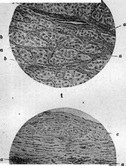
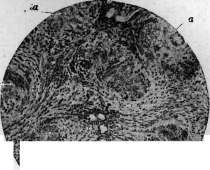
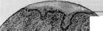

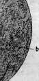
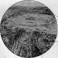
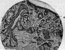
Figure 1. Rheumatic sclerosis; a-proliferation of connective tissue; b-hypertrophied muscle fibers. Figure 2. Rheumatic nodules (a) in the subcutaneous tissue in the stage of substrate disorganization. Figure 3. Rheumatic nodules in the myocardium (a); b-infiltrate in the myocardium; c-sharply thickened sclerosed endocardium. Figure 4. Rhinoscleroma of the larynx: a-epithelium (atrophic); b-diffuse inflammatory infiltrates. Figure 5. Rhinoscleroma of the nose: a-diffuse cicatricial sclerosis of the mucous membrane, epithelium absent; b-infiltrates in the depths. Figure 6. Rhinophyma of the nose: a-epithelium, b-proliferated sebaceous glands. For the most part, this involves the reverse collagenous transformation of fibrinoid-swollen connective tissue with the formation of coarse, collagenous strands. The continuing loss of cells and hyalinization of the connective tissue bundles leads to the formation of a scar. The direct transition of fibrinoid-swollen tissue, which has a structureless, seemingly necrotic appearance, into collagenous, cicatricial connective tissue is especially well manifested in large rheumatic nodules with a small cellular zone, as well as in so-called primary scleroses (see below). It is interesting that during relapses of acute rheumatism, the process is often selectively concentrated in the scars remaining from the previous attack of the disease. In addition to the above-described changes, which undergo three successive phases, in acute rheumatism, there are changes with a different course. First of all, it can be noted that small foci of mesenchymal disorganization, the changes in which correspond to the first phase, i.e., the early rheumatic infiltrate, can undergo reverse development and disappear without a trace. Furthermore, sometimes a complete absence of the second phase, i.e., the cellular reaction, is observed, and a direct transition of the first phase into the third; in this case, there occurs a renewed transformation into collagenous connective tissue of the disorganized, fibrinoid-swollen, and myxomatous edematous substance—a "primary rheumatic sclerosis" according to Talalaev. Finally, in various organs during acute rheumatism, focal and diffuse small-cell infiltrates, rich in vessels, can occur, subsequently turning into angiomatous scars (Talalaev). In acute rheumatism, the above-mentioned changes can be widespread throughout the body or affect only individual organs and systems. In each organ, in each tissue, these changes acquire their own special shade. The cardiovascular system is affected with the greatest constancy in acute rheumatism. In the myocardium, depending on the period of the disease, either small foci of fibrinoid swelling, or typical Aschoff nodules, or finally, sclerotic foci of a stellate type with rays penetrating into the surrounding muscle tissue are found. These changes are found in the interstitial connective tissue, more often near vessels, sometimes in the adventitia of the latter; sometimes the nodule involves the muscle tissue with its periphery, which in such a place undergoes death and disintegration. The posterior wall of the left ventricle near the bicuspid valve and the upper part of the interventricular septum are affected most frequently and intensely, to a lesser extent the anterior wall of the left ventricle, the region of its apex, and the lower parts of the atrial walls. In the wall of the right heart and the upper parts of the atria, changes are rare. In addition, small-cell infiltrates can be found in the myocardium. All this gives a picture of rheumatic myocarditis. With rheumatic involvement of the coronary arteries (see below), in connection with the narrowing of their lumens, atrophy and necrosis can occur in the muscle with the subsequent development of fibrous calluses (cardiosclerosis). In the endocardium, especially of the left ventricle and atrium, in acute rheumatism, one usually finds foci of myxomatous edema and fibrinoid swelling, as well as typical Aschoff nodules located in the subendocardial connective tissue. These changes lie at the basis of subsequent primary and secondary scleroses of the endocardium, expressed in whitish thickenings of it, sometimes wedge-shapedly penetrating into the underlying muscle tissue. The valves, mainly of the left heart, are affected very often in acute rheumatism, and in them, one notes the development of either simple verrucous endocarditis, which has nothing specific in it and is expressed in the deposition of thrombotic masses on the valve, which subsequently undergo organization, or diffuse rheumatic endocarditis, in which rheumatic disorganization, myxomatous edema, fibrinoid swelling of the valve tissue, and the formation of Aschoff granulomas in it occur; these degenerative-proliferative processes lead to the formation of protruding warts on the valves, and upon subsequent transition to sclerosis, they are the cause of the disfigurement of the valves, leading them into a defective state. A similar change also affects the chordae tendineae of the valves, which undergo thickening and sclerosis as a result. In the pericardium, as in other serous cavities (pleural cavity, less often the peritoneum), exudative serous and fibrinous inflammations (rheumatic serositis) develop, the peculiarity of which is the presence of foci of rheumatic disorganization in the subserous connective tissue. In rheumatic pericarditis, an abundant number of Aschoff granulomas often develop in the epicardium, sometimes merging into an almost solid granulation tissue of large basophilic cells, penetrating into the fibrinous effusion in strands. The processes of organization of the exudate leave behind thickenings of the serous membranes, synechiae, and obliteration of the cavities. The pathological anatomy of rheumatic lesions of blood vessels has been especially developed in recent years by Klinge and Vaubel and Rössle. They found that in acute rheumatism, foci of fibrinoid swelling, turning into sclerosis, and sometimes the formation of Aschoff nodules, can be observed in the wall of the aorta and arteries. Sometimes the changes resemble nodular periarteritis, others show a similarity to Buerger's vasculitis, differing from it by a low tendency to thrombosis and the development of aneurysms. Fahr believes that some of the cases of malignant nephrosclerosis are based on rheumatic arteriolonecrosis. Finally, rheumatic lesions of the arteries can have a similarity to those arteritides that are encountered in a number of septic diseases (in streptococcal sepsis, scarlet fever, typhus). The changes in the arteries in all these diseases are united by the common essence of the process of disorganization in the form of fibrinoid swelling with a cellular reaction. This gives Rössle reason to speak of a group of hyperergic rheumatoid diseases of the arteries, which includes rheumatic and streptococcal arteritides, Buerger's vasculitis, periarteriitis nodosa, and arteriolonecroses. As a result of rheumatic lesions of the arteries, sclerosis of the arterial wall develops; if it has a widespread character, this gives grounds to speak of rheumatic arteriosclerosis. In the veins, rheumatic changes are not rare (Rössle). They are expressed in the appearance in the walls of the veins of foci of fibrinoid swelling with proliferation of the inner lining and subsequent sclerosis; sometimes thrombosis develops (rheumatic thrombophlebitis). In large veins, especially in the region of their valves, verrucous growths—verrucous endophlebitis—are not rare. Rössle admits the possibility of rheumatic involvement of the veins of only one organ, e.g., the liver—rheumatic obliterating endophlebitis of the hepatic veins. In the affected joints during acute rheumatism, serous or sero-fibrinous effusion is observed. In the synovial membrane, foci of myxomatous edema of the connective tissue, hyperemia, and often plasma infiltrates and lymphoid cuffs around the vessels are found; true rheumatic nodules are rarely encountered here. In contrast to this, in the periarticular connective tissue, there is the development of various rheumatic nodules of different sizes, which in the early period present a picture of fibrinoid swelling of the connective tissue, and subsequently reactive proliferation of cells with an outcome in sclerosis. In aponeuroses, fasciae, tendon sheaths, and tendons during acute rheumatism, the formation of small and large nodules of typical structure and course can occur; sometimes in one place, e.g., in the galea aponeurotica of the skull, a large number of nodules are concentrated. In the tendon sheaths and mucous bursae, in addition, villous granulation growths can be observed. Skin involvement in acute rheumatism is expressed in the formation, most often near the joints, of large rheumatic nodules, characteristic of rheumatismus nodosus. In some cases of acute rheumatism, inflammatory changes of the erythema type (erythema exsudativum multiforme, erythema nodosum) are observed, as well as hemorrhages (peliosis rheumatica). The voluntary musculature likewise participates in acute rheumatism. Most often, the muscles lying close to the joints, the muscles of the fauces, pharynx, and the diaphragm are affected.
Here, foci of waxy degeneration and disintegration of muscle fibers are observed, and at the same time, fibrinoid swelling of the connective tissue is visible in such a place; this is followed by a cellular reaction, which leads to the formation of a rheumatic nodule that transitions into a scar. Large rheumatic nodules can develop in the tendinous center of the diaphragm. In the skeletal system, only the formation of rheumatic nodules in the periosteum is well known. Regarding the nervous system in acute rheumatism, pathological changes have been studied only in relation to the peripheral nerves; in the periphery, and sometimes in the endoneurium of peripheral nerve trunks, rheumatic changes of small vessels with lymphocytic infiltrates around them are observed: Köppen, in 4 cases of severe sciatica in rheumatic patients, found such changes in the sciatic nerve. In the central nervous system, and especially in the sympathetic ganglia, only degenerative necrobiotic changes of nerve cells are detected. However, in chorea minor (chorea rheumatica), vascular changes are observed in the brain that are close to the general vascular involvement in acute rheumatism (Santha, 1933). In the digestive organs, changes are encountered mainly in the upper part of the digestive tract; in the connective tissue under the mucous membrane of the tongue, tonsils, pharynx, and esophagus, as well as in the connective tissue surrounding the tonsils and extending along the pharynx and esophagus, rheumatic nodules are constantly encountered, either in the form of foci of fibrinoid swelling of the connective tissue, or in the stage of granulation nodules, or as limited scars. Focal changes of the type already described are also encountered in the muscles of the fauces, pharynx, and esophagus. Changes in the tonsils attract particular attention. The fact that rheumatic nodules are observed here with great constancy and already in the very earliest periods of the disease, in connection with the frequent onset of the disease with tonsillitis, led Graff to recognize the changes in the tonsils in acute rheumatism as a 'primary rheumatic infect,' which, however, is not accepted by the majority of researchers. In the liver, Klinge describes foci of necrosis of hepatic cells with an accumulation of leukocytes in the capillaries, and Rössle notes foci of fibrinoid swelling under the serous covering, as well as changes in the hepatic veins (see above). In the muscles of the larynx, nodules characteristic of changes in muscle tissue are encountered; in the mucous membrane of the nasopharynx, larynx, and also in the connective tissue surrounding them, typical rheumatic nodules are observed. In the lungs, no special rheumatic changes are known, except for the occasional presence of Aschoff bodies in the outer coat of the branches of the pulmonary artery and the already described involvement of small vascular branches. In the spleen, foci of fibrinoid swelling are sometimes noted in the trabeculae, in the capsule, and also in the walls of the arteries. In the lymph nodes, hyperplasia is observed; some point to the frequency of erythrophagia by the sinus endothelium. In the kidneys in acute rheumatism, focal lymphocytic foci near vessels and glomeruli are not uncommon; the picture of focal glomerulonephritis is detected more rarely. Finally, changes in small arteries of the arteriolonecrosis type are described, and sometimes the development of granulation nodules with giant cells around such arteries. Fahr, as already indicated, classifies some forms of malignant nephrosclerosis under acute rheumatism. All the above-described changes in acute rheumatism can be combined in the most diverse ways with a preference for certain organs and systems. This gives rise to various clinical forms of acute rheumatism (see below).
A. Abrikosov. Symptoms and course. The course of Acute Rheumatism and its clinical picture in various periods of the disease and in individual cases can vary greatly. And in this respect, Acute Rheumatism resembles those two most important social diseases, in which the allergic restructuring of the organism also has decisive significance—tuberculosis and syphilis. In contrast to these two diseases, we do not yet have sufficient data in Acute Rheumatism to divide the course of the disease into various phases, periods, or forms. This depends to a significant degree on the fact that the understanding of Acute Rheumatism as an allergic reaction of the organism to a streptococcal infection (or to a pathogen as yet unknown) is still in the stage of initial development. For now—as a hypothesis—one can outline only the following main periods: 1) a period of diseases preparing the allergic state of the organism in relation to the given (streptococcal) infection; 2) a period of initial allergy, manifesting for the most part, and especially in adults, as typical acute rheumatic polyarthritis and myocarditis, while in children and at a young age, predominantly as a disease resembling attenuated sepsis (endo-, myo-, pericarditis, pleurisy, polyarthritis); 3) a chronic period of relapses, resp. exacerbations of established allergy with a primary localization of lesions in the cardiovascular system, mainly in the endocardium and myocardium. In any case, in the course of each individual disease of Acute Rheumatism—if all relapses, resp. all manifestations of Acute Rheumatism in one and the same person are considered merely as separate attacks or manifestations of one and the same disease—one can outline certain regularities in the sense that 1) in adults, articular phenomena are initially more pronounced and more frequent, while subsequently, the manifestations of the disease from the heart gradually acquire a dominant position, whereas the involvement of the joints recedes into the background; 2) cases with visceral localization of the rheumatic process (so-called non-articular forms) proceed significantly more severely and leave behind (apparently even in relatively mild, so-called ambulatory cases) more severe consequences in terms of heart damage than cases with articular localization of Acute Rheumatism; 3) in childhood and, less pronouncedly, in adolescence, the disease proceeds more severely (see statistics), with damage to the heart and internal organs generally being both more frequent and more pronounced, while articular phenomena are either entirely absent or less pronounced; conversely, in persons of older age groups during the first attacks of Acute Rheumatism, joint involvement is usually much more pronounced, while visceral manifestations are both rarer and weaker; 4) within the limits of each attack of Acute Rheumatism, the most severe from a clinical point of view, and the one that gives the greatest number of clinical manifestations, is the period that corresponds to the first, so-called "exudative-degenerative" phase of pathological-histological changes in the tissues, in particular in the myocardium. These are so far only the first emerging regularities in the clinical course of Acute Rheumatism. In any case, the only scientifically correct division of Acute Rheumatism into phases and forms will be one that is based on the immuno-biological regularity of the given infection. Therefore, the existing divisions of Acute Rheumatism into various forms, being based on purely external signs, are little satisfactory. Thus, according to the clinical course, they speak of acute, subacute, and chronic forms; of primary acute and secondary chronic rheumatism; of mild, moderate, and severe forms; of a generalized form; of a relapsing, continuously relapsing form. Regarding clinical-anatomical manifestations, Talalaev speaks of polyarthritic, cardiac, ambulatory, and myocardial forms, with their complication sometimes by nodular rheumatism, chorea minor, erythema nodosum. Regarding the clinical localization of rheumatic lesions, they speak of peripheral or open and visceral forms (French authors, Buchstab), and within each of these forms, they speak of articular, nodular, or nodosum (in the peripheral form), or of cardiac, cardiovascular, pulmonary, cerebral, spinal rheumatism, chorea minor, hyperpyretic, typhoid forms (in visceral rheumatism). The Committee for the Fight against Rheumatism in the USSR has adopted the classification presented in the article Rheumatism (see). All these subdivisions, as they schematize—some to a greater, others to a lesser degree—the clinical picture of the disease according to purely external signs, are to a certain extent artificial. In the clinic, one can always find a mass of transitional cases from one form to another, and these cases, which perhaps constitute even the majority, can often with equal right be attributed now to one, now to another of the listed forms. In connection with this, the clinical picture of Acute Rheumatism will be set forth hereafter in its entirety, with the highlighting of descriptions of rheumatic lesions of individual organs or systems. Very often—one can even say in the majority of cases—Acute Rheumatism develops after a recently suffered acute inflammation of the palatine tonsils (angina) or catarrhal inflammation of the respiratory tract, significantly more rarely after other infectious diseases. These diseases precede the development of pronounced phenomena of Acute Rheumatism often by a few days, but not infrequently by 1-2, or even 3 weeks or more. Müller (Fr. Müller) notes that in persons who have never previously suffered from Acute Rheumatism, the interval between the preceding disease and acute rheumatism fluctuates between 2-3 weeks; if, however, the given person falls ill with angina again while already having Acute Rheumatism, then the period between each new angina and a new exacerbation of Acute Rheumatism becomes ever shorter and can reach a few days. A preceding disease to Acute Rheumatism can be, besides those mentioned above, conjunctivitis, sinusitis, otitis, and diseases in the area of the gums and teeth (Passler), and gastrointestinal disorders, etc. Most often, however, as already indicated, Acute Rheumatism is preceded by a lesion of the upper respiratory tract and, mainly, the palatine tonsils, the area of which is considered by the majority of authors as the main "portal of entry" for infection in Acute Rheumatism. The frequency of preceding tonsillar lesions in Acute Rheumatism, according to individual authors, reaches up to 70-80% of all cases of Acute Rheumatism with articular phenomena (Kingston, Fowler, Stewart, Garrod, Koritsky, and others); the majority of authors, however, give significantly lower figures (25% according to Garrod, no less than 1/3 of cases according to Saint-Germain, 35% according to our data, 2/3 according to Widal, etc.). The extreme frequency of changes in the tonsils, indicating the presence of chronic or frequently recurring tonsillitis, in rheumatic patients upon their admission to the hospital (according to our data, it reaches 84.9%) gives reason to think that many of these tonsillar lesions pass unnoticed by the patients themselves. Koritsky finds suppurative foci in the tonsils in 72% of all cases of Acute Rheumatism with protracted articular phenomena. According to the latter author, the suppurative process can nest, besides the tonsils themselves, also in the tonsillar bed, in the peritonsillar crevices, and in the peritonsillar tissue; the presence of pus in the peritonsillar space can sometimes be detected only with the help of rotation of the tonsils with retraction of the anterior pillar and squeezing of pus from the peritonsillar crevices. The same author draws attention to the almost regular presence (in 85% of cases of Acute Rheumatism with protracted articular phenomena) of inflammatory changes in the lymphatic vessels and glands draining lymph from the upper respiratory tract, in the form of the so-called "cervical lymphatic ridge"; these vessels are located along the deep jugular veins and are palpable along the upper third of the anterior border of the sternocleidomastoid muscle (gland. lymph. cervical. prof. super.); similar changes were found by him also in the retromandibular glands. Almost systematically, in the pus from the tonsils, this author found hemolytic streptococcus, and sometimes also Strept. viridans. In a portion of cases of Acute Rheumatism, tonsillitis develops simultaneously with the appearance of articular lesions, or after their onset, or even when the rheumatic process has already seemingly subsided. In these cases, usually, after a very short time after the angina, a new exacerbation of Acute Rheumatism occurs. It is undoubted that a portion of the anginas observed in Acute Rheumatism (first of all, anginas during Acute Rheumatism and, it must be thought, a portion of the anginas preceding the development of a repeated attack of Acute Rheumatism) are specifically rheumatic, i.e., are one of the many possible manifestations of Acute Rheumatism, as has already been proven by special histological studies (Graff, Klinge, Sarafoff). Clinically, anginas in Acute Rheumatism, especially anginas developing during Acute Rheumatism, usually have a catarrhal character; less often lacunar anginas, and relatively very rarely are the developments of Acute Rheumatism preceded by phlegmonous anginas. French authors (Saint-Germain, Lasègue, and others) note diffuse redness in this case not only of the tonsils but also of the soft palate, the pillars, and the posterior wall of the pharynx, with a gradual transition of the redness, sometimes quite intense, into the normal color of the mucosa. Lasègue and Fernet note some edematous swelling of the affected mucosa.
It can be considered with certainty that if not all, then at least many of the anginas preceding Acute Rheumatism are contagious, as has been proven with certainty by a number of observations of tonsillitis epidemics, followed by the development of Acute Rheumatism (Collis, Sheldon, Coburn, and others). Whether anginas that develop during Acute Rheumatism—i.e., anginas in the majority of cases in all probability specifically rheumatic—are contagious is unclear. The disease of Acute Rheumatism itself quite often begins acutely without prodromal phenomena: 2-3 weeks after one or another of the aforementioned preceding diseases, when the patient considers themselves already recovered, suddenly, often with chills, the temperature rises (up to 38-39-39.5°, rarely higher) and simultaneously with this, or more often very soon after, joint lesions appear with swelling, often redness, always with sharp pain in the affected joints and complete impossibility of active movements in them. As a rule, joint lesions, starting usually with large joints, more often of the lower extremities, from the very beginning involve several joints; subsequently, as the process subsides in one joint or group of joints, the lesion begins to spread to other joints without any order, giving the entire clinical picture of the disease an extremely peculiar course characteristic of Acute Rheumatism. Much more rarely, the disease, having begun acutely, proceeds for the first time—usually not for long (3-5-7 days)—with only general phenomena (high temperature, severe sweating, general malaise, loss of appetite, etc.) without a definite, clearly ascertainable localization, and can, by its course, lead the physician to think of typhoid fever—the typhoidal form of Gränet—or some other acute infection. Usually, subsequently in these cases, joint lesions soon join, but often they are less pronounced and less generalized than where the rheumatic process clinically begins with them. In other cases, joint phenomena are very delayed or do not appear at all, and instead, symptoms of damage to various internal organs develop—most often the heart, sometimes with phenomena of heart muscle insufficiency. In much rarer cases, however, damage to some other organs may be revealed, for example, serous membranes (pleura or pericardium), rarely damage to the lungs, and apparently even more rarely to the parenchymal organs of the abdominal cavity (kidneys, liver); sometimes, to the initial general phenomena, symptoms of damage to the central or peripheral nervous system are added. In children, severe abdominal pain in the initial period of the disease can often lead to thoughts of appendicitis. In such cases, only the further course of the disease with the appearance of joint lesions allows for a correct diagnosis. Where the disease proceeds from beginning to end without joint lesions or with very weak pain in the joints (this occurs more often in childhood), the disease may remain unrecognized altogether. In other cases, and these cases perhaps constitute the majority, Acute Rheumatism begins gradually, more often also after a preceding angina or catarrh of the upper respiratory tract; such a gradual onset is observed especially often in cases without joint lesions, with the main localization of the rheumatic process in the cardiovascular system. In these cases, the development of a pronounced clinical picture of Acute Rheumatism is often preceded by general vague malaise, often mild pulling pains in the limbs, a feeling of discomfort in the joints during movement, aching in the body, lethargy, weakness, rapid fatigue, loss of appetite, sometimes headaches, sleep disturbances, nosebleeds, sweating; children often lose their mobility, interest in games, become dull, and turn pale. When measuring temperature, in such cases, one can often detect a subfebrile temperature (according to Kahler, an elevated temperature can precede the development of distinct joint lesions by 7 or more days). After these prodromal phenomena, in no less than half of the cases, a characteristic picture of Acute Rheumatism develops with multiple acute joint lesions (see below). The temperature, with the appearance of an acute rheumatic joint lesion, rises immediately or gradually to 38-39.5° and holds with morning remissions the entire time that inflammatory phenomena are present in the joints. Its height is often in a certain correspondence with the number and degree of joint involvement. As the joint process subsides, if there are no distinct lesions of internal organs and in particular the heart, the temperature gradually falls. In some cases, the temperature falls in parallel with the decrease in joint effusions, and usually remains at subfebrile levels for some time (sometimes only a few days) after the complete clinical subsidence of joint phenomena. In mild cases, the rheumatic process ends clinically with this, sometimes taking only 5-7 days. Incomparably more often, however, following the first flare-up, usually even before all its clinical manifestations have completely disappeared, and sometimes even after their seemingly complete subsidence, new exacerbations of the rheumatic process occur with damage to new, or even previously affected, joints or with damage to internal organs, most often the heart. In this case, the temperature again shows a significant rise. A rise in temperature (usually, however, less sharp) can often be observed even without any distinct localization of Acute Rheumatism. In these cases, it should almost always, apparently, be attributed to rheumatic myo- or endocarditis. One should always think about the involvement of the heart also in the case of a discrepancy between the degree of temperature rise and its duration, intensity, and joint phenomena. In general, the temperature curve in Acute Rheumatism usually has an uneven course with individual rises corresponding to the exacerbation of the rheumatic process. As a rule, it does not exceed 38-39°, which can be partly explained by a sharp increase in heat dissipation in connection with the increased sweating characteristic of such patients. Cases with a temperature of 40-41° are currently extremely rare and belong to the so-called hyperpyretic form of Acute Rheumatism (see below). It is undoubted that the type of temperature curve is currently highly influenced by those medicinal (primarily salicylic) preparations that are used for Acute Rheumatism and which almost all reduce temperature to one degree or another. The duration of the temperature rise is extremely different in individual cases—from a few (usually no less than 5-7) days to many months. Even a complete return of temperature to normal does not yet speak of the complete subsidence of the process and does not guarantee against a new exacerbation of the rheumatic process in the near future. Only a stably normal character of the temperature for several (no less than 2) weeks in the absence of other symptoms indicating the activity of the process allows one to think about its subsidence. Practically important is the circumstance noted by Talalaev that temperature rises usually correspond to the first stage of "exudative-degenerative" changes in Acute Rheumatism and that subsequent stages of the organization of the process (lasting up to 4 months) can proceed without rises. No less, if not more important, is the circumstance that in cases of recurrent Acute Rheumatism, with the localization of the rheumatic process in the cardiovascular system without joint phenomena and without damage to the pleura and pericardium or with only insignificant phenomena in them, the temperature can be very low (in the range of 37-37.5°); in some cases of latent Acute Rheumatism, it may not exceed 37 at all, and only its uneven type with large daily fluctuations, in conjunction with other symptoms, leads to the thought of the presence of Acute Rheumatism. Nevertheless, it is necessary to note that of all the symptoms of this disease in its active stage, a rise in temperature is the most constant symptom. An extremely characteristic symptom for Acute Rheumatism, especially in the presence of joint lesions, is the already mentioned extreme sweating of patients, in some cases especially sharp in the second half of the night, when the temperature falls (Weintraud). The sweat is usually acidic, much more rarely—after special cleaning of the skin—of neutral reaction, and has a peculiar "mousy" odor. Sweating, which is apparently one of the manifestations of damage to the autonomic nervous system, is observed in almost all cases of Acute Rheumatism with joint lesions where there is a more or less significant rise in temperature. Severe sweating in the presence of only small rises in temperature, according to Weintraud, is prognostically unfavorable, being a sign of a particularly persistent course of Acute Rheumatism. Often, the sharp pallor of acute rheumatics is striking, often not corresponding to the degree of their anemia. Usually, from the end of the first week (rarely earlier) and especially from the second week of the disease and later, a number of symptoms often appear that speak for damage to internal organs, especially the heart. These include muffling of heart sounds, systolic apical murmurs, often unstable and variable, disturbances in the frequency and rhythm of the pulse, more rarely changes in the borders of the heart, enlargement of the liver and spleen, often the appearance of protein and erythrocytes in the urine, urobilinuria, phenomena from the lungs, sometimes the nervous system, etc.
As a rule, these lesions are very unstable; appearing quickly and increasing in their intensity, they for the most part pass quickly without a trace. Only symptoms from the heart often acquire a persistent character; if in the initial acute period of the disease they must be attributed mainly to rheumatic involvement of the myocardium, then the persistence of these symptoms in the later period of the disease, especially starting from its 8th-9th week, as well as their appearance in this period in the absence of a noticeable exacerbation of the rheumatic process, speaks for the presence of rheumatic endocarditis (see). It is necessary, however, to note that often symptoms from the heart can be very slightly expressed, as if all other manifestations of Acute Rheumatism are subsiding, and the doctor, considering the patient healthy, discharges him, and after a few months the patient is found to have a heart defect that was not previously present, as a result of an imperceptibly occurring rheumatic endocarditis. Often from the very beginning, Acute Rheumatism (most often in the form of endocarditis) proceeds almost imperceptibly and is endured by the patient on his feet (the so-called 'ambulatory' form of Acute Rheumatism by Kutyrin). In these cases, along with vague general symptoms (slight weakness, rapid fatigability, mild general excitability with a generally good state of health, subfebrile temperature), there are also noted slight shortness of breath, a 'sensation of the heart by the patient', significant lability of the pulse, which does not disappear even in a lying position, tachycardia, impurity of tones, at times a clear apical murmur; in the blood—slight leukocytosis with slight eosinophilia, sometimes with mild neutrophilic leukocytosis and a nuclear shift to the left. Very often one can observe how a patient, whose temperature has already subsided, whose symptoms of joint involvement have disappeared, and who has already begun to get out of bed, turns out to be struck by a new wave of Acute Rheumatism, forcing him to lie in bed again. In general, the shorter the duration of the rheumatic process and the milder it proceeds, the fewer such exacerbations; in very mild cases of Acute Rheumatism, they may not occur at all. Patho-anatomically, such clinical flare-ups of the process, as shown by the studies of Talalaev, apparently correspond to the appearance of new foci of rheumatic inflammation in the myocardium; it also seems quite probable that they appear in a number of other organs besides the heart (for example, in the periarticular tissue, etc.). In relatively rare cases (in childhood and adolescence), periodically occurring exacerbations follow one after another with very short intervals over the course of a number of months; these cases, always with more or less severe heart damage, as a rule, quickly lead to death. Talalaev defines the duration of the course of this 'continuously recurring' form of Acute Rheumatism from 5 months to 1 1/2 years. An intermediate position between these cases of Acute Rheumatism and mild cases, where after 3-5 weeks the rheumatic process clinically seems to end completely, is occupied by those fairly frequent cases where, after a series of exacerbations, the rheumatic process does not result in complete clinical recovery, where symptoms of heart suffering remain, and, less often, painful and functional changes in the joints (usually without any significant objective deviations from the norm). Here we are dealing with the transition of the rheumatic process into a subchronic and even chronic course (the so-called secondary chronic rheumatism). If in such cases we are talking only about joint phenomena, which is rare, then cautious (in the absence of elevated temperature) application of further physiotherapeutic methods of treatment as a rule gives a fairly quick complete return to normal. In cases of a rheumatic process that has passed into a chronic course with localization in the joints, but without heart involvement (first of all without endocarditis), usually, under the condition of proper treatment and prolonged bed rest, it is possible to achieve (easier in adults, harder in children and adolescents) a favorable clinical completion of the process. In those cases where the rheumatic process has caused endocarditis, the disease, upon the cessation of the activity of Acute Rheumatism, ends as a rule with a remaining valvular heart defect. Involvement of the myocardium by the rheumatic process affects the course of the disease unfavorably usually only in those cases where the myocardium, either by virtue of past diseases or by virtue of prolonged increased load, as is the case with a long-standing defect, is already significantly damaged or weakened. In these cases, a picture of increasing heart failure may develop, and the patient may die. Only in cases of severe damage to the conduction system of the heart can the patient quickly die from rheumatic myocarditis with an otherwise little-damaged myocardium and in the absence of valve involvement. Finally, in individual cases, the rheumatic process that has passed into a chronic stage gradually takes on a clearly septic course, clinically giving the picture of sepsis, or endocarditis lenta. The direct etiological and pathogenetic connection between sepsis lenta and the rheumatic process has not yet been generally recognized to this day, however, both clinical observations (Lang, Strazhesko, et al.) and bacteriological (Rosenow, Freund and Berger, Clawson-Bell, et al.) and morphological (Talalaev, et al.) findings in both of these processes point to the absence of any clear boundary between them and to the sometimes completely imperceptible transition of a typical rheumatic process with myocardial involvement into a typical endocarditis lenta. This gives reason to think that both these diseases represent two forms of one and the same infectious process, taking on only a different clinical and morphological course in connection with a change in the ratio of forces between the microorganism and the macroorganism; apparently, this change in the ratio of forces is caused primarily by a change in the reactive capacity of the macroorganism, i.e., the transition of its hyperergic state in relation to the given infection into a hyperergic, or anergic state (see more details under Endocarditis, Sepsis). Once suffered, Acute Rheumatism leaves behind an increased predisposition to repeated illnesses with it, which constitutes an extremely characteristic feature of Acute Rheumatism. Church determines the frequency of relapses at 19.057%; according to Hegler, out of 142 patients with articular rheumatism in the Virchow Hospital, 82 were admitted for the first time and 60 repeatedly. According to Ilyinsky, out of 401 cases of Acute Rheumatism, only 135 patients had their first attack, 129 their second, and 137 their third or more attacks. It is necessary to point out that, as noted by Pribram, cases in private practice, which are under long-term observation, show a greater frequency of relapses than data from hospital observations, because cases of Acute Rheumatism proceeding sluggishly, with indistinct clinical symptoms, may not end up in the hospital. Thus, Vilk and Rabinovich (1932), having re-examined 90 cases of Acute Rheumatism in Moscow after discharge from the hospital, were able to detect in 20 individuals (i.e., 22%) an exacerbation of the rheumatic process with an increase in temperature in the period immediately following discharge, and all these cases of exacerbation did not end up in the hospital. The time of onset of a relapse after the first attack can be very different—from a few days to many tens of years. More often, however, relapses occur after a more or less short period after the first attack, and the tendency to recurrence of Acute Rheumatism is especially great in patients who have suffered rheumatic endocarditis during previous attacks of Acute Rheumatism (Weintraud, et al.). The severity of painful symptoms during relapses can be very different; in general, however, each new relapse worsens the further course of the disease and its outcome, sharply increasing the frequency and degree of heart damage in acute rheumatics. Thus, according to the material of Vilk and Rabinovich, the number of heart defects among those primarily suffering from Acute Rheumatism upon re-examination turned out to be 40%, while among cases that had relapses of Acute Rheumatism, it was 74%. Pribram notes the severe character of relapses of Acute Rheumatism in terms of heart damage after a previously suffered severe acute infection; according to this author, relapses of Acute Rheumatism during pregnancy and in the postpartum period are severe, as well as after intercurrent gonorrhea; in the latter case, Pribram allows for the possibility of a mixed infection. It is practically important that during subsequent relapses of Acute Rheumatism, joint phenomena are usually less pronounced and even often absent, as a result of which relapses of Acute Rheumatism can proceed even imperceptibly, often under the guise of banal heart failure. Factors causing relapses of Acute Rheumatism can be of the most diverse character, including, apparently, both simple cooling, to which rheumatics are extremely sensitive, and all kinds of infections, most often, however, infections of the upper respiratory tract, in particular the tonsils. Physical overexertion, especially in a person who has not yet fully recovered from a previous attack of Acute Rheumatism, can also apparently contribute to the onset of a relapse; in connection with this circumstance, one must place the very frequent relapses of Acute Rheumatism in individuals who were discharged early from a medical institution and, most importantly, sent to work early.
Thus, Meerovich, having re-examined 90 workers of the Izhorsky Metallurgical Plant who had suffered from acute rheumatism, established relapses of the disease in 45% of individuals who were discharged to work 15 days after the disappearance of clinical signs of the disease (!). In some cases, however, no connection between the relapse and either infection or unfavorable external circumstances can be established. In acute rheumatism, the greatest significance lies in the involvement of the entire cardiovascular apparatus, since this localization of acute rheumatism is not only the most frequent but also leads to the most substantial disturbances of circulatory function. If acute rheumatism is a severe disease, often leading to death or disability, it is precisely due to the involvement of the cardiovascular apparatus. In terms of its practical significance in the sense just indicated, the first place among the localizations of the process in the cardiovascular system should be given to the involvement of the valvular endocardium, i.e., rheumatic endocarditis (see), since it almost always leaves behind valvular defects. Involvement of the heart muscle in acute rheumatism occurs even more frequently than involvement of the endocardium. It was first described by Itard (1824), but more or less frequent references to it in the literature begin to appear only from the end of the 19th and especially from the beginning of the 20th century (mainly after 1904, when Aschoff described the changes in the myocardium specific to acute rheumatism); recently, it has been attracting more and more attention from clinicians both in connection with its exceptional frequency in acute rheumatism and in connection with the significance that the state of the rheumatic patient's heart muscle has for his entire future fate. Both pathological-anatomical studies and clinical, and in particular electrocardiographic, observations speak to the extreme frequency of myocardial involvement in acute rheumatism. Individual pathologists (Thalheimer and Rothschild, Talalaev) even speak of the obligate nature of inflammatory lesions of the heart muscle in acute rheumatism; according to electrocardiographic data, the frequency of myocardial involvement fluctuates between 74% and 100%. With ordinary clinical methods of examination, it is also not infrequently possible to detect the presence of myocarditis in acute rheumatism, but in a significantly smaller percentage of cases. Clinical assessment of the significance of myocardial involvement is generally difficult, and in a disease that simultaneously very often causes severe involvement of the valvular apparatus as well, it is even more so. No less frequent in acute rheumatism is the involvement of blood vessels, especially arteries, but it is even more difficult to assess the clinical significance of this localization at the present time. Undoubtedly, acute rheumatism affects vessels of all calibers, but involvement of large arteries, in particular the aorta, relatively rarely gives rise to pronounced clinical phenomena; as for small vessels, the localization of foci of the lesion (infiltrates, granulomas) in their walls—in the adventitia and in the periadventitial tissue—is undoubtedly favored by acute rheumatism. Undoubtedly, these small foci often lead to a temporary or permanent disturbance of the vessel's function in the sense of narrowing its lumen or closing it, and this is the case more often the smaller the caliber of the vessel. The more numerous and densely packed these foci are, and the greater the functional significance of the tissue nourished by the affected vessels, the more substantial will be the disturbances of the functions of the corresponding organ or tissue. Thus, we are able to judge these lesions of small vessels only by the disturbance of the functions of the corresponding organ, and it is natural that we cannot clinically distinguish them from the involvement of the organ itself, or its connective tissue, by the same process. But insofar as acute rheumatism is characterized precisely by the involvement of the mesenchyme—interstitial tissue and vessels—we have the right to assume that the manifestations of this disease on the part of internal organs represent to a significant extent the result of the involvement of the small vessels of these organs. What has just been said applies primarily to the involvement of the myocardium, in relation to which it was established relatively long ago that rheumatic granulomas are localized in the walls of blood vessels or in the connective tissue surrounding them. Another feature of the localization of rheumatic foci in the heart muscle, which has clinical significance, is their predominant localization under the endocardium and in certain areas of the heart muscle (see pathological anatomy of acute rheumatism). The symptoms of rheumatic myocarditis are generally similar to the symptoms of myocarditis of other etiologies (see Myocarditis). From the point of view of the concept of acute rheumatism as a process that produces focal involvement of the myocardium, a certain part of the disturbances of heart muscle function observed in acute rheumatism and the clinical symptoms caused by them is understandable (see below). But besides the symptoms that can be considered without any strain as a manifestation of focal myocardial involvement, symptoms are observed in the heart in acute rheumatism that are most natural to interpret as manifestations of diffuse involvement of the heart muscle. This includes, first of all, those auscultatory and percussive changes in the heart that are so frequent in the initial acute, or alterative-exudative, phase of the rheumatic process, which usually proceeds with high temperature and multiple joint involvement, namely: muffling of heart sounds, a slight increase in its dullness, very often apical systolic murmurs (according to West, Coombs, Ilyinsky, and Nikolaeva, they are observed in 75% of all cases of acute rheumatism, and in children apparently even more often), and less frequently an accentuation of the 2nd sound over the pulmonary artery. This also includes a significant portion of the electrocardiographic changes so frequent in this same period, namely: low voltage, lowered or negative T wave. These early changes in the heart testify to a disturbance of the contractile function of the entire heart muscle; in other words, they speak of the frequent presence in the initial acute period of the rheumatic process of not only focal but also diffuse myocardial involvement. The fact that an exacerbation of the rheumatic process in the presence of an already pronounced valvular defect often contributes to the development of decompensation speaks in favor of the fact that the active rheumatic process, in addition to characteristic focal changes in the myocardium, also causes its diffuse involvement, which impairs its contractile function. Since focal and diffuse lesions are clinically seemingly connected with each other, it is simpler to consider the latter as caused by the former. The fact that manifestations of diffuse myocardial involvement in acute rheumatism are observed as a rule in the first phase of focal rheumatic lesions, i.e., during the time of focal exudative-alterative changes, also speaks in favor of this view. Focal changes in the myocardium are easiest to consider as a manifestation of the localization in the corresponding areas of an infectious agent penetrating via the hematogenous route. In the majority of cases, the symptoms of diffuse myocardial involvement in acute rheumatism, although observed very frequently, nevertheless do not have a sharply pronounced character and recede in their intensity into the background in comparison with the sharply pronounced joint phenomena. Of the manifestations of focal myocardial lesions in acute rheumatism, almost all relate to disturbances of the heart rhythm. This is quite understandable, since rheumatic foci, due to the fact that they are often located predominantly under the endocardium, affect the system that generates and conducts impulses. In addition, small rheumatic foci can easily disturb the function of various parts of this system, since the dimensions of these parts of the system are themselves very small. The most common disturbances of heart rhythm in acute rheumatism are tachycardia and bradycardia. The fact that these changes in pulse rate are caused not only by the general infectious state of the patient is evidenced by the circumstance that there is often no correspondence between the pulse rate and the rise in temperature. Systematic electrocardiographic study of acute rheumatic patients (Grotel, Lukomsky, et al.) shows that changes in the electrocardiogram, appearing in the early period of the disease, in most cases quickly return to normal as it subsides, but reappear with new exacerbations of the rheumatic process. In general, the duration of electrocardiogram changes is from a few weeks to 1-3 months, relatively rarely longer; at the same time, the intensity of the electrocardiogram changes, as well as their persistence, as a rule, correspond to the severity of the myocardial involvement in the given case in general. In relatively rare and usually more severe cases, the changes in the electrocardiogram do not have a tendency to regress and can be detected in a rheumatic patient many months and even years after his discharge from the hospital. Clinical symptoms of rheumatic myocardial involvement usually begin to appear from the 4th-7th day of the disease and especially from its second week. In some cases of acute rheumatism, the process begins almost directly with symptoms of myocardial suffering in the absence of or with the later appearance of joint lesions; in these cases, the recognition of the true nature of the pathological process is extremely difficult.
In rare cases, manifestations of cardiac muscle damage, proceeding with high temperature and a generally severe condition of the patient, stand in the foreground throughout the entire duration of the disease, accompanied by pronounced symptoms of heart failure; sometimes such myocarditis, especially in children with old rheumatic heart defects, leads to death. In general, however, as already indicated above, the severity of cardiac muscle damage in Acute Rheumatism is usually not great, and often even the intensity of clinical phenomena does not go beyond the limits of what is usually observed on the part of the heart during febrile states. Accordingly, the function of the cardiac muscle in rheumatic myocardial lesions in the majority of cases appears to be little impaired, and phenomena of any pronounced insufficiency are observed comparatively not so often. Symptoms of myocardial damage have a more pronounced character, as already mentioned above, in childhood, in persons with simultaneous endo- or pericarditis, and in persons with previously acquired rheumatic heart defects.
Differing in their rapid course and usually favorable outcome, rheumatic myocardial lesions can recur to no less, if not a greater degree than joint lesions, with the only difference being that while the severity and intensity of joint lesions generally tend to decrease during further recurrences and in more severe cases of Acute Rheumatism, the severity of symptoms of myocardial suffering during new recurrences of Acute Rheumatism generally increases, and the more so, the more severe this recurrence is and the more generalized the rheumatic process. However, this inverse parallelism in the intensity of joint and myocardial phenomena is far from absolute. A number of clinicians, mainly of the French school, consider the chronic course with periodic recurrences to be typical for rheumatic myocarditis. In any case, rheumatic myocarditis, recurring over many months and even years, often has a decisive significance for the fate of the patient, leading to heart decompensation and death.
Extremely important from a purely practical point of view are cases of latently proceeding rheumatic myocarditis, especially often observed apparently in persons with valvular rheumatic heart disease. These cases clinically proceed usually under the guise of banal heart decompensation of a valvular defect; and as a rule, the presence of an active rheumatic inflammatory process in them is not recognized; these myocarditis cases often develop as if without any visible cause, more often after minor infections—influenza, angina—and often (although not necessarily) proceed with subfebrile or even with "normal" temperature, i.e., at a temperature below 37°, but of an uneven, irregular type, with large daily fluctuations (by 1–1.5°). Joint lesions in such cases are often absent or are weakly expressed.
Usual heart therapy in such cases often does not give a significant effect. This lack of effect from cardiac agents in connection with an indication in the anamnesis of a previously suffered Acute Rheumatism and with subfebrile temperature often is the only thing that leads to the thought of the presence of active rheumatic myocarditis. Subjective sensations on the part of the heart in patients with Acute Rheumatism usually boil down to a sensation of palpitations, dull pain of a prolonged type, various unpleasant sensations in the heart region (a feeling of heaviness, pressure, constriction, stabbing, difficulty breathing, etc.). The intensity of these phenomena as a rule is not great, and by their character they do not go beyond the limits of those sensations which accompany acute and subacute inflammatory changes of the myocardium of other etiology and which are much more characteristic of so-called cardiac neuroses.
In some, comparatively rare cases of Acute Rheumatism, pains in the heart region reach significant strength and resemble pains of the angina pectoris type; occasionally they develop into attacks of severe status anginosus with all the typical clinical and electrocardiographic signs of myocardial infarction. These pronounced pain phenomena on the part of the heart must be attributed to the localization of rheumatic foci in the larger branches of the coronary arteries of the heart. It was already indicated above that vascular walls in general and the walls of the coronary arteries in particular are favorite places for the localization of rheumatic nodules or infiltrates.
Regarding the damage to the coronary arteries of the heart by the rheumatic process, there are already numerous pathological-histological data (Romberg, Aschoff-Tawara, Geipel, Coombs, Watjen, Talalaev, Klinge and Vaubel, etc.). These data, among other things, testify to the fact that damage to these arteries often leads to narrowing or even closure of their lumen. They fully explain the just-indicated pain phenomena of the angina pectoris type and the development sometimes of a picture of myocardial infarction. If clinical phenomena of this category are comparatively still rare in Acute Rheumatism, this is explained by the fact that rheumatic infiltrates and nodules disrupt patency apparently more often not of large, but of small branches of the coronary arteries of the heart, and this clinically gives not phenomena of angina pectoris or myocardial infarction, but phenomena of small-focal damage to the cardiac muscle and distinct clinical, and in particular electrocardiographic, phenomena only in the case if the nutrition of one or another part of the system producing and conducting impulses is disrupted, or perhaps in the case when these foci sit very densely.
With the localization of foci of rheumatic inflammation in the small branches of the coronary arteries of the heart, we will have, as already said above, the same clinical manifestations as with direct focal damage to the myocardium, i.e., with rheumatic focal myocarditis. Whether disturbances of blood supply to small areas of the myocardium cause these or those subjective and in particular pain phenomena, we do not yet know. It is necessary to add that clinical medicine until the very last time paid little attention to the manifestations of rheumatic coronary arteritis, and therefore there is no precise idea yet about the significance that Acute Rheumatism has in the origin of pathological phenomena on the part of the coronary arteries of the heart. If Kahn (Kapp) could establish the presence of Acute Rheumatism in the anamnesis in 20 out of 82 cases of angina pectoris and considers that it played an etiological role in these cases, then of course one cannot yet attach decisive significance to these data.
Undoubtedly, clinical medicine faces the task of more careful study of patients suffering from Acute Rheumatism or having suffered it, in relation to clinical manifestations of damage to blood vessels. Pathological-anatomical data of the very last time (Siegmund, Klinge, Vaubel, Rössle) give reason to believe that in the group of arteriosclerosis there is a certain part of sclerotic lesions of arteries as a result of the localization of the rheumatic process in them, that consequently from the group of arteriosclerosis it is necessary to isolate rheumatic arteriosclerosis, just as this was done at one time in relation to syphilitic. However, Hanot at one time expressed the thought about the possibility of the rheumatic origin of arteriosclerosis, especially in young people with Acute Rheumatism in the anamnesis.
Regarding rheumatic aortitis, besides pathological-anatomical data, there are already some clinical indications. Recently, cases of aortic aneurysms have been described that developed during Acute Rheumatism and, it must be thought, in direct connection with it in persons who had no grounds to assume the presence of syphilitic or any other infection. According to Widal and May, damage to the aorta itself in Acute Rheumatism is usually in connection with damage to the aortic valves. The diagnosis of rheumatic aortitis of course encounters great difficulties, especially if there is no aneurysmal dilation of the aorta. Still, in some cases it can be made with a certain probability.
In rare cases, one can clinically detect damage to other arteries; it is possible that attacks of acute abdominal pain (in connection with these pains, cases of laparotomy have been described), sometimes observed in Acute Rheumatism, usually at the beginning of the disease and more often apparently in children, are connected precisely with lesions of the mesenteric arteries. Damage to the arteries of the limbs can manifest itself, besides pain phenomena, also with a feeling of numbness and crawling of ants in the corresponding limbs with a decrease in pulsation more distally from the place of the lesion of the corresponding artery. Finally, a number of clinical cases of phlebitis during Acute Rheumatism have been described (Bouillaud, Trousseau and Peter, etc.), usually passing easily and quickly, but in individual cases multiple and bearing a severe character. As for places of particularly frequent damage to veins during Acute Rheumatism, Talalaev points to the area of venous valves.
Inflammatory damage to the joints in acute rheumatism (rheumatic polyarthritis, rarely monoarthritis) until recently was considered the main and mandatory manifestation of the disease. At the present time, it can be considered established that its absence in Acute Rheumatism is not rare (according to Talalaev, approximately in 40%), especially often apparently in childhood.
In adults, joint involvement in acute rheumatism is observed in more than half of all cases, although the intensity, prevalence, and persistence of the lesion can vary extremely in individual cases and even in the same person during different attacks of the disease. In general, however, it can be stated that the more attacks of acute rheumatism a given patient has suffered, the more the internal organs—and in particular the heart—are affected by the rheumatic process, and the more weakened the patient's organism is, the less frequently joint involvement is observed and the less pronounced its character is. Nevertheless, cases are not so rare where the disease begins with heart involvement and where joint suffering appears only during a new exacerbation of the rheumatic process. In children, complaints in acute rheumatism are often reduced to only vague aching or pulling pains in the joints in the absence of any pronounced objective signs of involvement, as a result of which their true character is very often not recognized. The time of appearance of joint lesions varies: more often, pronounced clinical phenomena in acute rheumatism begin precisely with joint lesions; significantly less often, rheumatic polyarthritis develops later. The main symptoms of rheumatic arthritis are: soreness, swelling of the affected joint, and often of the surrounding tissues, slight redness, an increase in the temperature of the skin covering it, and a disturbance of its function. Individual symptoms—more often skin redness, less often swelling—may be absent; there is always only soreness and the disturbance of function inseparably connected with it. With the development of rheumatic polyarthritis, pain and disturbance of function appear first, later swelling and an increase in the skin temperature over the joint, and finally redness; as the process subsides, the phenomena usually disappear in reverse order, although at times the pain may disappear while the swelling still continues to persist. The intensity and persistence of individual symptoms can be very different. Soreness is usually so intense that in the acute period of the disease it almost completely deprives the patient of the ability to move the affected joints; at times, even the touch of a blanket to these joints causes the patient great torment. The involvement of the joints of the spine is especially painful, forcing the patient into almost absolute immobility. Such a patient, in the words of Widal and May, lies motionless, like a statue, afraid to even attend to his natural needs, since any movements necessary for this cause him cruel suffering. The most painful places are the points of attachment of tendons and fibrous fasciae to the bones, which is in direct connection with the abundance of nerve endings in them, as well as with the very frequent involvement in the rheumatic process of the parts of the corresponding tendon sheaths adjacent to the affected joint (rheumatic tenovaginitis). Lasègue even claims that the pains in rheumatic polyarthritis are localized outside, and not inside, the joint and that the cause of the soreness is the involuntary tension by the patient of the muscles around the affected joint; this is easy to verify by forcing the patient to completely relax the muscles in the affected limb: in such cases, according to Lasègue, one can perform passive movements in the diseased joint without causing the patient any suffering. Complete immobilization of the affected joint and its protection from any external touches, according to Widal and May, instantly soothe the pains. In a number of cases, however, the pain phenomena may have such weak intensity that the patient completely retains movement in the joint. Often, for quite a long time after the passing of the acute phenomena of polyarthritis, patients complain of some soreness in the joints, usually during movement, but sometimes only at night. In the first period after an acute attack, it is most natural to associate these pains with residual, not yet fully liquidated inflammatory changes; in later stages, however, other factors apparently lie at their base, provided there are no other phenomena indicating the activity of the rheumatic process with its transition into a chronic course. Swelling of the joints is usually accompanied by simultaneous swelling of the periarticular tissues with their diffuse edematous infiltration, which in more pronounced cases extends to a fairly large distance from the affected joint. In some cases—usually in large joints—the presence of inflammatory fluid in the joint cavity can be detected by fluctuation. The affected joints of the limbs are in a semi-flexed position; when the fingers of the hands are affected, the fingers are usually spread apart; the patient usually lies on his back with semi-flexed, outwardly turned legs and arms thrown back. The skin over the affected joints is taut, smoothed, and shiny. Often there is some redness of it, more noticeable on the extensor surface; in individual rare cases, on the contrary, there is intense pallor. With pronounced rheumatic tenovaginitis, one can sometimes notice a red stripe running at a usually small distance from the joint, corresponding to the affected tendon sheath. The temperature of the skin over the affected joint is 0.5–1° higher than its temperature over a symmetrical healthy area. When an electric current is passed, the sensitivity of the skin of the diseased joint turns out to be reduced. The inflammatory process is localized more often in large joints, but unlike so-called rheumatoid arthritis, it is characteristic of acute rheumatism precisely to involve small joints in the process, up to the involvement in individual cases of the joints of the trachea and the inner and middle ear and even synarthroses (for example, the symphysis pubis). In general, joints that are subjected to greater mechanical trauma or prolonged thermal effects due to the professional occupations of a given person are affected more often and more intensely (joints of the hands in washerwomen, joints of the legs in infantrymen in armies, postmen, etc.); this also explains the predominant involvement of the joints of the right limbs noted by many authors. The rheumatic process often begins with these more heavily worked joints and persists in them most stubbornly. Table 10 can give an idea of the comparative frequency of involvement of individual joints. Table 10. Frequency of acute rheumatism involvement of individual joints (summary data of Haygarth, Hirsch, Monneret, Arch. Garrod, Stoll, and Roily). Joints: Ankle, Wrist, Elbow, Hip, Fingers of the hands, Toes, Spine, Sternoclavicular, Jaw, Pubic symphysis. Often several joints are affected at once, sometimes symmetrical, but more often asymmetrical; the number of joints affected simultaneously, but to varying degrees, can reach, in cases of moderate severity, according to Widal and May, often 8–10; single joint involvements, according to Roily, are observed in approximately 5% of cases. Having begun usually in several joints, the process subsequently begins to jump from one joint to another without any definite order (hence the term "migratory rheumatism"), and, ending in one joint or a group of them, the rheumatic process begins in others, often to return again to joints that have already been ill. Often, the rheumatic process also affects mucous bursae. The duration of rheumatic arthritis and the speed of its appearance are very different. Still, in general, inflammatory changes appear faster than they disappear; if a completely unchanged joint can turn out to be sharply swollen, reddened, immobile, and extremely painful after a few hours (sometimes even after 2–3 hours), then the subsiding of the inflammatory process, although faster than in arthritis of other origins, still usually proceeds more slowly and as a rule takes at least several days. Bouillaud, Widal, and May note a greater duration of joint suffering in monoarthritis than in multiple joint involvement. In approximately 2/3 of all cases of acute articular rheumatism, the joint process begins acutely, and the rapidly developed lesions disappear just as quickly with proper energetic treatment. In the remaining third of cases, the joint involvement develops gradually, imperceptibly intensifying; in these cases, the intensity of joint phenomena—even at their height—is usually less than in cases that developed acutely. Accompanying phenomena (fever, leukocytosis, acceleration of the erythrocyte sedimentation rate, etc.) in such cases, all other things being equal, are also less pronounced, but the duration of the process in gradually developing cases is on average longer. The benignity and "migratory" nature of joint lesions is an extremely characteristic feature of acute rheumatism, as is the usually complete restitutio ad integrum of the affected joints. The development of persistent joint changes on the basis of acute rheumatic polyarthritis, according to the unanimous opinion of clinicians, occurs only relatively very rarely; in particular, suppurative processes in the joints are almost never observed, and their appearance is usually considered as the result of the addition of another infection.
If in individual cases, after what appears to be a complete clinical subsidence of the active rheumatic process, certain joint changes still remain, they are, as a rule, completely eliminated in the future. Only joint pain, as indicated above, sometimes remains for a long time and may recur over the course of several years under the influence of unfavorable external conditions, mainly under the influence of cooling and fatigue of the corresponding joints. The transition to chronic fibrous or deforming arthritis in adults is observed clinically extremely rarely. Recently, the pathological-anatomical works of Klinge and Grzimek have pointed to the inclusion in the group of Acute Rheumatism of a number of cases from the group of so-called genuine chronic polyarthritis, with Grzimek noting a high frequency of typical rheumatic heart lesions in persons with genuine deforming arthritis. These observations stand in complete contradiction to well-known clinical facts and, in view of their importance, require the most careful and thorough verification. The centrifugate of joint effusion in rheumatic polyarthritis contains a significant number of formed elements, mainly neutrophils; there is always a certain amount of erythrocytes and degeneratively degenerated cells of the synovial membrane, and flakes of fibrin. The color of the exudate is rather turbid with a yellowish or reddish tint. In its character, it can be completely identical to the exudate in gonorrheal arthritis (Widal and Ravaut, Risak). X-ray examination of joints and bones in rheumatic polyarthritis revealed in the acute stage of the disease phenomena of epiphyseal osteitis, manifesting as diffuse lightening of the bone tissue and a somewhat unclear structure of the spongy part of the articular ends and unclear outlining of its contours. These phenomena undergo regression with the subsidence of the rheumatic process in the corresponding joint. Many old authors have described painful swellings with reddening of the skin above them in the region of the tibiae, clavicles, and other bones in acute rheumatic polyarthritis under the name of "acute rheumatic periostitis." Their relationship to Acute Rheumatism, however, is unclear. Serous membranes. The rheumatic nature of pericarditis, pleurisy, and rarely peritonitis observed in Acute Rheumatism, long recognized by clinicians, has recently received confirmation from pathologists; Coombs, Thorel, and Talalaev described Aschoff bodies in pericardial deposits that were in the stage of organization in cases of Acute Rheumatism. Lesions of the serous membranes in Acute Rheumatism are often combined with each other, giving a picture of polyserositis (see), and as a rule in these cases we have a combination of pericarditis with pleurisy; much less often, peritonitis is also added. Of the lesions of individual serous membranes, the lesion of the pericardium should be placed in first place in terms of importance and often in terms of its influence on the prognosis of the disease, as well as in terms of frequency (Talalaev, Rolly). The figures cited by individual authors (clinicians and pathologists) on the frequency of rheumatic pericarditis are extremely different, ranging from 3.2% according to Rolly to 75% according to Williams; we find average figures in Omerod (38.0%), Roth (36.8%), Bamberger, Schrotter (30%), and Talalaev (50%). Talalaev's figures refer to all (including non-articular) forms of Acute Rheumatism, whereas the figures of other authors take into account only articular forms. The reason for such a large discrepancy in the figures of individual authors, according to Rolly, lies in the difference in material regarding climate, race, lifestyle, etc.; to an even greater extent, the circumstance that many authors derive percentage ratios based on small material, the random selection of which can often distort the true state of affairs, has an influence. The figures for the frequency of rheumatic pericarditis indicated by clinicians are apparently lower than their true frequency, since in the clinic this diagnosis is as a rule made only in more or less severe, clinically expressed cases; in relation to childhood, Stolte and along with him Leichtentritt even believe that the majority of rheumatic pericarditis cases remain unrecognized, since in this age group joint phenomena are sometimes completely absent. For the majority of cases of rheumatic pericarditis, a number of authors assume direct infection of the pericardium through the bloodstream. Talalaev speaks in favor of the possibility that "an anatomically severe lesion of the myocardium... is one of the factors predisposing to the occurrence of pericarditis," although all his cases refer to the severe form of Acute Rheumatism, where the lesion of the pericardium may be connected with the general severity of the infection, which Talalaev himself admits. Most often, rheumatic pericarditis is observed, as, incidentally, are lesions of other serous membranes, at a young age, especially often in children. A summary of statistics by English authors notes the highest frequency of rheumatic pericarditis in cases with hyperpyrexia. There is no doubt about the significantly more frequent presence of pericarditis in cases of Acute Rheumatism proceeding with phenomena of endocarditis. Clinically, rheumatic pericarditis can develop before, during, and after joint lesions (Barbier et al.), as well as without joint lesions at all (Talalaev). Usually, rheumatic pericarditis is dry; in these cases, it can leave behind adhesions of the pericardial layers, sometimes reaching complete obliteration of its cavity. Most often this is observed in childhood, giving severe circulatory disorders in the child and a picture of the so-called Herzumklammerung (Volhard and Schmieden) with blood stasis in the area of the veins carrying blood to the right heart, and sometimes causing the need for an operation (Cardiolysis). Cheadle points to the special tendency of rheumatic pericarditis to a subacute course and fibrous induration. Effusive pericarditis poses a more immediate danger to the patient. The effusion, always serous-fibrinous, can increase with very great speed; true, it relatively rarely reaches very large dimensions requiring pericardial puncture. Having appeared in the pericardial cavity, which has already undergone partial or almost complete adhesions of the layers during a previous pericarditis, this effusion can quickly lead to death with pain simulating angina pectoris. In general, however, especially in adults, the course of rheumatic effusive pericarditis is nevertheless favorable: the rapidly formed effusion is also rapidly absorbed. The clinical symptoms of rheumatic pericarditis are common with symptoms in pericarditis of other origins (see Pericarditis). As a feature of it, Barbier, Talalaev, and some others point to the possibility of its relapses during an exacerbation of the rheumatic process. Sometimes the phenomena of pericarditis have a very short duration, so that in the absence of pain in dry pericarditis and compression symptoms (Ewart's signs) in effusive pericarditis, it may remain unrecognized, especially since the pericardial exudate, as shown by Lees and Poynton, who confirmed their clinical observations by autopsy, rarely reaches sizes that give corresponding symptoms. For the diagnosis of mild cases of rheumatic pericarditis, systematic auscultation of the entire heart region is important. Pain is most often localized in the region of the sternum and the precordium; according to Coombs, localization of pain in the epigastric region is also frequent, simulating an acute disease in the abdominal cavity and thereby sometimes even giving cause for laparotomy. Sometimes the pain is localized in the interscapular space or, intensifying during swallowing, radiates into this space. In exceptionally rare cases, the patient's only complaint, besides elevated temperature, may be dysphagia depending on rheumatic mediastino-pericarditis (the so-called "esophageal form" of Acute Rheumatism—Grenet, Buchstab). The presence of symptoms indicating the involvement of the mediastinum in the process (intense pain, swallowing disorders, heart rhythm disturbances, etc.) worsens the prognosis. Very often, pleurisy is added to rheumatic pericarditis, increasing the shortness of breath and pain caused by the pericarditis itself (pericardio-pleurisy, Duroziez). At first, this is usually only a left-sided process, then it can also involve the right pleura. Pleurisy in Acute Rheumatism is encountered less frequently than pericarditis. There are no exact data yet on their true frequency, because, on the one hand, undoubtedly not all of them are recognized, and on the other hand, all the figures cited so far refer only to the polyarthritic form of Acute Rheumatism. According to the data of various authors, the frequency of pleurisy in Acute Rheumatism ranges from 1.98% (English authors) to 14.7% (Mosler). Most authors indicate 3-6% (Senator, A. Garrod, Pribram, Fuller, Wunderlich, and others). The frequency of pleurisy in Acute Rheumatism, according to Pribram, has become lower in recent decades. As for the nature of these pleurisies, their development during Acute Rheumatism and their entire clinical picture give reason to think about their rheumatic nature. Like rheumatic pericarditis, pleurisy can be observed before, during, and at the end of rheumatic polyarthritis, and according to individual authors, also in its absence.
Almost always in this condition, pleurisy is combined with endopericarditis and, as a rule, is encountered in more severe cases of acute rheumatism. Thus, out of 19 cases of pleurisy reported by Pribram, 18 already had endocarditis and 7 had pericarditis. The left pleura is most often affected. Both pleural cavities are involved in the process in approximately 2/3 of all cases of pleurisy. As clinical features of rheumatic pleurisy, authors note frequent sharp initial pains, pronounced dyspnea, an almost complete absence of cough, pronounced weakening of breath sounds and egophony, which are most acute at the site of the initial localization of the process, usually a small amount of effusing fluid, and, what is especially characteristic, its rapid disappearance within 3–8 days; the appearance of pleurisy on the other side after a few days is not uncommon, with its course there being less turbulent. It usually does not leave adhesions behind. The exudate, which is usually sterile, contains endothelial cells, erythrocytes, and a few lymphocytes under the microscope. Only rarely does the exudate acquire a purulent character, which is usually associated with the addition of a secondary infection; in these cases, both the clinical course of the process and its outcome (development of adhesions) worsen. In some cases, the process may affect the diaphragmatic pleura predominantly. The time of development of pleurisy is most often at the beginning of the 2nd week. Peritonitis in acute rheumatism rarely manifests itself clearly clinically and is therefore recognized extremely rarely. Cases of it described in the literature number only a few and all relate to patients with a severe form of acute rheumatism. As a rule, peritonitis is encountered where there is already damage to the pericardium and pleura by the rheumatic process. Characteristic of it, according to Marmonier, are a serous, rapidly reabsorbing effusion, tenderness, and abdominal distension. The fact that they are observed in combination with other undoubted manifestations of acute rheumatism speaks for the 'rheumatic' nature of these peritonitides. Phenomena of suffering of the central and peripheral nervous system in acute rheumatism in adults are observed rarely, especially since the introduction of salicylates. In particular, the so-called 'cerebral rheumatism,' which was previously observed in 2.7–3.4% of cases of acute articular rheumatism, now represents an extremely rare phenomenon (according to Rolleston's material—0.32%). In terms of the relative frequency of central nervous system involvement, only childhood stands apart, specifically in relation to the so-called St. Vitus' dance (chorea minor) often observed here—a disease whose rheumatic nature can now be considered established (for more details, see Chorea). The so-called cerebral rheumatism, first described by Griesinger, Boerhaave, van Swieten, and others, is usually considered a manifestation of damage to brain tissue by the unknown causative agent of acute rheumatism or its toxins (Widal and May, etc.). Cerebral rheumatism usually develops when other manifestations of acute rheumatism are present, in particular acute rheumatic polyarthritis. The symptomatology of cerebral rheumatism is extremely diverse, in accordance with which a multitude of its forms have been described by various authors; thus, the following have been described: a comatose form, where from the beginning to the end of the disease process the dominant symptom is coma; forms with meningeal symptoms and with symptoms of bulbar involvement; rheumatic insanity (folie rhumatismale of French authors); Stoll's rheumatic apoplexy; Gubler's forme cephalalgique with attacks of unbearable headaches, etc. Bennie, Widal, and May distinguish three main forms of cerebral rheumatism: superacute with a duration of a few hours, acute with a duration of 2 to 12 days, and subacute or chronic, which can drag on for up to 3–4 months and longer. The most frequent acute form usually develops after certain prodromal phenomena, manifesting with a rise in temperature to 40° and higher, with the joint pains observed until then often disappearing suddenly; the patient is agitated, tosses and turns, tries to get out of bed, delirium develops, motor disorders and speech disorders appear, and in the majority of cases, with phenomena of impaired function of the circulatory and respiratory organs, the patient perishes in a comatose state. In some of these cases, it is still possible to save a seemingly hopeless patient through the energetic application of cool baths and other cooling procedures. The subacute, gradually developing, and usually ending in recovery form is manifested more often by insomnia, persistent, sometimes unbearable headaches, a depressed state of the patient with the appearance of gloomy thoughts, the development of apathy, at times a manic state, and the appearance sometimes of visual and auditory hallucinations. The superacute form, compared by Stoll in the suddenness of its development and the acuteness of its course to apoplexy, often proceeds with symptoms of extreme psychic and motor agitation of the patient and usually leads to death in a few hours, and sometimes even faster. The development of cerebral rheumatism is often preceded by a number of symptoms, such as: profuse sweating, abundant miliary rash on the skin (miliaria rubra, according to Debertraud), deterioration of the patient's general condition, transient sensory disturbances, headaches, insomnia, disturbance of psychic equilibrium, depressed mood with causeless thoughts of death, restlessness, delirium at night or after sleep, sharp rises in temperature without morning remissions; however, it is necessary to note that both hyperpyrexia and cerebral rheumatism can be observed separately from each other; according to Weber, increased frequency of urination is also observed. Simultaneously with the development of these prodromes, joint pains usually, sometimes suddenly, weaken or even disappear. Rolleston, who insists on distinguishing cerebral rheumatism from the hyperpyretic form of acute rheumatism, believes that cerebral rheumatism in the majority of cases proceeds with a normal or only moderate temperature, occurring more often in the period of recovery from acute rheumatic polyarthritis. As factors predisposing to cerebral rheumatism, individual authors point to heredity, intense mental work and psychic trauma, alcoholism, and gout. Vidal and May explain the fact that cerebral rheumatism is more common in men, and primarily in the most active period of life—between 20 and 50 years of age—by the influence of brain overwork. Of all forms of acute rheumatism, cerebral rheumatism has the most severe prognosis. Mortality in it can reach up to 40% (for all its forms taken together). At the same time, according to the combined statistics of Ollivier and Ranvier (127 patients), cases proceeding with comatose or convulsive phenomena inevitably end in death; the same, according to Fox, is observed almost always in patients in whom the temperature reaches 41.5°; cases with a delirious state of the patient give a less poor prognosis. At autopsy, only phenomena of hyperemia of the brain, punctate hemorrhages in the white matter of the brain, and a greater or lesser significant amount of often completely normal in its composition cerebrospinal fluid in the ventricles of the brain and in the arachnoid space are found. Incidentally, Gorshkov and Babkina, on material of over 100 banal (without cerebral phenomena) cases of acute rheumatism, found an almost regular, distinct increase in subarachnoid pressure, measured by lumbar puncture. Whether a specific rheumatic meningitis is observed in acute rheumatism is debatable. Along with cerebral rheumatism, where all symptoms point to damage to the brain, a number of authors, mainly French, also describe phenomena of myelitis (rhumatisme spinal) in acute rheumatism: after initial pain in the back and excessive fatigue, phenomena of paraplegia with increased tendon reflexes and paralysis, sometimes multiple, develop in patients; joint phenomena sometimes join only subsequently, and with their development, the symptoms of myelitis begin to subside and then disappear completely. Damage to the peripheral nervous system in the form of neuralgias, mono- and polyneuritis with the subsequent development in individual cases of muscle atrophy can also take place in acute rheumatism, developing, like cerebral rheumatism, more often during the first two to three weeks of the disease and usually ending in recovery. Their development during acute articular rheumatism and their clinical course give a number of authors grounds to think about the rheumatic nature of at least some of them; many authors to this day still consider this question open. Patho-anatomically, in a number of cases of sciatica that developed during acute rheumatism, many authors have described accumulations of cells in the nerve; Koeppen (1932) found in 3 out of 4 cases sclerotic changes in the vessels supplying the nerve and is inclined to think that the primary changes are vascular, which can only sequentially lead to changes first in the ganglion cells and only then in the nerve fibers. Older authors (e.g., Botkin) point to a decrease in skin sensitivity that is common in acute rheumatism. Undoubtedly, there is significant damage to the autonomic nervous system in acute rheumatism; unfortunately, until now, both the functional and morphological changes of this system in this disease have been almost completely unstudied.
Clinical facts providing grounds to think about the involvement of this system are, first of all, sharp disturbances in sweating in acute rheumatic patients, certain changes in the pulse and electrocardiogram, e.g., sinus and respiratory arrhythmias (Lukomsky, Pressmann), and the sharp changes in metabolism found recently by Leporsky and his collaborators (mainly water and chloride retention in the body during the acute period of the disease), etc. The facts already cited are sufficient to show how diverse and multifaceted the manifestations of changes in the autonomic nervous system can be in Acute Rheumatism and how urgently necessary it is to study its functional and morphological state in this disease. Among skin lesions in Acute Rheumatism, miliaria (miliaria crystallina) is often observed, associated with the profuse sweating of rheumatic patients and high temperature. Sometimes a rash also appears—erythema nodosum, less often erythema exsudativum multiforme, urticaria, etc. These skin lesions apparently do not have a direct etiological connection with Acute Rheumatism itself, with the possible exception of erythema nodosum, the position of which among other diseases is not yet entirely clear (see Erythema). Individual authors (Rolly, Strazhesko, and others) note a favorable clinical course of Acute Rheumatism with these skin lesions, especially with the presence of erythema exsudativum multiforme (Rolly), and the comparative rarity of endocardial involvement in such cases. The question of the relationship of so-called purpura, respectively peliosis rheumatica (see Purpura), to Acute Rheumatism is controversial; while Beck, partially Kaposi (v. Boeck, Kaposi), and a number of other old authors speak of its close connection with Acute Rheumatism, many deny this connection, considering purpura rheumatica to be one of the forms of hemorrhagic diathesis or an infectious disease sui generis. Glanzmann regards it as anaphylactic purpura and separates it from Werlhof's disease (see), pointing to the absence in purpura rheumatica of changes in bleeding time, blood clotting time, and clot retraction, platelet count, etc. It is usually observed in young and middle age (according to Rolly, mainly between 16 and 40 years, more often in men). Its usual localization is the lower extremities (mainly the shins, especially in the area of the ankle joint), less often the upper extremities, the trunk, and occasionally the face. Its eruption is observed more often after, sometimes simultaneously with, or even before the appearance of joint lesions and is often, but not necessarily, accompanied by an increase in temperature. Hemorrhages on the mucous membranes and in the internal organs are usually absent; heart lesions in the form of endocarditis are not uncommon. There is a great tendency toward recurrence. The average duration is about a month. One can speak with certainty for the rheumatic nature of the so-called noduli rheumatici—rheumatic nodules ('rheumatismus nodosus' of Hirschsprung, Barlow, Rehn, and English authors), first described by Hilliers and Jaccoud and studied in detail by Meynet (1875). Kisel designates them as an 'absolute symptom' of Acute Rheumatism, i.e., as a symptom pathognomonic for Acute Rheumatism. These nodules, lying subcutaneously, often erupt without being accompanied by a rise in temperature, as if in separate bursts, during the subsidence of joint phenomena, usually of moderate intensity in these cases; sometimes nodules are observed even in the complete absence of joint lesions. Their usual places of often symmetrical location are: areas near the elbow and metacarpophalangeal joints, at the spinous processes of the spine, ankles, on the lateral parts of the patellae, forehead, back-lateral parts of the head, fascia palmaris, tendons, mainly the extensor of the fingers, the flexor of the wrist, and also between the periosteum and the bone; their appearance has been described in the muscles, even in the heart—in the area of the right ventricle and in the pericardium. The size of the nodules is from a millet seed to a pea, rarely larger. Their number in each case is very different—from a few nodules to several hundred. The skin over them is unchanged and not adherent to them; the deeper-lying tissues, on the contrary, are often adherent; pressure on them is usually painless, sometimes there is slight tenderness. Their consistency is sometimes firm, sometimes softer; appearing quickly, they can disappear almost just as quickly, but usually they last quite a long time, and the speed of their disappearance, according to Merritt, is related to their size, being equal to 5-6 weeks for small nodules and 8-9 weeks for large ones. As a rule, they appear in more severe cases of Acute Rheumatism with involvement of internal organs, in particular the heart (according to Berkovitz, in 97% with other lesions besides the joints), almost always in persons younger than 20 years, more often in girls, especially often in weak children. Prognostically, the appearance of noduli rheumatici is an extremely unfavorable symptom. Since Brogsitter, Huzella, Graff, and Klinge have described the presence of morphologically specific changes in muscle tissue for Acute Rheumatism, the existence of 'muscular rheumatism' as a special form of Acute Rheumatism should be recognized. Leube, as early as 1894, described acute muscle pains of rheumatic origin, pointing out as their clinical features the frequent (in 1/3 of cases) presence of elevated temperature, sometimes generalization of muscle lesions, and the possibility of heart and joint involvement in this process. Typical cases of this kind are, however, very rare. In view of the almost complete lack of development of the clinical picture of rheumatic muscle lesions, it is extremely difficult to separate them from the collective group of muscle lesions united in common parlance by the term 'muscular rheumatism'. The pronounced, sometimes rapidly developing atrophy of individual muscles and muscle groups often observed in Acute Rheumatism is possibly connected with their specific inflammation, although here one can always think about their partial atrophy from disuse due to joint lesions and especially about a disturbance of their trophic innervation. Lung lesions, which were very frequent in the past, are observed much less often after the introduction of salicylate therapy (Bäumler). They are observed especially often (in 57-58% according to the old data of Fuller and Latham) in cases with rheumatic endo-pericarditis. These lesions are localized mainly in the lower lobes, especially on the left, and usually run a very benign course of the bronchopneumonia type; as a rule, they have little effect on the general condition of the patient and on the further course of the disease. In connection with this, they are often overlooked by the physician. The features of these lesions are usually their imperceptible onset, low temperature, moderate leukocytosis (up to 9-10,000, if there are no other significant rheumatic lesions simultaneously), sometimes with an increase in the percentage of eosinophils and lymphocytes, the insignificance and transience of physical changes in the lungs, and the frequent transition of the process from one part of the lungs to another; Lebreton also notes the pallor of such patients and their strong sweating. Sometimes pulmonary changes develop and run parallel to joint and other rheumatic lesions. The prognosis is usually favorable. In contrast to these mild forms of pneumonia, some authors describe the so-called 'edematous' form of lung involvement in Acute Rheumatism, clinically very reminiscent of acute or chronic pulmonary edema and, in cases that develop 'like lightning,' usually ending in death, despite the most energetic attempts to save the patient. It is most natural in such cases to think of a rapidly developed sharp weakness of the myocardium. In a part of the cases where there is myo-endo-pericarditis with a sharp enlargement of the heart or with sharp stretching of the pericardial sac by effusion with simultaneous significant effusion in the pleural cavity, etc., these pulmonary phenomena must be recognized as congestive, atelectatic, or considered as a manifestation of pulmonary infarction. But in another part of the cases, the uniqueness of the course of the pulmonary disease and sometimes the parallelism in its course and other undoubtedly rheumatic lesions (e.g., polyarthritis) forces one to recognize the rheumatic inflammatory nature of these acute pulmonary lesions. Pathoanatomically, the existence of specific rheumatic pneumonias has been proven by a number of works. Rheumatic pneumonias develop more often starting from the 2nd-3rd week of Acute Rheumatism; in individual cases, they can precede joint or heart lesions, and according to Benoist, even be the only manifestation of Acute Rheumatism. Cathala and Olivier point to the very good effect of salicylates on them as their distinctive feature. Relatively often in Acute Rheumatism, mild bronchitic phenomena are observed; laryngitis is encountered in general very rarely. Their relationship to Acute Rheumatism is unclear; in those cases where they precede Acute Rheumatism, one could think of them as diseases stimulating the development of Acute Rheumatism, perhaps as 'portals of entry' for the infection. Kidney lesions, which are very frequent in Acute Rheumatism, for the most part manifest themselves only as rapidly passing and small albuminuria, hematuria, and less often cylindruria; these symptoms usually appear at the height of the disease (more often in its first two weeks).
Kidney lesions in the form of pronounced glomerulonephritis (sometimes even its hemorrhagic form), nephrosonephritis, and, even more rarely, nephrosis are observed significantly less frequently (in 0.7% of cases according to Rolli, and in 1.1% according to data from the Zurich clinic); they usually relate to cases with simultaneous distinct heart involvement, more often in the form of rheumatic endocarditis, or to cases of recurrent acute rheumatism. Attempts to link all kidney lesions in acute rheumatism to the administration of salicylates are incorrect, if only because in individual cases these phenomena, having developed before the application of salicylate therapy, pass during it. This—along with their general clinical course—allows one to speak of their development, if not in all, then at least in a certain portion of cases, in direct connection with the acute rheumatism itself. For the most part, these kidney lesions are unstable and disappear clinically completely upon the subsidence of the general rheumatic process. But there are indications that multiple widespread involvement of renal arterioles by the rheumatic process can lead to the development of arteriosclerotic nephrosclerosis, specifically its malignant type (Fahr, Klinge). Liver lesions are encountered less frequently in acute rheumatism than lesions of other internal organs. For the most part, they are of secondary origin, being caused mainly by phenomena of heart failure. Nevertheless, in individual rare cases, one can observe the picture of distinct hepatitis (more or less pronounced jaundice, sharp urobilinuria, swelling and tenderness of the liver, and sometimes the spleen, transient monocytosis, etc.); it usually develops in the 2nd–3rd week of acute rheumatism, rarely later, and usually in cases of a more or less generalized rheumatic process. The phenomena of hepatitis are very unstable and pass quickly with energetic salicylate therapy, possibly in direct connection with it. Weakly expressed symptoms of liver suffering (in the form of urobilinuria, often very prolonged, usually short-term swelling of the liver, transient monocytosis, etc.) are apparently observed in acute rheumatism far from rarely. The simultaneous frequent enlargement of the spleen in such cases allows one to speak of a hepatolienal syndrome and of a lesion of that tissue which is common to the liver and spleen, i.e., the reticulo-endothelium. The similarity of the course of these lesions with other rheumatic lesions (their clinically complete regression, rapidity of the course, possible effect from the use of salicylates, their appearance during acute rheumatism) gives grounds to classify them as one of the many possible forms of manifestation of acute rheumatism (Weil, Ilyinsky). Przibram, Rolli, and other authors speak of the frequent enlargement of lymph nodes during acute rheumatism with joint lesions. It must be admitted, however, that in comparison with the intensity of the joint lesions, the changes discovered clinically in the corresponding regional lymph nodes are small. Regarding lesions of the lymphatic apparatus of the upper respiratory tract in acute rheumatics, see above. On the part of the gastrointestinal tract in acute rheumatism, the usual loss of appetite for febrile processes is noted, as well as a tongue coated with a whitish or yellowish film. Complaints of nausea are fairly common, significantly less often of vomiting, pointing mainly to intoxication of the organism and to irritation of the gastric mucosa by salicylates. Sometimes gastrointestinal phenomena precede the development of acute rheumatism, however, the question of their connection with the subsequent rheumatic process in these cases remains open. Pearson, McLean, and others point to the frequency in children of very characteristic pains in the upper part of the abdomen without nausea or vomiting; they are observed at the beginning of acute rheumatism and more often in girls than in boys. Coombs considers these pains to be of pericardial or muscular, less often gastric, character. It is also possible to assume a connection with the lesion of the mesenteric arteries sometimes observed in rheumatics. In the majority of cases of acute rheumatism, constipation is observed, sometimes taking on the character of persistent constipation, especially at the beginning of the disease. This is explained not only by the almost complete immobility of patients lying in bed with joint lesions and the small amount of food they consume, but—in more severe cases—also by the artificial retention of stool by the patients themselves due to the fear of pain during defecation. Regarding red blood cells, in cases of acute rheumatism with phenomena of polyarthritis, an early, rapidly developing hypochromic anemia is often observed, with its maximum generally in the 2nd–3rd week of the disease in acutely proceeding cases and approximately by the end of the 2nd month in cases of protracted course. Hayem notes a parallelism between the severity and duration of the acute rheumatic joint attack and the degree of anemia. In the majority of cases, it is not sharp; only in more severe cases with distinct lesions of internal organs, in particular the heart in the form of endocarditis, can the number of erythrocytes fall to 1.5–2 million, and in individual cases even lower than 1 million per 1 mm3. In rare cases, according to Türk, nucleated erythrocytes are encountered in the peripheral blood. The percentage content of hemoglobin falls faster during the development of anemia than the decrease in the number of erythrocytes, which leads to a decrease in the color index (usually to 0.80–0.65 and lower). Improvement of the rheumatic process is accompanied by a rapid rise and often a complete return to normal of the number of erythrocytes and hemoglobin. The pathogenesis of anemia in acute rheumatism has not yet been studied. In cases with heart involvement (in particular the endocardium) without acute joint lesions, hypochromic anemia is also common; in comparison with the acutely developing and high-temperature polyarthritic form, it develops here apparently more slowly, but its intensity and especially its persistence are greater. Regarding white blood cells in acute rheumatism during the period of elevated temperature and joint effusions, neutrophilic leukocytosis is observed, as a rule not exceeding 15,000 and usually staying within the limits of 8,000–12,000; only in isolated cases, more often apparently with widespread involvement of serous membranes, in particular with rheumatic pericarditis, does it reach higher figures (a maximum of 28,000 in Przibram's material). During exacerbations of the rheumatic process, leukocytosis, if it rises, does not usually reach the initial high figure, and in general, changes in the blood as a whole are less pronounced than at the beginning of the process. Along with neutrophilia, often not sharply expressed, and a shift of the leukocyte formula to the left, one can note in a portion of cases a small, usually increase in the number of monocytes; lymphocytes, reduced (according to Türk, both relatively and absolutely) during the acute rheumatic attack, increase with the subsidence of joint phenomena and with the fall of temperature due to the decrease in neutrophils, and their quantity, according to Türk, can significantly exceed the norm. Eosinophils, decreasing in number at the height of temperature and joint phenomena, disappear completely from the peripheral blood only in rare, and moreover usually fresh, cases of acute rheumatism; some authors even note at times an increase in them in the acute period of the disease; with the subsidence of joint phenomena and the fall of temperature, their quantity almost as a rule begins to increase, often exceeding the norm in both absolute and relative figures and reaching at times a colossal magnitude (up to 45% according to Ilyinsky and Nikolayeva). Türk considers cases with early eosinophilia as proceeding particularly favorably and giving a good prognosis. However, these changes in the white blood cells, being common in acute rheumatism, in particular in rheumatic polyarthritis, are nevertheless often expressed very indistinctly, especially in cases of acute rheumatism proceeding with poorly expressed manifestations (e.g., in cases of the so-called "ambulatory" acute rheumatism of Kutyrin). Schilling notes a sharp disproportion between the severe clinical picture of acute rheumatism, on the one hand, and the very insignificant reaction on the part of the blood, on the other. Individual authors attempt to use morphological changes of the white blood cells as an indicator of the activity or subsidence of the rheumatic process, considering in particular elevated leukocytosis with already normal temperature and subsided joint phenomena as a sign of a rheumatic process that has not yet subsided. It is necessary, however, to point out that even a complete return of the leukocyte formula and leukocytosis to normal does not yet provide a guarantee against the possibility of a relapse of acute rheumatism even in the coming days. In this regard, morphological changes of the white blood cells must be recognized as apparently much less subtle indicators of the activity of the rheumatic process than the erythrocyte sedimentation rate (ESR). The latter in acute rheumatism is highly accelerated (Hermann, Westergren, Kahlmeter, Slutsky and Kazanina, Meerovich, and others), reaching in the height of joint phenomena 100 mm and higher in the first hour when determined by the Westergren method (norm 3–7 mm for men and up to 10–11 mm for women). With the improvement of the rheumatic process, the ESR decreases in parallel with the decrease in clinical symptoms of the disease; upon exacerbation of the process, the ESR accelerates again, sometimes even earlier than the appearance of other symptoms of exacerbation; a persistent return to normal occurs only upon complete clinical subsidence of the rheumatic process. In connection with this, the ESR is currently given not only diagnostic but also great prognostic significance in acute rheumatism.
Most authors note a greater sensitivity of this reaction compared to other clinical symptoms in terms of detecting still-active rheumatic changes and consider the discharge of patients, as well as the application of physiotherapeutic treatment to them, possible only upon a stable return of the ESR to normal, pointing to the frequency of relapses when this rule is not observed. A stable return of the ESR to normal gives much greater chances that there will be no relapses in the near future, but, according to Wilk and Rabinovich, it does not completely guarantee against them. The acceleration of the ESR in acute rheumatism is, one must think, in direct connection with a significant increase in fibrinogen in the blood of acute rheumatics (Munk, Holzweissig, and others), indications of which can be found even in the works of older authors. There are also indications of changes in other blood protein fractions: thus, old observations (Becquerel and Rodier) speak of a decreased content of blood albumins; Starlinger and Bazilevich point to an increase in the globulin fraction. Among other changes in the blood in acute rheumatism, there are indications of an increase in its viscosity and an increase in the number of platelets. Among the immunobiological features of the blood of acute rheumatics, one should note the sharp drop in the complement titer found by Weil and Buchholz in rheumatic polyarthritis and rheumatic endocarditis (as well as in acute glomerulonephritis); finally, very recently, Coburn has noted a high titer of antistreptolysin in relation to hemolytic streptococcus in the serum of acute rheumatics and a close connection between the time of appearance of this antibody in the serum and the time of manifestation of the activity of the rheumatic process. Endocrine glands. Thyroid gland. Various authors have described cases of acute thyroiditis in acute rheumatism. Vincent even considers the thyroid gland as a site of common localization of acute rheumatism (in 65-69% of his cases), especially at its onset. Apparently, pronounced cases of thyroid gland involvement in acute rheumatism are rare; more often, it seems, the so-called 'signe thyroïdien' described by Vincent in 1906 is observed - a painful swelling upon pressure of one or several lobes of the gland, developing in parallel with joint involvement and rapidly undergoing regression under the influence of salicylates. With the appearance of this swelling of the thyroid gland, mild symptoms of Basedow's disease appear (tremor of the fingers, heart murmurs, pulsation of the neck veins, profuse sweating). Weintraud sees in the 'signe thyroïdien' a protective measure of the organism and thinks that cases where this symptom is absent are distinguished by their duration and persistence. Whether involvement of the thyroid gland during acute rheumatism really has any influence on the course of the entire rheumatic process cannot be said with certainty at the present time due to the small number of relevant observations. Apparently, rheumatic thyroiditis can subsequently cause the development of Basedow's disease; how often such rheumatic thyroiditis is the cause of this disease is still debatable. There are completely fragmentary reports regarding the involvement of other endocrine glands in acute rheumatism. Somewhat more data exists only regarding the involvement of the gonads. Thus, some authors have indications of orchitis, which is not uncommon in acute rheumatism, resembling orchitis in epidemic parotitis; it can affect both glands and sometimes leads to their atrophy, but more often it proceeds in parallel with articular acute rheumatism and disappears together with its other manifestations. Menses usually cease with the appearance of severe polyarthritis, sometimes for a long period. According to Eisenhart (1895), after severe articular acute rheumatism, atrophy of the uterus (as well as the mammary glands and ovaries) occurs; usually, this phenomenon is temporary, caused both by a decrease in general nutrition and by the toxic effect of the disease process on these organs. Weil and Buchholz (1932) speak of the possibility of involvement of the pancreas with the subsequent development of diabetes mellitus on this basis. Pancreatitis during acute rheumatism had been reported even earlier. Coburn (1931), reporting on two cases of obesity that developed after chorea, expresses the assumption of a possible connection between this obesity and changes in the region of the pituitary gland. These latest data require verification and confirmation. The metabolism of acute rheumatics has almost not been studied at all yet. Systematic attempts in this direction are beginning to be undertaken only very recently. Nevertheless, even those as yet completely isolated data in this regard that have already been obtained force one to think about a number of pronounced disorders of various types of metabolism. Thus, N. Ivanov (from Leporsky's clinic) found in the afebrile period of acute rheumatism, shortly after the subsidence of articular phenomena, a significant (up to 20% on average) decrease below the norm of the basal metabolic rate, in contrast to what is observed in infectious diseases in general. In accordance with these data are some observations on the increased content of O2 in the blood of rheumatics and the observations of A. Gefter and D. Okunev (from Kurshakov's clinic) on a sharp decrease in the afebrile period of acute rheumatism in the utilization of O2 by tissues during muscular work. The totality of these data allows one to think about a decrease in oxidative processes in the tissues, or, respectively, tissue acidosis in acute rheumatics. The latter is indirectly supported by old observations by Löwy on the decrease in blood alkalinity in acute rheumatism and later ones by MacMillan on the decrease in the alkaline reserve of the blood, and finally, data by Solntseva (from Kurshakov's clinic) on the retention of bicarbonates in the tissues of acute rheumatics after a loading test. It is interesting to note here also the some decrease in basal metabolic rate found by Pemberton, Swain, and Spear in some cases of rheumatoid arthritis. Nitrogen metabolism has not been studied in any systematic way. One can apparently think about an increased breakdown of tissue proteins. This is supported by the significant increase in the content of both total (on average by 35%) and residual nitrogen (up to 114%) in the blood found by Malyavin (from Leporsky's clinic); the latter confirms old data by Garrod and Quinquaud on a significant increase in urea in the blood of acute rheumatics; with the improvement of the rheumatic process, the content of total and especially residual nitrogen clearly decreases. One can also note Pemberton's data on the increase of creatine in chronic arthritis with its return to normal upon improvement of the process. Fat metabolism in acute rheumatism has not been studied at all yet. From the data on carbohydrate metabolism, there are indications of hyperglycemia in acute rheumatics and a decrease in their tolerance to glucose, which disappears upon recovery. From the available data on mineral metabolism, one can point to the increase in blood lime found by a number of authors during the rheumatic process; much greater interest, however, is presented by Leporsky's indications of a very significant retention of salts in the body and especially NaCl (up to 60% of it is retained); since no increase in NaCl is detected in the blood, it is obviously a matter of its retention by the tissues (mesenchyme, according to Leporsky). In parallel with this, Leporsky notes a very significant tissue retention of water (reaching up to 40% of the water introduced during the Volhard water test). Clinically, this corresponds to phenomena of hyposthenuria and (less often) isosthenuria in the urine; this also obviously explains the usual, sometimes sharp, weight gain in acute rheumatics with polyarthritic phenomena. This tissue retention of water, NaCl, and other salts was observed by Leporsky in the already afebrile period of the disease; subsequently, with the improvement of the process, it gradually decreases, and by the end of recovery, water excretion becomes even excessive. These disturbances of water and salt metabolism, coinciding in time with the development of 'exudative-degenerative' changes in the connective tissue, are attributed by Leporsky to mesenchymal metabolic disorders and he considers them a functional reflection of the morphological rheumatic process. Among other features of the biochemistry of acute rheumatics, Kurshakov notes a greater than normal increase in lactic acid and a decrease in carbonic acid in the blood during muscular work, especially after it. In very rare cases, eye lesions in the form of iritis and episcleritis are observed in acute rheumatism (Krückmann and others), which sometimes exacerbate with each attack of acute rheumatism and, one must think, are closely pathogenetically connected with it. Prognosis in acute rheumatism in relation to the near future, more precisely to the outcome of the active rheumatic process in terms of preserving life, is favorable in the vast majority of cases in adults; in childhood, the prognosis should be made more cautiously in view of the higher mortality of children from acute rheumatism (see statistics above). The least favorable in terms of prognosis are the fortunately rare cases of cerebral rheumatism, especially those proceeding with hyperpyrexia or with comatose phenomena, where mortality reaches colossal figures. Heart lesions are of very great importance for the prognosis, which ultimately determine the further fate of the patient in almost all cases of acute rheumatism.
In the absence of clinically significant cardiac lesions, the patient usually quickly recovers their ability to work, with the exception of those not-so-frequent cases where a prolonged articular process delays complete (clinical) recovery. With more or less pronounced heart lesions, and especially in the presence of signs of its insufficiency, the prognosis becomes doubtful: while patients rarely die directly from rheumatic endocarditis, death from heart failure due to recurrent rheumatic endocarditis and myocarditis in the presence of an old rheumatic defect is a common occurrence. In general, the prognosis regarding the outcome is more favorable the fewer attacks of Acute Rheumatism the given patient has suffered in the past, the less frequently these attacks follow one another, and especially, the less the patient's heart was previously damaged as a result of past intoxications and infections, and in particular rheumatic endocarditis and myocarditis, or as a result of the general conditions of their life and work; childhood and adolescence, the appearance of noduli rheumatici, profuse sweating at low temperature, severe persistent anemia, generalized lesions of internal organs, very widespread and long-lasting joint lesions with the involvement of even the smallest joints in the rheumatic process (e.g., joints of the trachea, sternoclavicular, etc.), late profuse nosebleeds, prolonged elevated temperature and accelerated ESR, the presence of pregnancy—all this also worsens the prognosis regarding the immediate future. The appearance of such skin lesions as erythema exsudat. multiforme and to a lesser1 extent, apparently, erythema nodosum according to Rolli, Strazhesko, and others, has a prognostically favorable significance; the same significance, according to Türk, has high early eosinophilia. As for the prognosis for the further future, respectively regarding the outcome in complete or incomplete recovery in terms of ability to work, respectively disability, then, according to Cadet de Gassicourt, in children only in 1/5 does complete recovery occur; a less numerous group has a mild heart lesion, and in 3/5 an incurable lesion; the prognosis is worst in children with noduli rheumatici. In adults, the further prognosis is better. Nevertheless, according to Miyoshi, the mortality of acute rheumatic patients exceeds the normal by an average of 15%, with the group of rheumatic patients with one attack of articular Acute Rheumatism by 9%, and in the group with 2 or more attacks by 25%. When forecasting for the future, no less, if not greater importance—along with the clinical course of the disease and the presence of cardiac lesions—is held by those external conditions of work and life into which the acute rheumatic patient will enter after discharge from a medical institution, and the general lifestyle that they will lead in the future. If strenuous work, especially physical, combined with sharp changes in temperature, with cooling and getting wet, poor living conditions, in particular housing conditions, all kinds of excesses, and all factors in general that weaken the body and create an increased possibility for the development of various kinds of infections, contribute significantly to the disease of Acute Rheumatism in general, then the influence of these factors is especially strong on persons who have already suffered from Acute Rheumatism and, by virtue of this, have acquired an increased predisposition to new diseases of it. The prognosis in terms of the ability to work of acute rheumatic patients in the future depends almost exclusively on the degree of heart damage. It is necessary to point out the frequently arising need for transfer to conditions of light labor, to other work, or to the group of partial or full invalids. To a significantly lesser extent, joint lesions affect the further prognosis both in terms of health and in terms of ability to work; stiffness and pain in the joints, which sometimes remain in rheumatic patients after a suffered attack of the disease, pass quickly with proper treatment, and if they do affect the ability of patients to work, it is in most cases only temporarily. Nevertheless, regarding articular disability after Acute Rheumatism, conditions of work and life play a very large role. Diagnosis. The presence of a febrile state that developed after an infection, usually of the upper respiratory tract, joint lesions of a multiple nature, shifting from one joint to another, appearing quickly and disappearing quickly and without a trace, severe sweating of patients, their pallor, the very frequent addition of heart and serous membrane lesions to articular suffering—all this gives in a large number of cases of Acute Rheumatism such a typical clinical picture that it cannot be confused with any other disease. Difficulties in diagnosis here can only be in the initial period, when only general phenomena are present or when the rheumatic process is localized in another organ besides the joints. In such cases, one can initially mistake Acute Rheumatism for typhoid fever, tuberculosis, or some other acute infection. The subsequent appearance of joint lesions usually quickly resolves the issue. Very similar to Acute Rheumatism in their clinical picture are cases of serum sickness occurring with joint lesions, as well as so-called "rheumatoids." The appearance of the disease after the administration of serum, anamnestic indications of its administration in the past, and the frequent presence of urticaria make it easy to recognize serum sickness. Joint lesions in such infections as scarlet fever, dysentery, sepsis (especially postpartum), syphilis, gonorrhea, tuberculosis, typhoid fever, etc., are recognized in general mainly on the basis of their connection with the underlying diseases, and also due to the fact that they in most cases do not have that generalized, migratory character as is usually the case with Acute Rheumatism, and often, especially in sepsis and gonorrhea, produce suppuration, which never happens with Acute Rheumatism. Besides that, they generally produce little effect or none at all when using salicylate therapy, and conversely—in such infections as syphilis or gonorrhea, very good results are obtained from appropriate specific treatment. Sometimes, however, in gonorrhea, lues, tuberculosis, dysentery, sepsis, and especially scarlet fever, joint lesions have a multiple, migratory character; in these cases, anamnestic indications of a previously suffered disease (indications of a typical scarlet fever rash, hemorrhagic dysenteric diarrhea, syphilitic skin lesions, purulent discharge from the urethra in gonorrhea, etc.) along with such symptoms as a positive Wassermann reaction, the presence of gonococci in the urine or urethral discharge, etc., allow for a correct diagnosis. Nevertheless, in individual cases (e.g., in gonorrhea in women, in tuberculosis, sepsis), recognizing the true nature of articular phenomena can become difficult, especially since in some cases of Acute Rheumatism, joint lesions do not subside with salicylate and other anti-rheumatic treatment. Shaking chills, significant swelling of the spleen, sharp characteristic fluctuations in temperature in sepsis, and the regular almost complete absence of cardiac lesions in other "rheumatoids" often help to resolve the issue even in these not-so-frequent cases. When making a diagnosis in cases with joint lesions, it is important to remember that sometimes in Acute Rheumatism, only one or several joints are affected. The main diagnostic difficulties, which have the greatest practical importance due to their greater frequency, are encountered when distinguishing slowly and sluggishly progressing cases of Acute Rheumatism with joint lesions from so-called primary chronic rheumatism, and especially when recognizing cases of Acute Rheumatism without joint lesions, with the localization of the rheumatic process in internal organs, mainly in the heart. To exclude primary chronic rheumatism, the presence of cardiac lesions in the corresponding patients is of exceptionally great importance, as well as an anamnestic indication of Acute Rheumatism in the past and sometimes the presence of noduli rheumatici; a tendency toward eosinophilia, greater acceleration of the ESR in Acute Rheumatism, and sweating of patients also speak more in favor of Acute Rheumatism. Recognizing the rheumatic nature of the disease in cases of Acute Rheumatism with localization of the process only in internal organs, mainly in the heart, represents the most difficult and still unresolved task of internal medicine. In the vast majority of cases, the question here boils down to the diagnosis of rheumatic myo- and endocarditis. Practically, it is very important to keep in mind that they must be considered in all cases of seemingly causelessly developing heart failure that responds poorly to conventional therapy, especially if it concerns a person who has suffered from Acute Rheumatism in the past, if there are elevations (even if insignificant) in temperature or an irregular type of it with large daily fluctuations (for more details, see Endocarditis). In acutely developing cases, proceeding with high temperature and pronounced signs of myocarditis, a good effect from the use of salicylates contributes to a correct diagnosis. Young age, the presence of an old rheumatic heart defect or one of unknown origin, the absence of indications of any specific infection that could have caused the corresponding phenomena, will also speak to a significant degree in favor of the rheumatic nature of the heart lesion. When suspecting an "ambulatory" form of Acute Rheumatism.
(rheumatic endocarditis) in its favor will speak—besides general symptoms, in particular subfebrile temperature and a more or less persistent systolic murmur—also tachycardia that does not disappear in a lying position, slight monocytosis and eosinophilia, and sometimes mild neutrophilia with a slight nuclear shift to the left. It is necessary, however, to be very cautious when making a diagnosis of Acute Rheumatism in such cases, because all these signs are not very characteristic, and the origin of prolonged subfebrile temperatures with various general phenomena and poorly expressed symptoms from the cardiovascular system can undoubtedly be very different. Only a clear connection of such states with typical manifestations of Acute Rheumatism gives significantly greater confidence when defining them as an ambulatory form of Acute Rheumatism. In conclusion, it is necessary to point out that the recognition of cases of joint lesions, lesions of internal organs, the nervous system, etc., which are atypical in terms of their clinical picture, as belonging to Acute Rheumatism, has encountered in the past and still encounters today the fundamental difficulty that the circle of manifestations of Acute Rheumatism was not previously and cannot now be considered clearly defined. Understanding Acute Rheumatism as an allergic reaction to a streptococcal or as yet unknown infection does not reduce, but rather increases these difficulties, especially since it is even possible experimentally to induce a typical patho-anatomical and clinical picture of Acute Rheumatism with characteristic lesions of both joints and the heart, not by specific infection and not even by streptococcal cultures, but by serum and other non-specific proteins (Klinge), provided that the experimental animals are appropriately sensitized beforehand. On the other hand, patho-histological works of the same school seem to smooth out the boundary between acute rheumatism and other so-called rheumatic diseases, for example, sciatica and chronic deforming arthritis. Treatment. For the treatment of acute rheumatics in the past, a huge number of agents and methods have been tested, which at the present time have only historical interest. Among them, abundant and frequent bloodletting (widely used, for example, by Sydenham and Bouillaud), which aimed to remove the inflammatory principle from the body, as well as alkaline therapy (Wright, Fuller, Chambers, and others), which aimed to neutralize the excess of acids in the body of the rheumatic patient (according to Prout, Fuller—lactic acid), enjoyed special success for a long time. The latter method of treatment is again gaining a certain interest at the present time in connection with the latest indications of acidosis in acute rheumatics, and it is interesting to note the indication of Pribram regarding the lower frequency of cardiac complications with alkaline therapy compared to treatment with salicylates. At the present time, the treatment of acute rheumatics consists of general measures, which are usual for all febrile diseases (they are mandatory for any method of treating Acute Rheumatism), and of medicinal, physiotherapeutic (in the broad sense of the word), and rarely surgical assistance to the patient; recently, attempts have also been made at dietary treatment of acute rheumatics, treating them with specific sera and vaccines, and using irritant therapy. Of the general therapeutic measures, bed rest is absolutely mandatory during the entire period of elevated temperature and in the presence of other symptoms of activity of the rheumatic process (in particular, phenomena from the heart and other internal organs, accelerated erythrocyte sedimentation rate) and for no less than 1-2-3 weeks after their subsidence. Patients with symptoms of rheumatic endocarditis and more pronounced myocarditis should be kept in bed for a particularly long time (often for months) and only very gradually accustomed to the transition to a normal way of life. Early rising, associated with physical exertion and sometimes with the patient becoming chilled, often leads to relapses of the disease. In view of the increased sensitivity of acute rheumatics to chilling and in connection with their severe sweating, the patient must be warmly (but not heavily) covered or dressed, completely protected from drafts and chilling in general, for example, during a medical examination, or when attending to natural needs (defecating on a bedpan), and must lie in a ward with an air temperature of about 18-20° on a soft bed, and in case of severe joint lesions—on a water cushion. Care of the skin (rubbing with, for example, aromatic vinegar), frequent changes of linen, and care for the cleanliness of the oral cavity (frequent rinsing, in case of tonsillitis, preferably with alkaline solutions) are necessary. Light, easily digestible, and sufficiently caloric food, preferably in liquid and semi-liquid form (see below for details). Drinking in moderate amounts. Monitor bowel movements. Of the medicinal therapy, which still remains at the center of attention in the treatment of Acute Rheumatism, in particular its joint forms, preparations of salicylic acid have occupied the main place since the end of the 19th century. The basic requirement when using them, as well as when using other preparations, is a sufficiently large dosage, and their earliest and most prolonged use possible; otherwise, frequent prolongation of the disease and often the absence of effect in the future even from large doses of the drug may occur. Salicylic acid itself (0.5-1.0 per dose up to 4.0 per day at the beginning of the disease and a gradual reduction to 2.0 per day as the process subsides) (doses are everywhere for adults), in view of its significant toxic properties, is now used internally relatively rarely. The most widely used is the relatively well-tolerated sodium salicylate (0.75-1.0 per dose orally, 6.0-8.0 in aqueous solution per day; in the absence of effect, a rapid increase to 10.0-12.0). To reduce toxic, mainly dyspeptic phenomena, especially from large single doses, and in connection with the rapid excretion of salicylic acid and sodium salicylate from the body, it is better to give them at 2-hour intervals throughout the day (preferably after meals, with milk or with correctives in view of their unpleasant taste); the drug should not be given at night so as not to disturb the sleep of the already tormented patient. The addition of sodium bicarbonate—up to double the amount of the salicylates given—reduces the toxic properties of the latter, but also increases the speed of their excretion from the body (Ehrmann and others). The usual method of administration is orally. If the drug is difficult to tolerate when given orally (nausea, vomiting, etc.), it can be administered via enema (Sodium salicylate 4.0; Distilled water 50.0; Tincture of opium 5 drops per enema) or, especially in persistent or hyperpyretic cases, intravenously (0.5-1.0-2.0 per injection in the form of a 5-10-20% aqueous solution) once a day. Treatment with salicylates orally with the simultaneous use of "towing" (buksir), according to Speransky (repeated extraction of cerebrospinal fluid with subsequent immediate reintroduction into the spinal canal), in view of individual cases of unfavorable outcome described by Leporsky—possibly in connection with the "towing" itself—forces one to treat this method very cautiously. When dyspeptic disorders, tinnitus, or dizziness appear, salicylic acid and sodium salicylate should be replaced with another drug; the same should be done for the sake of caution in the presence of pronounced symptoms of kidney suffering, if one can suspect a connection between their appearance and the administration of salicylates. S. P. Botkin generally advised changing drugs more often in Acute Rheumatism. To replace sodium salicylate, well-tolerated preparations of salicylic acid are used: aspirin (1.0 four times a day), less often salol (2.0 3-4 times a day; in view of its slower and weaker action, it can be used only when the rheumatic process is subsiding), salophen (1.0 from 3 to 6 times a day), salipyrin (6.0 in the acute period and 3.0 per day later; acts better than salol and salophen), diplosal (0.5-1.0 four times a day), melubrin (1.0 six times a day), etc. Good results have also been described with the use of such antipyretics as phenacetin (0.5-1.0 up to 4.0-5.0 per day—very well tolerated), antipyrine (0.5-1.0 up to 3.0-5.0 per day; idiosyncrasy to it is not uncommon; it should not be given for a long time due to its effect on the heart), antifebrin (0.25 per dose; often leads to vasomotor disorders), and especially pyramidon (0.2-0.5 up to 2.0-3.0 per day); many also recommend atophan (4.0-5.0 daily), lactophenin, citrophen, and other preparations. It is best, having started treatment with energetic doses of sodium salicylate, to give it until the first symptoms of poisoning appear (tinnitus, dizziness, nausea, vomiting, etc.) and then replace it with aspirin or such antipyretics as pyramidon or phenacetin. Large doses of salicylates (6.0-8.0) should be given for about a week, even if the rheumatic process has already begun to subside earlier; subsequently, the dose is gradually (every 3-4 days by 1.0) reduced, and it is necessary to give salicylates for another 2-3 weeks to avoid relapses of the disease. In case of exacerbation of the process, the dose is immediately increased to 6.0-8.0.
If there is no effect from salicylates (in the form of a drop in temperature, subsidence of joint phenomena, etc.), one should quickly switch to antipyretics, and if they do not help either, try other agents (e.g., atophan, etc.) or the intravenous method of administering salicylates. Simultaneously with the oral administration of these drugs, warming compresses and ointments containing salicylic acid are applied locally to the affected joints (Bourget's ointment: salicylic acid, lanolin, turpentine essence, 5.0 each, and lard 40.0; ointments with mesotan, methyl salicylate); in case of severe pain, immobilization of the joints by applying a splint bandage. The result of using such treatment is, in most cases, a rapid (within even a few hours, more often 1-2 days) gradual drop in temperature, sometimes a strikingly sharp improvement in joint phenomena, so that a patient who had previously been lying motionless can sometimes get up and walk after 2-3 days, a reduction in symptoms from internal organs, except for the heart, and a shortening of the clinical course of the disease. However, the effect of salicylates on the rheumatic process in the heart, if it exists at all, is far from being to the same degree as on joint lesions; of the heart lesions, only diffuse myocardial damage during periods of activity of acute rheumatism seems to respond to salicylate therapy. As for other forms of heart damage, there are even indications of a greater frequency of heart damage after the administration of salicylates (Pribram, G. Smith). The latter circumstance is more naturally linked to the early cessation of bed rest for patients after the rapid subsidence of joint phenomena under the influence of salicylates. In particular, in rheumatic endocarditis, the effectiveness of salicylate and other drug treatments is denied by most authors, although it should be noted that some authors who used salicylates, pyramidon, and other drugs for a long time and persistently (Potain, Strazhesko, B. Egorov, and some others) saw good results with this. In any case, this question is still unclear. The mode of action of salicylates and other drugs in the rheumatic process, even on joint lesions, is also still unclear: there are only indications of a significant accumulation of salicylic acid in a number of organs—joints (Jacoby), lungs (Jürgens), kidneys, liver (Tsvilikhovskaya), and of its antipyretic, anti-inflammatory, and antiseptic action, but the points of application of its action and the very mechanism of the latter are unclear. In a protracted course of the joint process, atophan sometimes gives good results, and, according to French authors, sulfur in the form of sulfur milk (0.05-0.1 pro die orally) or subcutaneously, as well as intramuscularly at 1/2 cm³ in the form of a suspension of colloidal sulfur in Provence or olive oil—every 2-4 days. French authors also speak of good results often obtained from the use of thyroid preparations in these cases. Sometimes the inflammatory process stubbornly lingers in one of the joints; here, in addition to general treatment, local treatment is especially recommended: Bier's congestive hyperemia (daily application for several hours of a rubber tourniquet or the cuff of a Riva-Rocci-Recklinghausen apparatus directly above the affected joint of the limb); thermal procedures (local dry-air, light, or water baths, diathermy, etc.), passive movements, and massage of the muscles adjacent to the joint. Mud therapy and balneotherapy are permissible only on the condition of complete and prolonged subsidence of the activity of the rheumatic process to eliminate residual phenomena in the joints after an acute attack of rheumatic polyarthritis; to avoid recurrence, their use must be very cautious and mainly local. General baths are contraindicated until the patient has completely recovered, and even more so in the acute period of the disease, both because of the possible cooling of the patient during them, and even more so because of the great load on the heart that they entail. Only in cases of hyperpyrexia, which is often an early initial symptom of cerebral rheumatism, is early, energetic use of cool (about 20°) baths necessary until the body temperature is reduced (with each bath) to 38.5°, with dousing of the head with cool water and subsequent rubbing of the body after the bath and the administration of hot tea or coffee orally. The frequency of preceding palatine tonsil lesions in acute rheumatism has led to attempts to treat acute rheumatism by cauterization, incision of the tonsils, or their removal (Passler and many others). While cauterization, incisions, and partial removal of the tonsils are as a rule pointless, since they do not guarantee the complete removal of the focus of infection, the complete operative removal of diseased tonsils is permissible only when acute inflammatory phenomena in them have subsided (Fr. Müller and others); otherwise, a worsening of the process is possible. A preliminary detailed determination of the localization of the suppurative focus is necessary, since, according to Koritsky, it can often nest in the peritonsillar tissues, which can make tonsillectomy not only pointless but also dangerous. If in individual constantly recurring cases of acute rheumatism, the removal of diseased tonsils apparently does have a good effect, then in general, this method of treatment and prevention of acute rheumatism has by no means justified the hopes that were placed on it. The same should be said regarding the removal of diseased teeth and the treatment of lesions of the oral cavity and paranasal sinuses (Passler and others); their treatment is, of course, necessary, but it is hardly possible to pin great hopes on it for the cure of acute rheumatism or for the reduction of its recurrences. According to Gurevich, rapid clinical recovery in joint forms of acute rheumatism is obtained from the early use of autohaemotherapy. In cases with very sluggishly progressing joint lesions, there are isolated indications of the good effect of other types of irritant therapy (for example, lactotherapy). In any case, this question is unclear, since in these cases it could have been a matter of chronic arthritis of a different etiology. In relation to rheumatic arthritis, the expediency of using irritant therapy in the form of the introduction of foreign protein substances raises great doubts and concerns in the sense of the possibility of exacerbating the heart damage that is always present here. Therefore, one cannot be certain of the expediency of autohaemotherapy in acute rheumatism either. The use of serum therapy and vaccine therapy for acute rheumatism has not yet emerged from the experimental stage and, until the questions of the etiology and pathogenesis of acute rheumatism are fully clarified, it cannot be used on any wide scale. Recently, attempts have been made at dietary treatment of acute rheumatism (Pemberton, Pevzner, Leporsky). The most interesting, both from the point of view of the results obtained and from the point of view of its theoretical justification, is the dietary treatment of acute rheumatism in the form proposed by N. I. Leporsky. Proceeding from Speransky's views on the role of secondary dystrophic tissue changes in the pathogenesis of acute rheumatism and extending these concepts to the pathogenesis of metabolic disorders in this disease, this author attempts to influence these metabolic disorders by dietary measures and thereby eliminate the rheumatic process. Since, according to Speransky, in acute rheumatism there is observed an increased breakdown of proteins, a significant decrease in basal metabolism and oxidative processes in the body with subsequent apparent tissue acidosis and a sharp retention of water and salts, in particular chlorides, in the tissues, Leporsky proposes prescribing to acute rheumatism patients a chloride-free diet and, for the first 8-10 days, a dry diet (or with 200-400 cm³ of fluid per day) with the complete exclusion of animal proteins and a restriction of their total amount (no higher than 0.5 g per 1 kg of weight), with a restriction of carbohydrates, a significant content of fats, and with a significant excess of bases over acids in it. In general, Leporsky's diet for acute rheumatism consists of bread, porridges, potatoes, vegetables, fruits, sugar, and butter; its total caloric content is no higher than 1,500 calories per day. After the temperature drops to normal, Leporsky repeatedly performs a flushing of the body with water (Volhard's water test). He notes with this treatment, in combination only with rest and the administration of valerian infusion, significant improvement as early as the 5th-8th day of admission to the hospital, a lower percentage of lung, liver, and kidney lesions than with salicylate treatment, a shortening of the hospitalization period for patients, and a sharp decrease in the duration of the febrile period; the frequency of heart lesions is apparently the same as with salicylates, although, according to Leporsky, the course of heart lesions is more favorable than with salicylate treatment. Similar to Leporsky's diet is the somewhat earlier proposed diet of Pemberton, with the difference that the latter does not speak of the need for a sharp restriction of water and NaCl and considers it necessary to administer vitamin-rich fish oil, vegetables, and fruits. Undoubtedly, attempts at dietary treatment of acute rheumatism deserve the closest attention and require verification on a large body of material. The prevention of acute rheumatism must proceed along the lines of individual prevention and along the lines of prevention carried out by state bodies and public organizations. In the implementation of the measures necessary for the prevention of acute rheumatism, the activities of both the individual and public and state organizations are closely intertwined and closely connected with each other.
The main task of all preventive measures of an individual and social-state order is, on the one hand, the creation of a healthy, physically developed and trained organism, accustomed to fluctuations in temperature, and an energetic fight against any manifestations of this or that infection in it, and on the other hand, the creation of healthy working and living conditions for it. In the direction of creating a healthy person, more or less protected from Acute Rheumatism, it is necessary to have gradual, reasonably conducted hardening and physical training, starting already from childhood. Broad involvement of the population in physical culture activities in this regard is an exceptionally important measure; it is only necessary to have maximum involvement of the child population in the ranks of physical culture organizations. It is necessary, however, even for a person accustomed to temperature fluctuations, to avoid sharp cooling, especially after significant physical exertion (for example, not to remain in a sweaty state in the cold or in the wind, to quickly change underwear in such cases and wipe the body with a dry towel), immediately upon the appearance of chills during cooling to perform a series of energetic muscular movements, rubbing the body, taking hot drinks internally, etc.; it is also necessary to avoid severe overwork, both mental and physical, and sharp, one-time physical overexertion, especially in the absence of preliminary training. Sanitation of the organism in the sense of fighting foci of infection is very important, especially energetic in the presence of symptoms speaking of the impact of these foci of infection on the organism as a whole (subfebrile temperature, neuro-vasomotor and general nervous symptoms, etc.); in the latter cases, one should not stop even before radical surgical intervention if conservative measures for treating the corresponding foci do not help. It is necessary in general to eliminate all other factors that in one way or another weaken the organism, all kinds of intoxications, excesses, irregular and insufficient in terms of caloric content and qualitative composition of food nutrition, damp and cold dwellings, heavy and poorly protective clothing against the cold, etc.; finally, in a preventive regard, such factors as the correct choice of profession, isolation of patients with tonsillitis and other infections of the upper respiratory tract, especially in families of rheumatics, as well as, apparently, not too early a transition to the usual way of life and work after suffered infections, in particular after influenza and tonsillitis, play a very important role. The implementation of these measures is exceptionally important for persons hereditarily burdened with a predisposition to Acute Rheumatism, and especially for persons who have already previously suffered from Acute Rheumatism and are therefore particularly predisposed to contracting it. It is only necessary to emphasize that, for example, hardening of the organism, accustoming it to physical exertion here must be carried out especially gradually and that in case of a poor reaction to them, the corresponding procedures must either be reduced in their dosage or even temporarily completely stopped. The large-scale removal of palatine tonsils and teeth carried out in the USA and England in the presence of even insignificant changes in them or even (in relation to tonsils) without them has completely failed to justify itself as a preventive measure in relation to further illnesses and relapses of Acute Rheumatism (Kaiser et al.); these measures can only make sense in the presence of special indications for them. Isolation in apartments where there are persons who have suffered from Acute Rheumatism, of patients with infections of the upper respiratory tract, is absolutely mandatory, since rheumatics are especially sensitive to them and since after such infections outbreaks of Acute Rheumatism are not uncommon, especially strong (with the character of an epidemic) in places of concentration of persons predisposed to Acute Rheumatism or who have already suffered from it (e.g., in hospitals, in rest homes for rheumatics). In individual cases, with a great tendency of a given person to relapses of Acute Rheumatism, the question of changing residence should even be raised. Coburn cites striking examples of the complete disappearance of relapses of Acute Rheumatism in persons who moved from New York, where the incidence of Acute Rheumatism is very high, to the subtropical region of Puerto Rico, where Acute Rheumatism is generally not observed; upon moving back to New York, these persons again developed (usually after suffering an infection of the upper respiratory tract) relapses of Acute Rheumatism. Coburn links these phenomena to the fact that in the upper respiratory tracts of such persons, with the move to Puerto Rico, the hemolytic streptococci previously located there disappeared, which reappeared in these persons upon their return to New York. In relation to persons whose work is connected with constant cooling, with staying in the wind, in conditions of sharp fluctuations of the environment, in damp and cold premises, requires a lot of walking or generally significant physical exertion, it is necessary to raise the question of transfer to lighter working conditions or even a change of profession. For youths and children who are rheumatics or from rheumatic families, the correct choice of profession and the creation of a school regime that is not too burdensome for them acquires exceptionally great importance. A very important measure for the prevention of relapses of Acute Rheumatism is the discharge of patients from a medical institution only after a certain period (in mild cases of Acute Rheumatism, it is desirable to wait no less than 1 1/2-2 weeks) after the clinically complete subsidence of the rheumatic process (a stable return to normal temperature, ESR, leukocytosis; disappearance of articular and internal, especially cardiac lesions); in persons with endocarditis or with a suspicion of it and with more pronounced myocarditis, this period should be significantly lengthened, and its duration should be placed in direct correspondence with the clinical course and general severity of the rheumatic process. It is especially important not to send persons who have suffered from Acute Rheumatism to work too early (see, for example, the data cited above by Meerovich on the frequency of relapses in Acute Rheumatism). When determining the time of discharge from the hospital and the time when the patient should start work, the widest individualization is necessary with full consideration of the working and living conditions of the given patient (one approach, for example, should be for a clerical employee working in the conditions of a closed warm room, and a completely different approach for a loader engaged in heavy physical work in the open air). Pathological-anatomical data by Talalaev determine the duration of the rheumatic process in general at 6 months; on the basis of them, this author speaks of the necessity of sparing the patient during this entire period. General clinical data, which are guided in practice when determining the moment when a patient can start work, generally indicate a significantly shorter period (according to Vilk and Rabinovich, 57.4 days). The basis for deciding the question is the functional state of the cardiovascular system; insignificant pains in the joints with a long absence of symptoms of activity of the rheumatic process are not a contraindication to starting work. Since, however, it is undoubtedly the case that the granulomatous-proliferative stage of Acute Rheumatism and the stage of sclerosis very often proceed in the absence of any clinical symptoms of activity of the rheumatic process, and since, on the other hand, a priori the lifestyle, nature, and amount of work of a given patient must undoubtedly be reflected in the pathological-anatomical course of the rheumatic process, it is necessary to recognize as highly expedient the provision to all acute rheumatics after discharge from the hospital of the opportunity to live for a long time—for a period of up to several months—in conditions of a special regime (e.g., in special sanatoriums for rheumatics modeled after the sanatoriums already existing in the USA, England, and Scandinavian countries). A more precise study of the degree and nature of the influence of food composition on the incidence and course of Acute Rheumatism may lead to the possibility of appropriate persons conducting a special anti-rheumatic dietary regime with the aim of preventing both illnesses with acute rheumatism in general and outbreaks of its relapses. The implementation of a significant part of the just-mentioned measures is possible only with the broad assistance of the state and public organizations. The task of creating healthy cultural conditions, both domestic (meeting all requirements of housing hygiene, rational organization of nutrition, rest, development of a network of physical culture organizations with broad involvement of children in them, bringing medical institutions closer to the population, etc., etc.) and working conditions (rationalization of production, labor protection, safety engineering) with the elimination or reduction to a minimum of all those harmful factors that in one way or another contribute to the disease of Acute Rheumatism, lies mainly with them. All these are measures of a general order. If in the implementation of measures for the general improvement of working and living conditions in the USSR (and only in the USSR), tremendous work is being unfolded, then in the implementation of measures necessary for fighting specifically against Acute Rheumatism, until now both here and abroad, extremely little is still being done, and in essence, all measures in this direction are reduced almost only to hospital treatment of usually acute cases of this disease.
The figures of Zimmer, despite all their inaccuracy, are very indicative in this direction, comparing for Germany the expenses for the treatment of tuberculosis patients and "rheumatics" (under the latter term the author understands, it is true, not only cases of acute rheumatism, but also a number of other "rheumatic" diseases): 17.28 times more funds are spent on spa treatment for one tuberculosis patient than on spa treatment for one "rheumatic"; moreover, the majority of "rheumatics" using spa treatment, one must think, do not belong to patients with acute rheumatism. If one takes the costs for hospital treatment (here the majority of "rheumatics" obviously falls to the share of articular acute rheumatism), then the expenses for a tuberculosis patient exceed the expenses for one "rheumatic" by 65.76 times, although for every 100 cases of tuberculosis in hospitals there are 61 cases of "rheumatism." The ratio of "rheumatic" diseases to tuberculosis, according to data on temporary disability, is 383 : 100, while according to data on disability for 1895–1925 it is 73 : 100, and for 1924–1927 it is 87 : 100. The situation of medical aid for acute rheumatics in our country differs little from this. The best confirmation of this can be the comparison of the quantities of anti-tuberculosis and anti-rheumatic scientific, medical, and sanatorium-resort institutions in our country in the USSR: while the former are numbered in the hundreds, the latter are only in the units. All this points to the necessity of a corresponding turn in the direction of strengthening the fight against acute rheumatism (regarding the work being carried out in our country and abroad on the study of and the fight against "rheumatism" in general, and its organization, see Rheumatism). Here it is necessary only to point out that in relation to acute rheumatism, it is absolutely necessary to study it separately from other so-called "rheumatic" diseases and to account for the morbidity from it, disability, mortality, etc., since acute rheumatism is a disease sui generis and affects mainly the heart and vessels, while other so-called "rheumatic" diseases affect mainly the organs of movement. Furthermore, in the fight against acute rheumatism, it is necessary to shift the center of attention to childhood and adolescence, which accounts for not only the maximum of the most severe rheumatic lesions, but in which the prerequisites necessary for the appearance in the future (under the influence of new additional factors) of acute rheumatism are often laid down and developed. Hence, among other things, follows the special importance of the widest possible information about acute rheumatism and the involvement in the fight against it, first of all, of both doctors working in children's institutions and in secondary schools (since they are still little informed about modern views on the etiology and pathogenesis of acute rheumatism) and the corresponding pedagogical staff, and through them, the parents. It is further necessary to have the widest possible deployment and popularization of the fight against infections, mainly of the upper respiratory tract, since, according to modern concepts, they are the most frequent starting point for the subsequent development of acute rheumatism. Finally, extremely important are: 1) immediate hospitalization of acute rheumatics, since the duration of the rheumatic process, its course, and to a certain extent its outcome are most closely connected with the time of hospitalization; 2) organization of subsequent observation of acute rheumatics (by way of their mandatory dispensary registration) after their discharge from a medical institution, with the provision of the opportunity for them to spend the first time after discharge from the hospital under conditions of a special regimen in special sanatoriums for acute rheumatics (and the first time after starting work, in night sanatoriums); and 3) the creation of an especially careful attitude toward acute rheumatics in terms of their working and living conditions in the future, since they are not only especially sensitive to various unfavorable influences, but these influences in them lead especially quickly to tragic consequences in the sense of their rapid and early invalidization and even death. In the direction of studying acute rheumatism, joint work of representatives of different specialties is extremely necessary—pathologists, clinicians (mainly pediatricians, therapists, and to a certain extent otorhinolaryngologists), bacteriologists, and occupational pathologists with the wide application, among other things, of the experimental method of research. Carrying out in a planned order the measures just noted, together with measures common to the entire collective group of "rheumatism" (see Rheumatism), will help both in the maximum reduction of morbidity from acute rheumatism and the mitigation of the severity of its consequences and the national economic losses from it, and in a more complete and deepened study of this, until now still far from sufficiently studied, disease. G. Lang, B. Ilyinsky. Acute rheumatism in children. If so-called chronic rheumatism is encountered in the overwhelming majority of cases in mature age, then so-called acute rheumatism not only does not spare children and adolescents but even predominates in these periods of life. English authors (Poynton and Schlesinger) believe that it is most correct to call acute rheumatism "childhood rheumatism," since its most diverse and vivid picture unfolds in childhood. Data on the morbidity of acute rheumatism by age are contradictory. According to Kurkin and Chertov, "morbidity from acute articular rheumatism steadily increases with age," meanwhile, according to the figures cited by Prinzing, the maximum morbidity of acute rheumatism falls on the ages of 10–20 and 20–40 years. Apparently, both the data of Kurkin and Chertov and the old data of Southey (see art. 462, table 1) partly include diseases that, according to our modern concepts, have nothing in common with acute rheumatism. According to the data of London school doctors (Dobbie), out of 10,000 examined children, heart disorders were found among children under 5 years old in 0.1%, under 7 years old in 0.2%, and under 12 years old in 0.7%. Information regarding the place occupied by the manifestations of this disease in children's hospitals also gives an idea of the morbidity of acute rheumatism in children. In Moscow, in the therapeutic department of the Filatov Hospital (formerly Sophia Hospital), on average over 25 years, rheumatism constitutes 6–7% of the total number of patients. As for the mortality of children from the consequences of rheumatism, it is necessary to take into account cases of death of children from heart diseases, since, if one does not count congenital defects at a very early age, their cause is predominantly acute rheumatism. According to the report of the English Ministry of Public Health for 1927, in England and Wales, about 25,000 people die annually from heart diseases, with 2/3 of this number falling on the age of 5–15 years. According to Whipple and Novoselsky, heart diseases occupy the 1st to 4th place in the series of causes of mortality in the age group of 5–9 years (after diphtheria, scarlet fever, and pneumonia), and in the age of 10–14 years, the 2nd place (after tuberculosis). The lethality of rheumatism, as can be seen from table 1 of the main article, is especially high in childhood. Up to approximately 3 years, cases of acute rheumatism, according to the unanimous assertion of all authors, are encountered extremely rarely; then, starting from preschool age, morbidity increases right up to the adolescent period. No special difference between the two sexes is noted in general, although some point to the predominance of more severe heart lesions in girls. In addition, a proven fact is the significant predominance of chorea (see) diseases in girls. From the complex of factors to which a role in the disease of children with acute rheumatism is attributed, many put forward "familial" influences. In rheumatic families, many established a more frequent morbidity compared to non-rheumatic ones (4 times or more). It remains unclear whether the genotype plays a role in this [partly against this speaks the extreme rarity of cases of acute rheumatism in infancy (Rauchfuss saw rheumatism in only two out of 15,000 children, Widerhofer in only 1 out of 7,000) or other identical predisposing moments]. Also, for the time being, assumptions about the significance of climatic or housing conditions remain unproven. There are, it is true, indications of certain seasonal maximums of the disease in the autumn and winter months. Although the connection of the frequency of children's diseases of acute rheumatism with poor economic and other living conditions is not subject to doubt, attempts to isolate one or another specific negative factor (such as dampness, cold, inappropriate nutrition, etc.) have not yet been crowned with success. Thus, a thorough work carried out by the British Medical Association could not establish a convincing difference in the frequency of one or another factor in the environment of rheumatic children and control children. It is impossible, however, not to admit that there are still few special works in the field of studying the exogenous factors of acute rheumatism in children. Clinical manifestations of acute rheumatism in children. So far, only the clinical picture of attacks of acute rheumatism is more or less well studied. There is still no complete idea of the state of the child between attacks or after their cessation, and almost nothing is known about the state preceding the attacks. Meanwhile, according to the modern doctrine of acute rheumatism, it is a question of a long process occurring mainly in the cardiovascular system and affecting the entire organism as a whole, with exacerbations being only episodes against the background of a little-studied picture of the entire process as a whole.
An acute attack of rheumatism in children, as in adults, is usually manifested by swelling of one or several joints, accompanied by pain; the skin over the affected joint is tense, red, and hot to the touch. The pain is sometimes very significant, but children usually react to it less than adult patients; furthermore, more often than in adults, the pain, swelling, and other joint changes do not last long and soon cease even without any intervention. Lesions of individual joints last from a few hours to a few days, and the 'jumping' also usually ceases within about a week. The knee and ankle joints are most often affected, less often the wrist, shoulder, hip, and elbow joints, and even more rarely the synarthroses and joints of the spine, lower jaw, and sternoclavicular joints. Usually, several joints are affected; monoarticular lesions are rare. There are exceptions to the general rule of the migratory nature of joint diseases, when changes in one or another joint remain for a longer or shorter time, but in the end, they still disappear without a trace. The question of the possibility of acute joint changes in rheumatism in children progressing to chronic ones must in any case be considered open (see below). The particular frequency of cardiac disorders in cases of rheumatism in children has been noted for a long time. However, more or less persistent cardiac changes in children are not always confirmed during the first attack. Sometimes they are noted several days after the onset of the disease, and sometimes only later (months and even years after the first attack). If, according to data provided by therapists (Rolly, Przibram), the frequency of clinical heart lesions in rheumatics is 30-40%, then pediatricians indicate much higher figures (according to Ibrahim 60-80%, according to Feer 80-90%). We now know that, pathologically and anatomically, the involvement of the cardiovascular system must be assumed from the very beginning of the disease in 100% of cases. This is, among other things, confirmed by cases where, upon death from an unrelated cause in children who had suffered attacks of acute rheumatism but without clinical symptoms from the heart, significant changes in the endocardium were found at autopsy. This is also confirmed by the fact that although the first attacks sometimes pass as if without a trace, subsequent attacks with cardiac symptoms reveal that the process has not died out but continues to exist latently. As already mentioned, symptoms from the heart can sometimes be detected without preceding joint symptoms. All recent work concerning the etiology of acquired organic heart lesions in children and adolescents comes to the same conclusion that the predominant, if not the only, cause of endocarditis and the heart defects caused by it in children is acute rheumatism. Other previously assumed causes (angina, various other infections—pneumonia, scarlet fever, septic processes) either have no significance at all or lead to chronic heart disease only in exceptionally rare cases. In addition to myocarditis and endocarditis during an exacerbation of acute rheumatism, one can sometimes confirm clinical manifestations of pericarditis, which often appears to remain unrecognized and sometimes ends favorably. The most severe picture is caused by rheumatic pancarditis with simultaneous involvement of all layers of the heart. Pleurisy, sometimes observed simultaneously with pericarditis, belongs to the rarer manifestations of acute rheumatism. One should usually be cautious in diagnosing rheumatic pleurisy, as independent serous pleurisy as a rule has a tubercular origin. Pneumonia and lesions of other internal organs (liver) in rheumatics are also described. In very severe cases, a picture of general sepsis with the involvement of many organs unfolds. Various skin lesions have been described in rheumatic children. According to Lehndorff and Leiner, erythema annulare is characteristic of acute rheumatism in children. There is no consensus regarding the connection of erythema nodosum with acute rheumatism. Very characteristic of acute rheumatism in children are the so-called rheumatic nodules, which have been mentioned in the literature for a long time (Meynet, 1875) and have since been described by many authors. Chorea (see) also belongs to the manifestations of acute rheumatism in children. Changes in the blood during acute rheumatism have been studied mainly during acute manifestations. They do not present anything characteristic, but one can sometimes note moderate leukocytosis, neutrophilia, and a shift to the left. The acceleration of the erythrocyte sedimentation rate is more sharply expressed. Some authors highlight the value of the erythrocyte sedimentation rate for determining the end of the acute inflammatory period, but this value of the erythrocyte sedimentation rate still requires in-depth study. When diagnosing acute rheumatism in children, it is necessary to keep in mind acute arthritis, which is encountered much less frequently than acute rheumatism, is often purulent, and is due to another infection (pneumococcal, dysenteric, gonorrheal) or arthritis as a local manifestation of general septic-pyemic processes. According to Molchanov, arthritis arising in the first acute period of scarlet fever does not belong to acute rheumatism; later ones, often complicated by cardiac symptoms, belong, according to the same author, to rheumatic manifestations. Treatment of acute rheumatism in children until recently concerned almost exclusively periods of exacerbation. The most commonly used remedy—salicylate preparations—is currently not considered by a number of authors to be a specific remedy for acute rheumatism. Already Rahn in 1878 and Menzer in 1901 pointed to the inability of salicylates to prevent cardiac complications and relapses. Salicylate preparations are evaluated in the same way in the latest monographs by pediatricians (Poynton and Schlesinger, Leichtenstern). However, in parallel with this, the majority of authors consider salicylate treatment to be the most effective and continue to adhere to more or less massive doses of salicylate preparations. In the Breslau clinic, Prof. Stolte gives per day 1/5 to 1/2 of a gram of sodium salicylate or aspirin for every year of the child's age; other authors give much more, seeing in them an 'unsurpassed remedy' against acute toxic symptoms. However, one must take into account the aforementioned relatively rapid spontaneous subsidence of acute symptoms in acute rheumatism in children, so that if symptomatic remedies are required to alleviate the patient's condition, it is hardly justified at the present time to prescribe 'massive doses' approaching the toxic limit. Kisel and a number of his students have been treating acute rheumatism in children for a number of years without salicylate treatment. In many cases, without visible harm to the patients and without worsening the consequences, one can limit oneself to bed rest, warmth, and in extreme cases, moderate doses of aspirin or other painkillers. In any case, energetic salicylate treatment requires new justifications and methods, as previous hopes for salicylates have not been fully justified. The new remedies being proposed (tolysin-neocinchophen, nirvanol, etc.) have also not yet received sufficient recognition. Attempts at specific treatment (vaccination) are also still very shaky. In general, therapeutic measures for acute rheumatism in children boil down to the same search for paths as in adults. The question of scientifically grounded prevention of acute rheumatism is currently quite indefinite in view of the insufficient clarification of the etiology. First and foremost, observation of members of rheumatic families and measures to improve housing conditions and to strengthen the health of children, especially those with signs of the aforementioned general disorders, are necessary. A large share of attention must be paid to children in whom manifestations of acute rheumatism have already been detected, and especially to those with cardiac disorders. The hospital treatment currently being carried out for a limited period during exacerbations is clearly insufficient. Even if a patient is discharged with a normal temperature, this is not equivalent to the end of the underlying inflammatory process. There is reason to believe that if treatment, primarily in the form of a proper regimen, is started early enough and continued until complete subsidence, this will lead to a reduction in that huge percentage of relapses, disability due to heart defects, and mortality that currently characterizes acute rheumatism in childhood. This is evidenced by the small amount of experience that currently exists regarding the rational fight against acute rheumatism in children in some countries of the West and America. The question of organizing health-improving measures for rheumatic children was first raised in England by Poynton even before the war. However, this idea began to be implemented only during the war in America, where in 1915 the first 'Clinic for Children with Heart Disease' was opened with the goal of long-term treatment and observation of them. In 1923, in the city of Glasgow, Findlay organized a 'center' for the fight against acute rheumatism in children. Since then, this cause has expanded somewhat through the initiative of individuals and medical-philanthropic societies. There are special departments for heart patients and rheumatics at some children's hospitals in England and America. Several sanatorium institutions have been opened. These institutions are in direct contact with children's hospitals and 'centers for the fight against rheumatism in children.' The elements of sanitary-hygienic influence in institutions for rheumatics are in general identical to those for tuberculosis.
Their separate existence is motivated by the difference in regimen, temperature, and other conditions, which for rheumatic patients require much greater individualization and a gradual transition from strict bed rest to an increasing degree of physical exertion. Great importance is attached to pedagogical work with children, taking into account the excitability and instability of their nervous system. The influence of abundant use of fresh air has proven to be very beneficial for rheumatic patients. The duration of stay is on average about 6 months. The precise determination of the moment of subsidence of the active inflammatory process of the cardiovascular system is a task for future scientific research. Judgment on the state of the process is based on data from: 1) objective examination of structural changes in the heart (percussion and auscultation); 2) X-ray examination; 3) instrumental examination of the cardiovascular system (electrocardiography); 4) blood examination; 5) functional tests; and 6) what is recognized as most important—the assessment of endurance in the sense of restoring normal working capacity. The results obtained so far are quite encouraging, as in children who have stayed in a sanatorium, based on further observations, the percentage of relapses and invalidism decreases compared to average data (Poynton and Schlesinger). In other countries, similar initiatives have apparently received even less development than in England and America (Fürsorgestelle for children with heart disease in Vienna; Hecht). Very essential is further dispensary observation of rheumatic children with the aim of timely intervention at signs of the beginning of a new outbreak (pain, weight loss, rise in temperature, blood changes). Until full recovery of health, it is necessary to stay in differentiated groups for weakened children in mass children's institutions. The recognition of the social-hygienic significance of acute rheumatism in children and the necessity of organizing a state-coordinated system of measures in the Soviet Union to combat childhood acute rheumatism served as the reason for the creation, within the All-Union Committee for the Study of and Fight Against Acute Rheumatism, of a special children's section, which sets as its goal: 1) the study of the prevalence of various manifestations of acute rheumatism in children, 2) the planning and coordination of scientific research work, 3) the development of a plan for the fight and assistance in its implementation, 4) the dissemination of correct ideas about acute rheumatism in children among broad medical circles and the population.
V. Markuzon. Lit.—see lit. to the article Rheumatism.
Related articles
Mentioned in
Cite this page
“Acute Rheumatism.” Soviet Medical Encyclopedia. English translation of Bolshaya Meditsinskaya Entsiklopediya, 1st ed. (Moscow, 1928–1936), ed. N. A. Semashko. https://sovietmedicalencyclopedia.pages.dev/article/acute-rheumatism/