Pericarditis
Historical document, translated for reference. It reflects medical knowledge of the 1920s–30s and is not medical advice.
Summary
Pericarditis is the inflammation of the serous membrane covering the heart. This 1930s Soviet medical article describes the various forms of pericarditis, their causes, pathological anatomy, and clinical manifestations.
Encyclopedia article (1928–1936)
PERICARDITIS (pericarditis), inflammation of the serous membrane covering the heart. Pericarditis was first described by Senac in 1749. P. can be acute or chronic in its course, dry or accompanied by the accumulation of exudate of various nature in the cardiac sac: serous, fibrinous, purulent, or hemorrhagic. With the absorption of the liquid components of the exudate, hyalinization of the fibrin, and the development of granulation tissue, the exudate may undergo fibrous transformation, adhesion of the pericardium to the epicardium, and even the deposition of calcium salts in the pericardium (chronic P., cardiac adhesions, obliteration of the cardiac sac, so-called obliteration or concretion of the pericardium, calcification of the pericardium). According to the American statistics of Norris, covering 979 autopsy cases, serous-fibrinous P. was observed in 29%, purulent in 8.8%, hemorrhagic in 5.2%, tuberculous (tbc pericardium) in 4.6%, calcification of P. in 1%, fibrous P. and adhesions of the pericardium in 51.4%. Acute P. rarely occurs before the age of 5-6 years. This disease is predominantly of juvenile age and adulthood, with the male sex suffering significantly more often than the female. According to some authors (Zinn), acute rheumatism is the cause of acute P. in 77%. Indeed, Coombs and Talalaev found typical rheumatic nodules in the deposits in the pericardium in rheumatic pericarditis. P. is extremely often a complication of endocarditis and myocarditis (according to Duchek in 34.4%, according to Romberg in 13%), pulmonary tbc (Duchek-13%, Romberg-11.8%), pneumonia (Romberg-2.35%, Hirschfelder-17%, Grisolle-5%), pleurisy (Duchek-51.2%), and azotemic uremia of various origins. It should be definitely emphasized that rheumatism and tbc (especially the first) are the most important etiological factors in the development of pericarditis. Acute forms were most often observed in uremic conditions. Any septic disease (angina, postpartum sepsis, subphrenic abscess, etc.) and generally any infectious disease can give rise to the development of acute P. The role of trauma in the origin of P. deserves mention. A strong blow to the chest, wounds, constant pressure on the sternum (e.g., from a poorly applied plaster corset) can give rise to the development of both limited and more or less extensive P. Infarction of the myocardium, developing on the basis of occlusion of the branches of the terminal artery or embolism of it, is also a cause of P. This is so-called pericarditis epistenocardica (see Angina pectoris, pathogenesis and symptomatology). Inflammation of the pericardium after trauma or after infarction in most cases occurs without the participation of bacteria; other forms of P. develop after infection of the pericardium by microbes, for example, due to the spread of infection by continuity or proximity (in endocarditis, pneumonia, pleurisy, subphrenic abscess) or metastatically (in tbc, influenza, angina, postpartum sepsis, gonorrhea, etc.).-Bacteriological studies by Norris gave the following results: out of 54 cases of P., he did not find bacteria in only 10 cases, in the other 44 microbes were found in the exudate, most often the lancet-shaped micrococcus (14 cases), pneumococcus (10 cases), and purulent streptococcus (6 cases). Acute P. in nephritis occupy a special place. Here the exudate is usually sterile, and the cause of P. must be recognized as the irritation of the pericardium by chemical substances retained in the blood in kidney failure during the uremic period of nephritis and excreted by the pericardium. Pathological anatomy. P. can be limited, partial, or involve the entire serous membrane (general diffuse P.). Limited P. is most often found at the base of the heart, probably because the vast majority of P. begins at the base. The changes in acute P. consist either of increased blood filling of the serous membrane with the formation of a plastic effusion (dry P.) or the formation and accumulation of a liquid or semi-liquid effusion in the cavity of the pericardium (exudative P.). In dry P., the first phase of changes consists of increased blood filling of the serous membrane, an increase in the number of visible vessels, and sometimes the formation of ecchymoses. The serous membrane gradually loses its smoothness and mirror-like appearance - it becomes cloudy, dry, and dull; under the microscope, cellular proliferation and desquamation of the endothelium are found in places. Soon both layers of the serous membrane are covered with islands of fibrinous exudate of yellowish-gray color, which then becomes denser and thicker due to new layers. Due to the continuous movement of the heart, the surface of the exudate becomes uneven, and both layers of the pericardium are covered with grooves, elevations, papillae, and depressions - villous heart (cor hirsutum, villosum), first described by Pliny. The blood or lymph vessels of the serous membrane are increased in number and size; the formation of new vessels is noted. At this stage, the process may stop (dry P.). However, in many cases, the production of liquid serous exudate then begins, which begins to accumulate in the cardiac sac - pericarditis becomes effusive. Initially accumulating at the base of the heart, as it increases, the exudate begins to stretch the pericardial sac. The amount of exudate is on average from 100 to 150 g, however, cases have been described where it reaches 2,200 g (Berard et Pehu). In rare cases, the exudate may be separated by separate partitions (mesh-like P.), and in separate cavities there may be effusion of different nature. In other cases, the effusion accumulates only in the posterior parts of the pericardial sac, and, being encapsulated, presses the heart forward. With a large exudate that surrounds the heart on all sides, the main mass of the effusion accumulates on the left, so that the heart appears as if pressed upward and somewhat backward, and its apex is located slightly above the lower level of the effusion. Later, the effusion can be absorbed through the lymphatic system, and the serous membrane returns to its previous normal structure, or only the liquid components of the exudate are absorbed, while the false membranes become dense and organized, and both layers of the pericardium stick together and grow to each other to some extent, leading to partial or complete obliteration of the pericardial cavity. The heart itself may undergo hypertrophy. Sometimes its muscle tissue undergoes some atrophy, fatty degeneration, and then the cavities of the heart expand, which leads to circulatory insufficiency. In some cases, dense warty growths form on the serous membrane, mainly on the epicardium, which can become cartilage-like in density; sometimes calcium salts are deposited in them, and the heart is enveloped in a shell over a more or less extensive area (petrified, or armored heart, coeur en cuirasse, Panzerherz). Partial ossification of the pericardium is occasionally noted. In most cases, the exudate remains serous-fibrinous. In other cases, the effusion can be purulent, which is most often observed in septic-pyemic processes in the body, especially those located near the cardiac sac, for example, in purulent myocarditis, in empyema, subphrenic abscess, in contamination of gunshot and stab wounds of the pericardium, etc.; purulent P. is also found in pneumonia, influenza, very rarely in gonorrhea. In purulent P., the exudate is rarely purely purulent; in most cases it is serous-purulent. The amount of exudate varies considerably (200-400 g). Purulent exudate can be partially absorbed, and its remnants then turn into a mastoid-like mass, sometimes divided by partitions due to the adhesions formed.-Putrefactive P. is observed in 39.6% of cases of rupture into the pericardium from ulcerated cancer of the esophagus and various putrefactive processes from the mediastinum, ribs, stomach, liver. Hemorrhagic exudate is most often found in tbc, rheumatism, and in the growth of tumors into the pericardium. A hemorrhagic tinge often occurs in epistenocardiac uremic P., in the presence of a hemorrhagic diathesis in general. The clinical picture of acute P. is extremely diverse depending on the form and extent of the pericardial inflammation and the nature of the exudate, and consists of symptoms of the underlying disease, of which P. is a complication, general phenomena depending on intoxication, and symptoms from the heart and vascular system. Dry and effusive P. differ significantly in their clinical manifestations. In individual cases, both forms of P. can occur without symptoms and be easily overlooked by the doctor, especially in cases where P. complicates a chronic disease of the heart valve apparatus, accompanied by functional disorders, significant hypertrophy of the heart, and pronounced acoustic phenomena.-In dry P., the symptoms consist of functional disorders and physical signs. In approximately half of the cases of dry P., the patient feels pain in the heart area in the form of heavy pressure or cutting pain, localized in the sternum or under the xiphoid process, often spreading to the neck or left arm and resembling the pain of angina pectoris. In rare cases, the passage of food is also accompanied by painful dysphagia, especially in posterior P.
Sometimes, especially at the beginning of P., there is a feeling of difficulty breathing, which in some cases occurs paroxysmally. Among other symptoms, it is necessary to mention the particular pallor of the face during paroxysms of palpitation, sometimes also hiccough, depending on the involvement of branches of the thoraco-abdominal nerve in the inflammatory process, as well as vomiting, observed mainly in children. In mild dry P., the temperature may be normal or may rise only for a few hours, as for example in P. following an attack of angina pectoris. In more severe and extensive P., it may remain within the subfebrile range and be of an irregular type for several days. The physical symptoms of dry P. are more characteristic than the functional disorders. The diagnosis of dry P. is made mainly on the basis of physical symptoms. Inspection of the precordial region gives no indications. However, percussion sometimes reveals a slight increase in cardiac dullness. Palpation in many cases of P. allows one to notice the friction rub of the pericardium, which is "better diagnosed by auscultation. The characteristic features of the pericardial friction rub are its superficiality, its spread from the base of the heart along the left edge of the sternum, its intensification when pressure is applied with the stethoscope, its poor lateral spread, and its incomplete coincidence with the phases of cardiac activity. The friction rub may be quiet, soft, or rough, harsh; it may be limited to a particular area or be audible over the entire precordial region. This rub is intensified in some during inspiration, in others during expiration, but it is always louder when the patient leans forward and when pressure is applied with the stethoscope in the fourth intercostal space to the left of the sternum, which is explained by the closer contact at this time of the inflamed, rubbing pericardial layers. The rub weakens or disappears entirely when exudate accumulates or when the fibrinous deposits on the pericardial layers are resorbed. In some cases, the friction rub is broken into three separate sound phenomena corresponding to the moments of greatest displacement of the heart, and consequently of greatest friction of the inflamed pericardial layers, namely - during atrial systole, at the beginning of ventricular systole, and in protodiastole. This friction rub then resembles the gallop rhythm, differing only in that it consists of three short friction rubs, not three tones. The true gallop rhythm is encountered in dry P. in those cases where myocardial changes are also observed along with P. This gallop rhythm differs from the pericardial false gallop not only in the nature of the sound phenomena but also in their localization: whereas the gallop rhythm caused by P. is better heard at the base of the heart, the gallop rhythm in myocardopathies is clearer in the middle part of the precordial region or at the apex of the heart. Sometimes in the region of the heart in P., accentuation of the second tone of the pulmonary artery is also heard, as well as endocardial noises, either of functional origin or depending on a concurrent endocarditis. The pulse in dry P. shows no special changes, but in severe forms of P. it is often rapid and even arrhythmic. Acute dry P. may remain dry throughout the illness, up to recovery; it can pass from the acute to the chronic form. Exudative P. When the exudate becomes abundant and liquid, and especially when it passes from sero-fibrinous to purulent, all the described symptoms of dry P. change, and some disappear entirely; in addition, new clinical signs appear, specifically characterizing the effusive P. Pain remains or it turns into a feeling of heaviness and pressure in the region of the heart, and to it, if the exudate reaches large sizes, is added dyspnea, depending on the difficulty of heart action and the decrease in the respiratory surface of the lungs. A large exudate, mainly compressing the atria, hinders the outflow of blood through the neck veins and the hepatic vein system, as well as the outflow of blood into the left atrium through the pulmonary veins; it also compresses the posterior mediastinum and mainly the left lung, and hinders the proper filling and free diastolic relaxation of the heart. Difficulties in the circulation of the lesser circle, decrease in the volume of blood flow, and stagnant phenomena in the veins of the greater circle result in the development of pallor of the skin with a cyanotic tint, the appearance of edema, mainly of the face and neck, enlargement of the liver, and the appearance of constant dyspnea, which often forces the patient to remain sitting in bed - he cannot lie on his back or on his left side, because under these conditions the most severe suffocation develops. Periodically to the described severe condition are added attacks of dizziness, sometimes reaching fainting and complete loss of consciousness. The activity of the heart is weak, the pulse is small, often arrhythmic; during deep inspiration it decreases or disappears entirely (p. paradoxus). Vomiting and intractable hiccough, depending on the pressure of the exudate on the vagus and thoraco-abdominal nerves, as well as difficulty in swallowing, caused by compression of the esophagus and irritation of the esophageal plexus, are often added. As for the temperature, it, being of an irregular type, is significantly higher than in dry P., and lasts much longer. With purulent exudate, it is of a constant type or more often intermittent. More characteristic data are obtained on physical examination. Inspection, especially in children, already makes it possible to notice smoothing and bulging of the intercostal spaces to the left of the sternum, from the third to the sixth rib; sometimes a true precordial hump is found. Palpation usually reveals a sharp decrease or complete disappearance of the apical impulse; in other cases, a slight pulsation is noted to the left of the sternum in the third intercostal space. More characteristic indications are given by percussion. As the exudate accumulates, the area of cardiac dullness increases, which gradually takes the form of a triangle with a rounded apex, forming a proper trapezoid
with a rounded top
with a rounded top
with a rounded top
with a rounded top
with a rounded top
of the pericardium, then the dullness of the heart is also greater to the left toward the armpit and partially upward, and the curved line limiting the area of cardiac dullness on its upper part has a slight indentation, which allowed Potain to compare the shape of the dullness area with a large exudate to a brioche (matite en brioche). Characteristic is the approximation of the borders of absolute and relative cardiac dullness; the former increases more strongly and with its borders almost approaches the borders of the relative dullness, so that in places both dullnesses almost or completely cover each other. Pathognomonic for pericardial exudate is the presence of the apex beat near the middle of the cardiac dullness, as well as its expansion to the right in the lower parts with the dullness filling the cardiophrenic angle. X-rayoscopy gives even more characteristic signs: the cardiac shadow becomes wider and rounder and acquires the appearance of a silhouette of a stretched bag tied in the area of large vessels; at the same time, due to the pressure of the liquid on the diaphragm, the latter in the standing position appears lowered compared to the norm, and the change of position and deep breathing have little effect on the position of the borders of the cardiac shadow silhouette, the edges of which appear lighter, and the center (heart) darker [see separate table (to the article Heart Defects), fig. G]. Auscultation makes it possible to be convinced that the previously heard friction noise of P. as the exudate develops gradually becomes less distinct and then disappears altogether; simultaneously with this, the normal tones of the heart become muffled. At the same time, especially in children but also in adults, due to compression of the left large bronchus and the entire left lung by a large exudate, muffling of the percussion sound appears in the lower part of the left half of the chest with a band of tympanitis, bronchophony and strengthening of fremitus over the dullness, and sometimes a clear pleuro-pulmonary friction noise at the bottom of the left lung, disappearing when the patient is sitting or in the knee-elbow position (Potain's sign). The pulse, only slightly increased in frequency, for a long time retains its normal properties, but then can become arrhythmic, weak and paradoxical. This is observed with large exudates and with the onset of heart failure, from which the patient may die. However, if the disease takes a favorable turn, the exudate is gradually absorbed, all signs gradually fade, and the friction noise appears again; at the same time P. passes into a chronic course; in the end, in most cases, it ends in the formation of partial or total adhesions of the pericardium. Not always acute P. proceeds with the clinical picture just described. First of all, asymptomatic P. (hidden P.) are encountered, when fever is not expressed, there is no difficulty in breathing, there is no pain in the area of the heart and the characteristic signs of acute P. do not appear, such as friction noise, changes in cardiac dullness. In such cases P. is diagnosed only at autopsy. To this group belong first of all P. with a small exudate, especially when it accumulates in the posterior parts of the cardiac sac (posterior P.), as well as secondary P. in endocarditis, myocarditis and infarction of the myocardium, when the symptoms of the main heart disease mask the signs of P.; secondly, so-called hydrophobic form of P., when due to irritation of the esophagus, each time water or food is swallowed, a spasm of the pharynx and esophagus occurs. This form is observed in the case when acute P. is complicated by inflammation of the thoraco-abdominal nerve and pleurodiaphragmitis. Finally, it is necessary to mention the features of the clinical picture in tbc of the pericardium and those cases when next to the exudate in the cardiac sac there is air or gas, which gets there as a result of rupture of the cardiac sac in injury, opening of a pulmonary abscess, resp. cavity, or due to the formation of gases by bacteria (B. coli commune) that have entered the exudate (pneumo-pericardium). - Tuberculous infection is a fairly frequent cause of development. Tub. P. develops either primarily, i.e., without clinical manifestations from another tuberculous focus, or more often secondarily, with a clearly expressed pleural, pulmonary, glandular or bone tbc. Usually however, even with primary tub. P., the process develops in connection with the spread of infection from the affected glands of the mediastinum, especially in adolescence, as a product of lymphogenous generalization from the primary complex. Often tub. P. is only a partial manifestation of tbc of the serous membranes-tuberculous polyserositis (Strumpell). It manifests either in the form of acute dry P. or in the form of acute exudative P. or (more often) immediately acquires a chronic course, causing almost imperceptible and gradual development of adhesions, as well as the development of powerful (up to 1 cm or more in thickness) granulations of a specific nature. In dry acute tub. P., from a pathological point of view, it is a matter of seeding of the pericardium with tuberculous miliary tubercles without special phenomena of diffuse inflammation of the serous membrane itself; in the exudative form of acute tub. P., there is diffuse inflammation of the serous membrane, as a result of which sero-fibrinous or hemorrhagic exudate more often accumulates, and in rare cases even sero-purulent. Clinical symptoms in acute tuberculous P., especially dry, are extremely unclear expressed, as a result of which such P. often remain unrecognized during life (according to Rousseau's statistics out of 55 cases of P., 19 times the disease was not recognized). But even in the exudative form, due to the fact that the amount of exudate often does not exceed 50-100 cm3, recognition is difficult and the doctor treating the tuberculous patient easily misses the initial period of P., noticing manifestations from the heart only when there are already symptoms of functional weakness of the heart or clear signs of adhesions, which often occur under the flag of false cirrhosis of the liver-Pic's disease. The main symptoms of this condition are ascites and signs of cardiac adhesions. Pic believed that ascites develops exclusively as a result of a decrease in the activity of the right heart due to difficulty in its work with adhesions, with subsequent stagnation in the liver and the system of the portal vein. Hence he named this symptom complex 'pericarditic false cirrhosis of the liver'. However, such an interpretation cannot be agreed with at all. The accumulation of fluid in the abdominal cavity occurs not only as a result of stagnation in the right heart; here a whole group of factors plays a role, such as sclerosis of the peritoneum with subsequent atrophy of the lymphatic clefts, weakening of the movements of the diaphragm due to its adhesions with the liver, pressure of the dense capsule of the liver on its veins and hence stagnation in the system of the portal vein, decrease in peristalsis of the intestine due to adhesions, etc. (Strazhesko). Recognition of acute P. in some cases is difficult, but in general, with a certain clinical experience and thought about the possibility of P., an experienced doctor in most cases still manages to make a diagnosis. The heard friction noise of P., differing by the above-mentioned characteristic features, the change in the accumulation of exudate in the shape of cardiac dullness and the properties of the cardiac impulse and tones, as well as the characteristic shape of the cardiac shadow silhouette on X-ray examination in the vast majority of cases ensure the correctness of recognition. The most difficult to recognize is limited or encapsulated posterior P.-its presence can only be assumed on the basis of painful dysphagia and data from oblique X-rayoscopy. Also difficult to recognize is P. in the exacerbation of endocarditis and myocarditis.-The prognosis in acute P. is determined by the severity and nature of the main suffering, of which P. is a complication, as well as the properties of the exudate and the state of the myocardium. According to Louis's statistics, 66% recover, and according to Bamberger's statistics-58%. In children, the prognosis for large P. is significantly worse than in adults. Rheumatic and tub. P. give much better prognoses than purulent, in which the prognosis is severe. However, even a person recovered from P. is less resistant and less capable of work due to the fact that the consequence of acute P. are adhesions of the epicardium with the cardiac sac, which in many cases hinder the function of the heart and often lead to insufficiency of blood circulation. The treatment of acute P. consists of the use of various therapeutic agents and of surgical intervention (paracentesis, pericardiotomy) for special indications. Abundant accumulation of exudate, causing a decline in cardiac activity and the development of dyspnea with attacks of syncope, or the purulent nature of the effusion force the patient to be handed over to the surgeons. The patient with acute P. should be in bed with a wedge placed under the pillow in case of dyspnea. On the precordial area, it is useful to apply for 2-3 hours a light ice bag or else heat (in rheumatic P.). The food should be sufficiently nutritious but digestible and should be given to the patient in small portions to avoid overfilling the stomach and bloating of the intestine, which hinders the activity of the heart and increases dyspnea.
One must ensure a proper regular bowel movement, which is achieved by prescribing fruits, mild laxatives, or enemas. Insomnia, pain, and shortness of breath are moderated by administering small doses of narcotic (morphine-0.01-0.015) or hypnotic agents (Adalin-0.5, one to two times); veronal 0.5, luminal-0.1, etc. In the rheumatic form, vigorous treatment with salicylate preparations is beneficial (6.0-8.0 Natri salicylici per day, 4-5-6 times 0.4 Aspirini). In the presence of a septic process, silver preparations are administered intravenously (Argoflavin 0.03-0.05; Electrargol 5.0-10.0; Argochrom 0.01-0.05, etc.). For heart weakness, cardiac agents are given (Digitalis 1.0-200.0, every two hours by 1 tablespoon; Digipurat 0.1 x3; Inf. Adonis vernalis ex. 8.0-180.0 every two hours by 1 tablespoon). For acute heart weakness, camphor, digalen, cardiazole, caffeine, etc., are injected. Chronic P. is either a consequence of acute pericarditis in cases where the acute process does not yield to treatment and gradually transitions into chronic P., or the inflammation of the pericardium immediately has a tendency to chronic development, which is observed in Bright's disease, in cachectic individuals, in gouty patients, or in cases of tuberculosis and syphilis. In these cases, upon autopsy, the heart is found covered with fibrous strands in the region of the apex, where in these places the pericardium is adhered to the epicardium, and in the cavity of the pericardium 5-10 cm3 of turbid fluid is found. In other cases, dense fibrous masses cover the heart over a large extent; finally, the heart may be covered with a calcareous shell (see above). The disease sometimes proceeds without definite symptoms; in other cases, periodic slight increases in 40° temperature, unpleasant sensations of pressure and pain in the region of the heart are observed, and sometimes an increase in the area of cardiac dullness and an indistinct pericardial friction rub is noted. Functional disturbances are weakly expressed and often even absent.
Treatment consists of a general strengthening regimen, reduction of cardiac load, administration of cardiac agents in small doses, salicylates, small doses of iodine, and local counter-irritants, such as rubbing the area of the heart with tincture of iodine, periodic blisters and cauterizations of the skin in the region of the heart, etc. Adhesions of the pericardium [synechia pericardii, symphysis cordis, fibrechia cordis (Sacconaghi), obliteratio pericardii] are a consequence of pericarditis and arise partially or totally from the 8th to the 25th day from the beginning of the inflammation. In many cases, adhesions are simultaneously observed in other serous cavities (see Polyserositis). Adhesions of the pericardium are found quite often upon autopsy (according to Leudet in 5%, and according to Norris in 3.5% of all autopsies). They may be partial or extensive, not sometimes limited to the pericardium, but extending beyond it outward, as a result of which adhesions, sometimes quite dense and powerful, may form between the parietal layer of the pericardium and the sternum on one side, or the spine or mediastinal pleura on the other side. These are all various types of mediastino-pericarditis. The heart in such cases is either hypertrophied or, conversely, atrophied. If adhesions occur in a normal heart muscle and there are special conditions (heart defect, nephritis, compression of the aorta or pulmonary artery by adhesions) requiring increased work of the heart, then in most cases hypertrophy of its musculature and some stretching of its cavities are observed; in the same case, if extensive adhesions and deposition of salts interfere with the filling of the heart and hinder its work, especially if there is a concurrent myocarditis, an atrophic condition of the heart muscle with degeneration and dilation of its cavities is observed in the end.
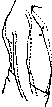
The clinical picture is characterized more by various clinical objective signs than by subjective symptoms. The latter are reduced to general symptoms of cardiac insufficiency, palpitations, a feeling of heaviness in the region of the heart, shortness of varying degrees, and congestive phenomena in the small and large circles of blood circulation. However, in some cases not only partial but even complete obliteration of the pericardium proceeds without symptoms (hidden form of heart adhesions), being only a finding at autopsy. Upon examination, retraction in the precordial region (Bouillaud's sign) is more often observed than bulging, as well as the absence during inspiration of forward movement of the lower part of the sternum (Wenckebach's sign); Wenckebach's sign is especially sharply expressed in those cases when there is simultaneously anterior mediastino-pericarditis and powerful adhesions between the heart and diaphragm; as a result, during inspiration the inspiratory profile of the abdominal wall crosses with the expiratory one at the level of the xiphoid process (Fig. 2). Greater importance is attached to retraction in the region of the cardiac apex during systole (Williams-Scoda's sign) (Fig. 3) and significant systolic retraction in several intercostal spaces (Jaccoud's sign) or systolic retraction of the lower part of the sternum and ribs simultaneously with the epigastric region (Heim's sign). In some cases, a rocking movement of the chest wall from front to back (mouvement de roulis-Jaccoud) is observed, with the upper part moving forward during systole and the lower part moving backward, and during diastole the opposite direction.
A solid line - expiratory phase; dashed line - inspiratory phase. Finally in other cases, especially in children, during systole retraction is observed on the left behind and to the side of one or several ribs (Broadbent's sign), and in the region of the apical impulse at the beginning of diastole a positive impulse is observed, which is not only visible but also easily palpable (Potain's sign). Percussion usually gives an increase in cardiac dullness in all directions and its immutability, neither depending on the phases of respiration nor

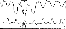
Figure 3. Negative apical impulse (above); below - sphygmogram.
from the position of the patient. This sign, in connection with the immobility of the apical impulse when the patient changes position (Potain's sign), indicates that the heart is fixed, i.e., adhered. But even more indications of the presence of adhesions can be obtained upon X-ray examination of the heart, when, in addition to the absence of heart mobility, the adhesions themselves and the presence of residual exudate in the cardiophrenic angle are sometimes discovered, and a decrease in the downward movement of the diaphragmatic dome during inspiration (Achelis-Wenckebach's sign).- Upon auscultation, in most cases the findings are indefinite and vary in different cases; sometimes the tones are clear and frictional murmurs are heard alongside them, in other cases, on the contrary, the tones are dull and soft. Sometimes characteristic of adhesions is the rhythm of protodiastolic gallop that is heard, where the additional tone coincides with the described protodiastolic impulse, resp. the rebound of the thoracic wall, which was drawn in during systole, at that moment (Bard-Potain's sign).- The arterial pulse in most cases does not present any characteristic changes. In most cases it is normal, but sometimes the pulse wave is small and decreases or completely disappears during deep inspiration - the so-called paradoxical Kussmaul pulse, which he considered pathognomonic for mediastinopericarditis (see Pulse). More characteristic are the changes on the venous side. Here, constant engorgement of the neck veins, accompanied by cyanosis of the face, is often observed, in other cases engorgement of the veins occurs only during inspiration, and finally sometimes sudden engorgement of the veins during diastole (Schoda-Friedreich's sign) is noted. All these venous phenomena depend on the compression of the superior vena cava by adhesions and strands, which intensifies during inspiration and also during diastole, and in addition on the descent of the diaphragm, especially in cases where there are dense adhesions of the heart with it. The course of the disease is extremely diverse in time and manifestations. In some cases, patients live for many years without special suffering, in others they die from phenomena of severe asystole within a few weeks. The course depends mainly on the condition of the myocardium and in the presence of myocarditis is rather short. An aggravating prognostic factor is the accompanying lesion of the lungs and kidneys, as well as widespread adhesions in the thoracic and abdominal cavities (periviscerites).- The diagnosis of heart adhesions is difficult in many cases, especially when the adhesion is partial. But even total adhesions can be asymptomatic. In other cases, the characteristic symptomatology described above allows for a confident diagnosis of heart adhesions. However, in general and now, despite the detailed development of the symptomatology of heart adhesions and the introduction of the X-ray method into clinical practice, diagnosis is difficult, and Stokes was right, who at one time said: 'I very much doubt that there is any, even one, certain sign of heart adhesions.'- Treatment consists of a strict regimen aimed at reducing the work of the heart and strengthening the heart muscle through careful loading with work and the administration of digitalis preparations for a long time. When signs of heart failure develop, the usual regimen for this condition and drug therapy are applied. In some cases, surgical treatment undoubtedly brings benefit by reducing the mechanical obstacle to the heart's activity.
N. Strazhesko. Surgical treatment of P. Available experimental and clinical observations allow us to consider surgical treatment of certain forms of P. as a rational method. The most developed technique is for the treatment of effusive P. Precise clinical diagnosis (especially X-ray diagnosis) must precede surgical treatment, since only the presence of an effusion in the pericardial sac positively decides the question of the possibility of surgical intervention. The essence of surgical treatment consists in eliminating (reducing) the inflammation of the pericardial sac and the accompanying disorders of cardiac function by the most complete and prolonged removal of the effusion from the pericardial cavity. In individual cases, indications for surgical intervention are those severe disorders of cardiac function which are caused by a rapidly progressive increase in intrapericardial pressure and find their expression in the clinical signs of so-called "cardiac tamponade." With rapid accumulation of fluid, sometimes only 150-300 cm3 are sufficient for the signs of cardiac tamponade to develop. In these cases, emergency emptying of the pericardial sac can save the patient's life. With gradual accumulation of effusion in the pericardial cavity, patients can tolerate large amounts of fluid (1-2 liters) for prolonged periods. The choice of surgical method for removing fluid depends on the pathological features and clinical course of the inflammatory process. For serous and serofibrinous P., either repeated punctures of the pericardial sac or its incision (pericardiotomy) are used. Therapeutic puncture, which is usually preceded by a trial (diagnostic) puncture, is performed with a syringe with a thick needle or a trocar. During puncture, it is necessary to avoid damaging the pleura, lung, a. et v. mammariae int., and the heart. In each case, the choice of site for puncture depends on the amount and character of the spread of the effusion. With small effusions (during repeated drainings), the danger of damaging the mentioned anatomical structures is greatest. With large effusions, Kurchman recommends puncture in the V or VI intercostal spaces along the linea mammaria sin. or to the outside of it. Delorme, Mignon and others (Delorme, Mignon) performed puncture at the left edge of the sternum. With atypical location of the exudate, Frenkel and Shaposhnikov performed puncture at the right edge of the sternum. A common method is puncture of the lower part of the pericardial sac in the angle between the base of the processus xiphoidei and the attachment of the VII left rib to the sternum. This puncture is safe for the pleura, lung, and heart and is favorable for the outflow of fluid. The pericardial sac is emptied slowly to avoid complications from the heart (often pathologically altered), which may occur under changed conditions of its function during puncture. With new accumulations of exudate, repeated punctures are performed. Favorable treatment results are rapid and in many cases are persistent. Improvement or cure of the process is noted in 50-60% [Schroetter on 100 cases had 47 cures and 53 deaths, Venus on 93 cases-29 cures, 6 improvements and 58 deaths]. To prevent new accumulation of fluid, Wenckebach proposed the use of pneumopericardium (introduction of filtered air, resp. nitrogen, oxygen into the pericardial cavity). -Pericardiotomy as a method of treatment for serous P. is used to achieve complete and prolonged emptying of the sac in cases where a single or double puncture did not give favorable results. In these cases, in the opinion of Schmieden and Fischer, pericardiotomy should be performed either with the use of short-term drainage (for 1-2 days) or with the use of suturing the edges of the incised sac to the muscles (according to Eiselsberg). The danger of suppuration in this case is not great. To completely eliminate the danger of infection and ensure constant resorption of the effusion, Schmieden and Fischer separate the subcutaneous tissue for a considerable distance in the wound, fix the edges of the incised sac to the muscles, and then tightly suture the skin wound. To establish constant resorption, in addition to "diversion into the subcutaneous tissue, diversion of the exudate toward the peritoneum and into the pleural cavity is proposed. In the treatment of purulent P., punctures and pericardiotomy are used. Repeated punctures at the present time have retained their significance only in connection with the use of irrigation of the pericardial cavity with solutions of rivanol. Most clinicians recommend more radical intervention - pericardiotomy. Rehn even proposes performing trial (diagnostic) pericardotomy in doubtful cases (regarding the nature of the inflammation). Pericardiotomy with drainage, carrying out the general principle of treatment for purulent processes, is the method of choice for treating purulent P. Drainage of the pericardial sac is best achieved by pericardiotomy with resection of a rib. Old methods - simple incision in the intercostal space, trepanation of the sternum - are imperfect in this respect. Of the various methods of pericardiotomy (Gussenbauer, Ollier, Delorme, Mignon, Kocher and others), at present the best is considered to be the opening of the sac with resection of the VII left costal cartilage. This method satisfies the necessary requirement of obtaining the greatest possible drainage and simultaneously draining both halves of the sac at its lowest point. Postoperative treatment of the patient is carried out in a sitting position. Irrigation of the opened sac with rivanol, as well as humanol, iodipinum, and ol. Sesami (Rehn, Klose) is recommended to prevent adhesions. Irrigation of the sac with physiological solution to remove fibrinous clots is considered advisable. The latest acquisition of modern surgery is the surgical treatment of chronic adhesive (adhesive) P. In cases where chronic adhesive P. occurs with a more or less severe clinical picture, indications for surgical intervention arise, and here we can speak of cases of mainly two kinds: 1) the heart is adherent to the anterior chest wall, whereby its systolic work is mainly impaired; 2) the heart is completely walled in in callous scar tissue, sometimes calcified. In both categories of cases, a picture of right heart insufficiency develops, severe congestive phenomena appear as an expression of impaired outflow from the area of v. cavae sup. In these cases, Cardiolysis (see) is indicated. V. Bogolyubov. Pericarditis in children. In childhood, P. occurs less frequently than in adults: according to Cnopf - in 5% of all autopsies, according to Edelman and according to data from the Aleksandrovskaya Hospital in Kiev - in 6% (in adults in 10%); it is believed that P. occurs relatively more frequently in infancy, although according to autopsy data of the Scientific Institute of Okhmatmlad NKZdr. on 340 autopsies it was encountered 6 times (about 2% of cases). In infancy, almost exclusively acute, purulent (resp. serous and fibrinopurulent) P. of secondary nature occur as one of the manifestations of sepsis; P. can complicate any infection, most often erysipelas, pneumonia and empyema, less often scarlet fever, omphalitis and others. Accordingly, strepto- and pneumococci are most often found in the exudate. In exceptional cases, P. is primary of embolic origin. TBE is extremely rarely a cause of P. in infancy. In older children, both acute and subacute and chronic P. occur. The most frequent cause is apparently rheumatic infection, then tuberculosis, less often scarlet fever (about 2% of all cases) and even less often other infections (measles, typhoid, gonorrhea). Pneumonia and empyema in older children lead to P. much less often than in infants - almost never per contiguitatem, but usually by the hematogenous route. According to Putzig and Langstein, rheumatism, measles and scarlet fever account for 50-60% of all cases of P. in older children. Clinical picture. Both dry and exudative forms are encountered. Due to the fact that in early childhood P. is only one manifestation of severe general infection, it is not easy to establish it upon examination, especially if it developed during pneumonia or pleurisy; here its symptoms are lost in the general severe picture of the disease. One can suspect the presence of P. if against the background of a severe infection, symptoms of cardiac insufficiency develop: cyanosis, especially during movement and excitement, and "cardiac" dyspnea (frequent restless breathing with short inspiration and expiration). A characteristic feature is the sitting position of the child; when attempting to lay him down, cyanosis, increased dyspnea, and restlessness appear; further, the development of P. is indicated by edema of tissues in the area of the sternum, swelling of the neck veins and a certain puffiness of the face (mainly in older children) and finally an increase in cardiac dullness upon percussion (in the presence of left-sided pneumonia or pleurisy this sign is obscured). The boundaries of cardiac dullness upon percussion can greatly expand and reach the II rib from above. In very small children, however, there is no sharp increase in the boundaries of the heart. A typical symptom - cardiac impulse within the boundaries of cardiac dullness - is also very rarely established in them. The pulse is usually rapid, small, and soft.
Heart tones are difficult to hear (sometimes better heard from the back) (Maslov). In older children at rest, the friction rub of the pericardium can sometimes be heard; in small children this symptom is almost never present (the rub is masked by louder respiratory sounds). In very large and prolonged P., a cardiac hump may form. A distinctive feature of P. in infancy, sometimes the most prominent, is in some cases aphonia, as well as a cough of a special character: dry, with persistent and distressing attacks, resembling the cough in enlargement of the mediastinal glands. On X-ray examination with a small exudate, blunting of the cardiac contour arches on the left and weak pulsation of the resulting single arch are found. With a larger exudate, the cardiac shadow may take the form of a sphere, and sometimes the heart itself is outlined as a darker shadow within the shadow caused by the exudate. In some cases of large effusions, an important symptom is the cessation of pulsation of the edges of the cardiac shadow. Prognosis. Purulent P. in small children, being a manifestation of sepsis, almost always ends fatally; the prognosis is somewhat better* for pneumococcal P. during pneumonia or pleurisy and generally in older children. Tuberculous N. runs less acutely; being a partial manifestation of generalized tbc, it is accompanied by emaciation and other manifestations of tbc and most often leads to a fatal outcome. The prognosis for rheumatic P. is better-the exudate is absorbed more or less quickly. However, complete recovery after P. is not often observed. Occasionally P. is observed in older children in polyserositis.-In differential diagnosis, one should keep in mind: myocarditis with secondary heart dilation and in early childhood-miliary tbc. The diagnosis of fibrous P. is very difficult, especially when the acute stage was missed; here the picture of the disease consists of the following phenomena: immobility of the heart with change of position, good pulse with large cardiac dullness, presence of systolic retractions in the area of the apex or xiphoid process or upper epigastrium, enlargement of the liver in the absence of other signs of congestion. Treatment in general is similar to the treatment of P. in adults; one should only stop at the technique of P. puncture. Many methods of pericardial puncture have been proposed; it is difficult to say which one should be preferred in childhood, as all of them have shortcomings; however, the most dangerous is puncture at the very edge of the sternum (injury to a. mammariae int.). Marfan proposed in children to make the puncture along the midline upward under the xiphoid process. Ternovsky advises performing the puncture with the patient lying down in the V-VI intercostal space on the left, away from the sternum. Recently Sutton proposed to puncture the pericardium from behind in the VII-VIII left intercostal space, closer to the spine than to the axillary line,-on the grounds that the exudate generally spreads to the back.
Related articles
Mentioned in
Cite this page
“Pericarditis.” Soviet Medical Encyclopedia. English translation of Bolshaya Meditsinskaya Entsiklopediya, 1st ed. (Moscow, 1928–1936), ed. N. A. Semashko. https://sovietmedicalencyclopedia.pages.dev/article/pericarditis/