Angina Pectoris
Historical document, translated for reference. It reflects medical knowledge of the 1920s–30s and is not medical advice.
Summary
This article provides a historical overview of angina pectoris, tracing its clinical description from William Heberden's 1768 report to the various theories regarding its pathogenesis developed throughout the 19th and early 20th centuries. It highlights the evolution of medical understanding, from the coronary theory to various neural and aortic hypotheses.
Encyclopedia article (1928–1936)
ANGINA PECTORIS (syn. Heberden's asthma) is, in its essence, primarily a subjective syndrome manifesting as severe retrosternal pain, accompanied by a feeling of fear and a sensation of the immediate proximity of death. History. On July 21, 1768, William Heberden read a report in London in which, based on 20 cases he had observed, he established a specific clinical picture that he named angina pectoris. "There is a disorder of the chest," he said, "which is characterized by peculiar symptoms, primarily the most severe pains. Due to the danger associated with the attacks, this disease must be given special significance. It is encountered not infrequently. Nevertheless, I have never once encountered a description of it in other authors. The localization, the feeling of the most severe pain, and the fear for one's life make, it seems to me, the name angina pectoris most appropriate for this disease. In people subject to the named disease, while walking, especially while walking after eating, such strong pains arise in the chest that it seems to the patients that they are about to lose their lives if the pain intensifies or lasts for some time longer. As soon as the patients stop, the pain attack ceases. Outside of the described attacks, the patients are completely healthy. This disease has nothing in common with dyspnea." The description given by Heberden remains classic in its main features to this day. It points out with amazing observational skill the difference between cardiac asthma and angina pectoris. Here, too, the paroxysmal nature of the phenomena in the absence of other symptoms of heart disease outside of attacks in typical cases of angina pectoris is quite correctly highlighted. In the name of historical justice, however, it is necessary to note that Erasistratus in ancient Greece, and later Ballonius, F. Bartolettus (1628), and Morgagni (1715) gave individual descriptions resembling the picture of angina pectoris. However, none of the listed authors isolated the syndrome they saw into a separate disease entity as Heberden did. It is therefore entirely fair to give the named disease the name Heberden's disease. French authors, among them Huchard, call the syndrome the "Rougnon-Heberden" disease, believing that Rougnon, independently of Heberden and even somewhat earlier, gave an accurate description of the affliction. However, historical research by Kohn has proven the error of the French assertion. For all the correctness of the clinical description, Heberden did not link any anatomical substrate to it. Moreover, observing a good pulse in his patients during an attack, Heberden did not link the attack to any anatomical changes in the heart. A few years after Heberden's report, an "unknown person" who suffered from attacks of angina pectoris bequeathed his corpse to Heberden for autopsy. At autopsy, no anatomical changes were found in the heart. However, at the autopsy, as Edward Jenner notes, the coronary vessels of the heart were not examined. For the first time, the significance of changes in the coronary arteries for the occurrence of attacks of angina pectoris, based on comparisons of autopsies with the clinical picture during life, was recognized by the same Jenner in a private letter. Confirmation of this view was provided by Parry, Fothergill, and Black (1795). To this day, despite the century-and-a-half history of the issue, there is no unified point of view in understanding the pathogenesis of angina pectoris. Huchard, in his classic monograph published in 1899, cites 80 theories of angina pectoris. Leaving most of them aside, it is necessary here to dwell only on those that are still viable and have not lost their significance. Historically, the coronary theory of angina pectoris must be placed in the first rank, put forward for the first time, as indicated, by Jenner and Parry and substantiated in 1776 by Fothergill. Its most prominent supporters at the end of the 19th and beginning of the 20th century are Potain, Huchard, Romberg, G. See, Gallavardin, and others. From 1811, a series of nervous theories appeared: neuralgia of the abdominothoracic nerve (Desportes), neuralgia of any cardiac nerve (Laennec), neuralgia of the cardiac plexus (Peter), neuritis of the cardiac plexus (Corrigan, Lancereaux), and other theories of nervous origin, the number of which, according to Huchard, equals 23. In 1814, Blackall, in contrast to coronaralgia, put forward the theory of aortalgia, according to which the pain arises in the root of the aorta. Corrigan adopted this point of view, and in more recent times, Clifford Allbutt, Wenckebach, and others. Cruveilhier in 1849, later Quain and Stokes, and in more recent times Shaposhnikov, Deutsch, Danielopolu, and others defend the theory of cardiac weakness caused by various reasons. In 1846, Latham expressed the assumption that the pain in angina pectoris is caused by a spasm of the heart muscle itself. In 1877, Gairdner, later Balfour, Leyden, Willius, Brown, Perfiliev, and others described a syndrome named by Gairdner as angina.
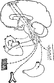
_4_.-t_.-l }_ psychomotor impulses to the muscles and bulbar centers of the heart and vessels. "*" ~*~ "+" + Effector impulses from the bulbar centers to the heart and vessels. ___________Primary reflex excitations, proceeding from the stomach, skin, heart, etc., to the bulbar centers and to the cerebral cortex. ....................... Secondary excitations originating from the heart. Figure 1. Diagram of the occurrence of an attack of Angina Pectoris sine dolore, at the basis of which lie deep changes in the coronary arteries of the heart. Such patients die with phenomena of thrombosis of the coronary arteries, but without pain. Mackenzie suggested that Angina Pectoris is not a manifestation of anatomical changes in the heart itself. According to Mackenzie, Angina Pectoris is the result of the sum of a whole series of viscero-sensory (sensitive), viscero-motor (motor), and other diverse viscero-sympathetic and viscero-parasympathetic reflexes originating from the heart. Irritation of a series of centripetal nerves leads to their strongest excitation and to the centers perceiving pain (see Figure 1). Hence the sensation of an attack of Angina Pectoris in the heart. Such are, in general terms, in historical sequence, the stages of various views on the pathogenesis of Angina Pectoris. Etiology. It is necessary to distinguish predisposing factors, which include dyscrasias: obesity, gout, frequently recurring emotions, in the physical manifestation of which a substantial role belongs to the play of vasomotor nerves, and factors directly causing changes, predominantly vascular. These include vasotropic infections, primarily Treponema pallidum, and vasotropic poisons: nicotine and lead. The vasoconstrictive effect of the latter on the coronary vessels can be easily observed in an experiment. The connection of Angina Pectoris with syphilis was noted by Zimnitsky and Josserand in 29%, Vaquez and Gallavardin in 30%, Pletnev in 40%, E. Romberg in 50%. Gallavardin, in his 450 cases of Angina Pectoris, broken down by groups, noted that in cases of: a) the presence of aortic dilation, Angina Pectoris had a syphilitic origin in 100%; b) aortic valve insufficiency, in 87%; c) the absence of aortic lesions, in 21%. Pletnev's material in 350 cases, arranged by the same groups, gave in round figures 92%, 81%, and 28%. D. Pletnev. Pathological anatomy. In the overwhelming majority of cases of Angina Pectoris, one finds arteriosclerotic changes in the coronary arteries of the heart, mainly the left; often the process is most intensely expressed in the anterior descending branch (ramus descendens ant.). In relatively rare cases, especially in young subjects (up to 30 years), changes in the coronary arteries may be absent, and such cases are customarily designated as angina pectoris vasomotoria, a term somewhat unfortunate only in that for ordinary cases of Angina Pectoris, the vasomotor moment also plays a very important role; the latter follows from the fact that cases of arteriosclerosis of the coronary arteries, and even with pronounced cardiosclerosis without Angina Pectoris in the anamnesis, do not represent a particularly rare phenomenon in pathological-anatomical practice. The indicated changes in the coronary arteries in Angina Pectoris usually coincide with their thrombosis and the development of a heart infarct (more often ischemic), with subsequent softening—myomalacia of the heart. In the absence of sclerosis and thrombosis of the vessels, their occlusion may be caused by emboli, for example, warty masses from the aortic valves in endocarditis. Both the volume of the infarct and the place of its development are subject to significant fluctuations; in the majority of cases, the focus of necrosis reaches a size of several cm in area and about 0.5–1 cm in depth, with the corresponding section of the muscle often bulging slightly outward (acute aneurysm of the heart); more rarely, in the area of such a protrusion, one finds a slit-like or zigzag rupture of the heart muscle with the development of hemorrhage into the cavity of the pericardium (so-called cardiac tamponade); in general, however, rupture of the heart is a rather rare phenomenon. Sometimes several foci of softening are observed simultaneously. The most frequent localization of myomalacia is the left ventricle, especially the papillary muscles, the region of the apex, as well as the anterior and posterior walls; the ventricular septum, right ventricle, and atria are affected more rarely; in the majority of cases, the necrosis encompasses mainly the inner layers of the myocardium, which causes the relative frequency of simultaneously developing parietal thrombosis of the heart; more rarely, the necrosis also encompasses the endocardium and epicardium. One often observes malacia of those sections of the heart that lie near old, already scarred softenings from previously occurring attacks of Angina Pectoris. If Angina Pectoris is caused by the closure of large, and even more so the main trunks of the coronary arteries (rapid closure of the main trunks of the coronary arteries usually causes sudden death without a preceding picture of Angina Pectoris) and an insignificant period of time has elapsed from the moment of their closure until death, for example, only a few minutes or hours, then macroscopically and microscopically the heart muscle may not have bright changes; in these cases, one notes only general flabbiness of the heart, uneven blood filling of the myocardium, and under the microscope one finds some edema, loosening of the tissue; the general structure of the fibers and their transverse striation may be preserved; it is important, however, to examine various sections of the myocardium in such cases, and it is often possible to detect initial phases of their necrosis in the muscle fibers. Pronounced changes characteristic of myomalacia can be observed under the microscope apparently no earlier than 5–6 hours from the moment of the development of the attack. In individual cases, Angina Pectoris is accompanied not only by myomalacia but also by encephalomalacia, i.e., softening of the brain; this presents the theoretical interest that vasomotor-spastic (usually, however, there are also arteriosclerotic) phenomena in the vessels can spread simultaneously to the vascular systems of other organs. As a result of Angina Pectoris, insofar as it is accompanied by a heart infarct, there usually remains a scar (“callus”) of the myocardium of one size or another; the expansion of such calloused sections sometimes gives a picture of a chronic aneurysm of the heart (see). See also Heart.
I. Davydovsky. Pathogenesis and symptomatology. At the present time, there is also no unified interpretation of the pathogenesis of the anginal syndrome, which is quite understandable. Cardiac pain, being fundamentally a clinical retrosternal pain, radiates in various directions, most often to the left side of the neck, under the left scapula, and into the left arm, sometimes into both arms, under the right scapula, less often along the tract of the esophagus into the stomach region (vomiting), into the lumbar region, the lower extremities, etc. (visceromotor, viscero-sensory, viscero-secretory, etc., reflexes). Usually, the patient turns pale, and the face becomes covered in sweat. In classic cases of angina pectoris, dyspnea is absent. Consciousness is clear. The picture of cardiac activity is variable. It can be normal, with the pulse being entirely satisfactory. In other cases, heart activity accelerates to 100-150 or, conversely, slows down to 40-30 beats per minute, and the pulse becomes barely perceptible. Severe attacks of angina pectoris can occur with apparently normal heart activity, just as the severity of the attack is not always parallel to the intensity of the pain. Attacks can last for minutes or hours. Arterial blood pressure, both during an attack of angina pectoris and outside of it, can be completely normal, but it can also be elevated and, conversely, can fall. If the pain syndrome is caused by thrombosis of the coronary arteries, peripheral blood pressure falls. Often, 1-3 days after an attack, especially in cases where the latter was caused by thrombosis of a coronary vessel, the temperature rises to 37.8-39.0°, and a pericardial friction rub appears. Post-anginal pericarditis was first described by Kernig, later by Josserand, and named by Sternberg pericarditis epistenocardica. If the pericarditis appears in the region of the left ventricle facing the posterior mediastinum, the friction rub is absent, and only a rise in temperature is present. Attacks of angina pectoris occur most often during physical exertion, after a heavy meal, under the influence of emotions, after sexual intercourse, and in the cold. It is possible to experimentally reproduce an attack accessible to analysis only in humans. Several such experiments have been conducted (see below). Their small number and the lack of uniformity when comparing antemortem symptoms with autopsy results explain the diversity that still reigns in views on the pathogenesis of angina pectoris. In a number of fatal cases of angina pectoris, no significant changes were found anatomically in either the heart or the aorta, and conversely, in the presence of serious and diverse pathological-anatomical changes, including those in the coronary arteries, anginal attacks were absent. Genetically, all attacks of angina pectoris can be divided into two large main groups: 1) cardiac angina pectoris and 2) extracardiac. The first group should include: 1) coronaralgia, 2) aortalgia, and 3) spasm of the heart muscle. One cannot pose the question as it has been posed in the literature until now: coronaralgia or aortalgia? Both forms of algia can exist. The prototype of anatomical coronaralgia is thrombosis and embolism of the coronary arteries. Thrombosis causes a very strong, prolonged attack of angina pectoris—status anginosus—with a decline in heart activity (meiopragia cordis). The outcomes of the attack are varied, up to and including death. Altered coronary arteries, as is observed in relation to all altered arteries, respond more easily than healthy ones with a spastic state to various influences. Pain is caused by the spasm of the coronary arteries. The coronary arteries are under the influence of extracardiac nerves, although the experiments of physiologists regarding the presence and influence of vasomotor nerves largely contradict each other. In addition to the purely nervous influence on the lumen of the coronary vessels, it is also necessary to take into account the excitability of the vessels themselves. Clinically, we must admit the possibility of spasm of the coronary vessels under the influence of the nervous system. The view held by some authors, according to which the spasm of peripheral arteries is primary, and the pain in the heart arises secondarily, depending on it, is incorrect. Peripheral spasm can cause an acute rise in arterial pressure, and in connection with it, an attack of dyspnea, but not pain in the region of the heart. The relaxation of spasmodically constricted coronary arteries explains the beneficial effect of nitroglycerin and other agents analogous to it in their effect on vessels. This effect is also observed in cases where blood pressure is completely normal. Where there are sharply pronounced changes in a portion of the coronary vessels, one must admit the relaxing effect of the named agents on collateral coronary circulation. Acute spasm of not only sharply altered but even apparently healthy coronary arteries can lead to death. Eppinger reported in 1924 on a patient with repeated attacks of angina pectoris who died suddenly in the clinic during an attack. At autopsy, no changes were found in either the aorta or the coronary vessels. The possibility of experimentally reproducing spasm of the coronary vessels (nicotine, lead), and most importantly, the comparison of the pain syndrome of angina pectoris with pains arising in other organs on the basis of arterial spasm and the ischemia caused by it, is the fundamental point in the theory of coronaralgia. It is known that spasms of peripheral arteries in Raynaud's disease are accompanied by pain, that Charcot-Erb intermittent claudication is extremely painful, that in migraine a spastic contraction of the retinal arteries is observed, accompanied by scotoma, etc. In contrast to the coronary theory of pain, the aforementioned authors (recently Allbutt and Wenckebach) energetically defend the theory of aortalgia. They point out that the initial part of the aorta, the so-called supra-sigmoid part of it, is abundantly supplied, predominantly in the adventitia, with Pacinian corpuscles, which give rise to the sensory nerve of the aorta, the depressor nerve (see Figure 2). The sensation of pain in the region of the heart arises, according to Allbutt, in cases of increased blood pressure in the aorta, or rather in the root of the aorta, under the influence of various causes (cold, physical and mental exertion). By virtue of the change in pressure in the aorta, its initial part is stretched. The onset of pain, however, is observed not in a healthy aorta, but only in cases of an aorta pathologically altered due to rheumatism, syphilis, arteriosclerosis, etc. The endings of the sensory nerves (and perhaps their centers) are, due to one reason or another, in a state of increased susceptibility. Wenckebach, who is close in his views to Allbutt, distinguishes two forms: 1) an acute form, caused by thrombosis or embolism of one or another coronary vessel, and 2) a chronic form, having its source in the initial part of the aorta. Allbutt and Wenckebach put forward two objections against the coronary hypothesis: 1) sclerosis of the coronary vessels has been found many times at autopsy, whereas during life the possessors of it did not suffer from attacks of angina pectoris, and 2) a number of cases have been described where, at the autopsy of people who died during an attack of angina pectoris, no changes in the coronary vessels were found. Wenckebach adds a third argument to this: when the lumen of the coronary vessels changes, cardiac weakness should occur, whereas in the majority of so-called chronic cases of angina pectoris, this is not observed. Since arterial blood pressure during an attack of angina pectoris is far from always elevated, Wenckebach expresses the assumption that the direct cause of the attack is not an increase in arterial blood pressure, but an increase in the work of the heart and an increase in the systolic volume of blood. According to Wenckebach, an attack of angina pectoris arises during increased work of the heart (physical exertion, mental excitement, cold, etc.) without its connection to an increase in peripheral arterial blood pressure. Wenckebach's hypothesis does not explain attacks of angina pectoris arising during complete rest. Likewise, many cases of chronically elevated arterial pressure are observed in which, despite an increase in the work of the heart, attacks of angina pectoris do not arise. In addition to what has been indicated, Wenckebach's assertion that pain in the aorta (chronic angina pectoris) and in the ostia of the coronary vessels (acute angina pectoris during thrombosis of the coronary vessels) always depends on the stretching of the vessels is extremely vulnerable, whereas pain in hollow organs, including vessels, in some cases is the result of their stretching, and in others—of their spasm. R. Schmidt, who also stands on the ground of aortalgia, narrows the question of the pathogenesis of angina pectoris even further, reducing the essence of the latter to a disease of the aortic plexus, to which the pathological process spreads per continuitatem from the walls of a rheumatically, syphilitically, or atheromatously altered aorta. As a result, neither the theory of coronaralgia nor the theory of aortalgia fully explains the attacks of angina pectoris.
Pletnev considers it more correct to explain the attacks of Angina Pectoris as being due either to the separate influence of the coronary and aortic factors, or to their combined action.
The pain itself may in some cases arise as a result of the aforementioned vascular spasm; in other cases, it is necessary to assume that during ischemia of the heart muscle, some active process of a physico-chemical nature occurs in the bloodless muscle itself, the expression of which is a spasm of the heart muscle.
The possibility of this process follows from the experiments of Walther with muscarine and Gaskell with alkaline solutions.
With the acceptance of this assumption, cases of death during an attack of Angina Pectoris, in which no anatomical changes were found, become understandable.
Under the influence of spastic contraction of the coronary vessels—an unfortunate localization of the spasm (spasme mal placé according to Gallavardin)—a spasm of the heart muscle occurs, clinically recorded by Ohm's apparatus, which transitions into ventricular fibrillation of the heart, as a result of which death occurs during an attack of Angina Pectoris.
Besides the spasm of the coronary vessels, spasms of the heart muscle, and stretching of the aortic root, convulsive contractions of the intercostal muscles participate in the clinical picture of the Angina Pectoris pain syndrome, as pointed out by Mackenzie—a viscero-motor, or, perhaps more correctly according to Danielopolu, a viscero-myotonic reflex.
The feeling of horror, of impending doom, and sometimes a feeling of inexpressible anguish, of which many patients with Angina Pectoris complain, deserves special analysis.
Upon looking at the patient, one can sometimes immediately see that something terrible is happening to them: the eyes are wide open, the entire face has an expression of some kind of horror, and the patient, experiencing retrosternal pain with various irradiations, is afraid to breathe.
The pulse often drops during this.
Heberden's "unknown" patient says that during an attack he felt a temporary cessation of all vital processes.
The patient described by Morgagni, like many others, cried out: "I am dying."
This feeling of horror, the angor of the old authors, is not identical to pain.
Furthermore, its degree is not parallel to the intensity of the precordial pain.
It is observed even independently of the latter (angina sine dolore).
The described angor is characteristic of the most severe cases of Angina Pectoris, in which a decline in cardiac activity is usually observed.
To a lesser extent, such sensations of the patient are associated with extrasystoles.
It is possible that in some cases the latter, and in others direct dropouts of heart contractions, are the sources of such fear of death.
Pain itself as such, wherever and however strongly it may manifest, does not cause such angor.
While considering the heart as the source of the latter, one must still remember that the described fear is a psychic phenomenon and is therefore connected with the central nervous system.
In addition to the described mechanism of Angina Pectoris, one must admit the possibility of extracardiac causes triggering the attacks, where the primum movens for the occurrence of coronary artery spasms and heart muscle cramps lies outside the heart itself.
Proof of this is provided by experiments on humans by Leriche, Pletnev, and Khesin.
On a neck exposed under local anesthesia for surgery, Leriche performed electrical stimulation of limited areas of the left stellate ganglion (gangl. stellatum).
Upon electrification of the lower pole of the ganglion, pain appears in the heart region, resembling an attack of Angina Pectoris in its character.
Upon electrification of the upper pole of the stellate ganglion, pain appears in the left arm.
Novocainization of the ganglion immediately stops the pain.
Upon electrification of rami communicantes, patients experience pain in the corresponding arm and shoulder.
Mechanical irritation (pinching) of the ganglia leads to the same results.
Over the last two years, Pletnev, together with Khesin, has performed over 80 paravertebral injections of novocaine and alcohol into the region of the lower cervical and three upper thoracic ganglia in severe anginal patients.
In many cases, upon the insertion of the needle into various of the mentioned areas, patients stated that the needle caused them a "prick directly in the heart."
Upon the introduction of novocaine through the same needle, without removing it, the painful sensations of the heart immediately disappeared.
In one case, upon the introduction of alcohol into D1, the patient suffered a severe attack of Angina Pectoris with pallor of the face and a drop in the pulse.
The attack lasted several hours and required repeated injections of cardiac and narcotic agents.
Three weeks later, the patient left the clinic "completely healthy."
In another case, the attack of Angina Pectoris was short-lived, about 20 minutes.
The pains quickly yielded to narcotics, but cardiac activity continued to be weak for several hours.
In a third case, after two paravertebral injections, the patient's anginal pains ceased, but both times, several hours after the paravertebral injections, persistent hiccups and bloody vomiting appeared.
The latter must be interpreted as the result of an ischemic infarct due to irritation of the corresponding nerve roots, and the hiccups as irritation of the phrenic nerve.
In a fourth patient, after repeated paravertebral injections of novocaine and alcohol into CVII-VIII, DI-II (intervertebral foramina—VII-VIII cervical, I-II thoracic), the pains in the heart region ceased, but the previous pains in the lower jaw, pains in the larynx, and hoarseness remained.
The observations of Pletnev and Khesin are analogous to the observations of Leriche.
They suggest that the primum movens for the appearance of painful attacks in the heart, and sometimes even the decline of cardiac activity itself, can come from the ganglionic nervous (thoracic and perhaps cervical) apparatus.
The case where, after repeated paravertebral injections, cardiac pains ceased but pains in the larynx and jaw remained, suggests that not all pains in Angina Pectoris should be attributed only to radiating pains.
Some of them arise in parallel, and not secondarily, according to the laws of irradiation.
Both types of pain are an expression of functional disorders of innervation due to sympathetic ganglionitis.
Changes in the sympathetic ganglia were found in operated and deceased patients by Staemmler and Ormos.
However, in one of the cases operated on by Brüning, as well as in the case of Benda (autopsy of a young woman of 20, who died from Angina Pectoris), the sympathetic ganglia turned out to be unchanged.
Taking into account these cases, as well as the fact that in persons who died from various infections, the sympathetic ganglia are almost always found to be changed (Mogilnitsky), and likewise considering the various types of degenerative changes in the sympathetic ganglia found in those who died from various causes starting from the age of 8 (Abrikosov), one must be extremely cautious in interpreting the connection between the occurrence of Angina Pectoris attacks and sympathetic ganglionitis.
Apparently, the center of gravity of the influences coming from the ganglia to the heart lies in some disturbance of the biotonus of the ganglia.
This phenomenon of anatomical and functional dissociation is analogous to others occurring in the animal organism.
As for the other extracardiac theory put forward by Mackenzie, the theory of increased sensitivity of the perceiving centers, it is theoretically easy to admit, but practically impossible to prove.
It is possible that, just as there are photographic plates that react more weakly or more strongly to light stimulation, there are people with centers that react differently to the impressions flowing into them.
To the group of angina pectoris whose starting point lies outside the heart, those cases should be assigned where painful cardiac symptoms disappeared following the removal of neuromas of the arm (Huchard, Mackenzie, Gubergrits).
Here, apparently, a reflex spasm of the coronary vessels took place—reflex angina pectoris.
Tumors of the nerves were found not only in peripheral nerves.
Thus, Blondin found in a subject who suffered from Angina Pectoris during his lifetime, a tumor the size of a large pea in the vagus nerve.
Blondin genetically links the patient's anginal attacks with the tumor he found.
The mechanism of death in Angina Pectoris is twofold.
Death occurs either quickly following the appearance of the pain syndrome or during a long-lasting attack (i.e., during the attack itself).
Sometimes, some time after its end, cardiac weakness develops, and the patient dies from the latter outside of a pain attack.
Death during an attack occurs due to ventricular fibrillation, both in cases of thrombosis of the large branches of the coronary vessels and in cases of spasm of the latter.
If death does not occur suddenly, in the first moment of the attack, then simultaneously with the pain syndrome, phenomena of acute heart failure (complete or partial) develop, in connection with which pulmonary edema and acute swelling of the liver are observed—one or the other symptom develops depending on the predominant anatomical and functional involvement of the right or left coronary artery.
If the patient survives the attack of Angina Pectoris and death nevertheless occurs soon, it is the result of subacutely developing heart failure.
The prognosis, more than in other branches of pathology, is individual.
In no case of an attack of Angina Pectoris can one say how it will end.
This applies equally to both thrombotic and spastic closures of the coronary vessels.
The long-term consequences of Angina Pectoris are just as varied.
After thrombosis or prolonged spasm of a coronary vessel, an ischemic infarction of the heart muscle develops. The muscle tissue is replaced by connective tissue. In the case of its significant size, an aneurysm of the ventricular wall may develop. The energy of contraction of such musculature is naturally weakened, and the hearts of severe angina pectoris patients therefore easily become insufficient. Upon replacement of muscle tissue with connective tissue in individuals who have suffered a serious attack of angina pectoris, the attacks may cease altogether; there is no active musculature capable of spasm, there are no vessels capable of spasms due to their thrombotic occlusion—the patients have "recovered" from angina pectoris (Buchstab), although the "cure" is achieved at the cost of a decline in energetics. Treatment. When treating attacks of angina pectoris, one must keep in mind the following main points: pain, at the center of which in most cases lies a spasm of the coronary vessels, sometimes a convulsive contraction of the heart muscle, and in some cases, mainly during thrombosis of the coronary vessels, a decline in heart activity. Therefore, agents that dilate coronary vessels are indicated: theobromine preparations (pure theobromine, diuretin), Euphyllin orally and intravenously 0.5-1.0 per dose, nitroglycerin from 0.0006 to 0.0015 pro dosi, or in the form of drops of a 1% alcoholic solution, 1-5 drops per dose, sodium nitrite orally and intravenously (2% solution up to 5 cm3), amyl nitrite (inhale on cotton wool 2-5 drops), etc. If these agents do not help with a single dose, they are repeated 3-4 times a day. Narcotics: morphine, pantopon, holopon, papaverine, and various cardiac agents in case of a decline in cardiac activity. In individual cases, Pletnev saw benefit from prescribing atophan at 1.0, three to four times a day. Among external remedies, mustard plasters or hot poultices are applied to the heart region and the interscapular region. Sometimes electric deep heating of the heart (diathermy) helps. To the hands and feet, heating pads or hot baths. If all the above-mentioned agents do not help, one should inject paravertebrally during an attack into CVII-DI-II a 1/2% solution of novocaine, 5 cm3 into each ganglion, in pure form or in combination with epinephrine. In the intervals between attacks, patients must primarily observe a dietary and work regimen, avoid physical exertion, cold, and emotions. Arteriosclerotics must treat their sclerosis, neurotics their nervous system, gout patients their gout, syphilitics their syphilis, etc. If the attacks are repeated and painful for the patient, then several repeated paravertebral injections of 75-85% alcohol should be made into the aforementioned ganglia, 5 cm3 into each ganglion. Alcohol causes necrotic changes in the ganglia and conducting nerves of the heart, thanks to which the agonizing anginal pain and the reflex and parallel phenomena associated with cardiac pain cease. Pletnev had occasion to observe the complete cessation of severe attacks of angina pectoris and the restoration of working capacity for a period of up to two years after such injections. Cases followed up for longer have not yet been seen. If ordinary therapeutic measures and the chemical operation (alcohol) do not help, one must resort to surgical treatment: one or another type of sympathectomy or ramicotomy (see below). Abdominal angina. In some cases, pains of the same character as angina pectoris are localized predominantly in the epigastric region, accompanied by various symptoms from the gastrointestinal tract: vomiting, abdominal distension, or (conversely) its retraction, persistent constipation. Bacelli proposed the name abdominal angina (angina abdominalis) for these attacks, and Huchard called it pseudogastralgic angina pectoris. Upon autopsy of such patients, in some cases, changes are found in the thoracic aorta and coronary vessels, while the arterial vessels of the stomach and intestines are apparently healthy; in other cases, conversely, the latter are sclerotically altered. At the basis of an attack of abdominal angina lie spastic contractions of the abdominal vessels. The latter can arise as a reflex in connection with attacks of angina pectoris, as well as depending on changes in the solar plexus—a picture described by Laignel-Lavastine under the name "acute syndrome of solar plexus excitation" ("syndrome solaire aigu d'excitation")—a phenomenon analogous to the above-described sympathicoganglionitis. For the treatment of these attacks, Jaboulay even proposed surgical intervention: traction of the solar plexus with a probe inserted between it and the abdominal aorta. Having begun as attacks of abdominal angina, they may subsequently entirely transition into attacks of angina pectoris. In sclerosis of the nerve branches of the abdominal viscera, acutely occurring pains in the abdomen are periodically observed, accompanied by sharp distension of the intestines and loud movement of gases within them. These symptoms are caused by periodically occurring spasms of the mesenteric vessels, in some cases by their thrombosis, and resemble the above-described abdominal angina. Schrotter calls the syndrome the "intestinal picture of arteriosclerosis," and Ortner calls it dyspragia intermittens angiosclerotica intestinalis, by analogy with Charcot's intermittent claudication.
D. Pletnev. Surgical treatment of angina pectoris. François Frank in 1899 expressed the opinion that centripetal conductors from the heart and aorta pass predominantly through the inferior cervical and superior thoracic ganglia of the sympathetic nerve and are transmitted through the sympathetic trunk to the spinal cord. Therefore, in Frank's opinion, resection of the sympathetic nerve ganglia could stop the pain in angina pectoris. Only 17 years later was this idea utilized by Jonnesco, who performed the first sympathectomy for this disease. According to the most accepted opinion, sensory fibers from the heart pass along the sympathetic nerve through 3 cardiac nerves to the three cervical ganglia and through them via the rami communicantes to the spinal cord, and then to the brain. Wenckebach, Eppinger, and Hofer believe, however, that painful sensations are transmitted along the depressor nerve of the vagus. The radiating pain into the upper limb during angina pectoris, which can be conducted there only through the stellate ganglion, speaks against the latter opinion. Transection of the depressor nerve during a severe attack of angina pectoris in two cases by Hesse, operated on under local anesthesia, did not stop the painful sensations; however, after transection of the sympathetic nerves, the pain stopped immediately. In 135 cases collected by Hesse in 1927 from the literature, and 8 of his own, 65% favorable results were obtained from the operation. A poorly satisfying result was obtained in 17%. Immediate mortality was 13%. In the majority of cases, death is explained by increasing insufficiency of the heart muscle. Late death was observed in 10 cases. Failure after an operation for angina pectoris becomes apparent almost always immediately after the operation. A good postoperative result for 1-2 months usually guarantees a further good result. Jonnesco at one time recommended total sympathectomy. A whole series of disadvantages of total sympathectomy forced many authors to abandon it completely and switch to partial sympathectomy. Cessation of attacks with total sympathectomy occurred in 56%, with partial in 62.6%. Unfavorable result with total sympathectomy was 18%, with partial 9%. Immediate mortality with total sympathectomy was 16%, with partial 12%. These data incline us in favor of partial sympathectomy. In total sympathectomy, the stellate ganglion is removed, through which pass the centrifugal branches for the heart and lung (accelerator nerves of the heart). On the basis of experimental studies and clinical observations, it has become clear that total sympathectomy is contraindicated in a diseased heart. From the group of partial sympathectomies, preference should be given to superior cervical sympathectomy—from the middle of the superior ganglion, slightly above the origin of the superior cardiac nerve, to the middle ganglion inclusive. Complete disappearance of attacks was observed with superior cervical sympathectomy in 80%, with excision of only the superior ganglion in 62.5%, with simple transection of the sympathetic nerve in 50%, and with depressorotomy in 60%. The best results are therefore obtained with superior cervical sympathectomy. Primary mortality with superior cervical sympathectomy is 10% (with total, 16%). Superior cervical sympathectomy does not produce microscopically detectable changes in the myocardium (Dock, Hartmann). The disadvantages of resection of the superior ganglion consist of postoperative neuralgias of the face, neck, and head. Excision of only the superior ganglion gives 36%, superior cervical sympathectomy 28.6%, and total sympathectomy 18.9% of these complications (Fontaine). Hesse points out a way to avoid facial neuralgia—resection of the superior ganglion at the level of approximately the middle of the ganglion, slightly above the origin of the superior cardiac nerve. Depressorotomy should be considered as a partial sympathectomy. But a whole series of circumstances does not incline one in favor of isolated depressorotomy. Mainly the variability of the depressor nerve, the difficulty of finding it, and the not entirely satisfactory results of the operation speak against depressorotomy. The aforementioned observation by Hesse also speaks against isolated depressorotomy. After depressorotomy, the heart, in the opinion of Brüning and Sauerbruch, is deprived of a sort of safety valve. Flörcken, Borchard, Kaufmann, Fedorov, Dobrotvorsky, and Hesse recommend combining superior cervical sympathectomy with depressorotomy, since in this way the maximum number of sensory pathways is undoubtedly severed. For the same reasons, one can add resection of the descending ramus of the hypoglossal nerve (Schumacher, Hofer, S. P. Fedorov, Hesse). Leriche, wishing to be as conservative as possible, recommends ramicotomy of Cvi-Dii. Danielopolu and Hristide proposed transection of the second left intercostal nerve, and then resection of the posterior roots of Cviii-Div, thanks to which all sensory pathways from the aorta and heart to the spinal cord are interrupted. The effect of sympathectomy in angina pectoris consists of the disappearance of pain, subjective unpleasant sensations, and a lowering of blood pressure, which lasts for a prolonged time. In connection with this, the general condition improves. How to explain the favorable effect of sympathectomy? Painful sensations do not reach the central nervous system. Thanks to the lowering of blood pressure, a certain economy in the expenditure of energy of the heart and vessels is obtained. Jonnesco believes that after sympathectomy, the coronary vessels dilate and the blood supply to the myocardium improves. This is possible, but not yet proven. Failures are explained by non-observance of indications and contraindications for the operation. Part of the failures is explained by an incorrect choice of side. According to Hesse, there exist, depending on the signs of manifestation of irritation of the right or left sympathetic nerve, so-called angina pectoris sympathicotonica dextra et sinistra, for which sympathectomy on the corresponding side is indicated. Objections against the expediency of surgical treatment of angina pectoris have been expressed as follows. In sympathectomy, important motor fibers (accelerator nerves of the heart) are severed. But this objection relates only to total sympathectomy, which is rejected by the majority of authors. With resection of the stellate ganglion, the development of a dromotropic effect of the vagus nerve is possible, but this theoretical objection is not confirmed in experiment and practice. In view of the removal of the vasomotor pathways of the pulmonary vessels, phenomena of pulmonary edema are possible. This objection has no relation to superior cervical sympathectomy and applies only to total sympathectomy. Mackenzie sees in painful sensations a "signalman's red light." Thanks to sympathectomy, the heart is deprived of this regulator, but one must not forget that the heart during attacks can be driven to absolute exhaustion. In the majority of cases, unilateral sympathectomy provided cessation of attacks, and only as an exception was it necessary to additionally resort to sympathectomy on the other side. The question of the necessity of an operation arises only after the entire arsenal of internal medicine has been applied correctly and has not led to success, and if there is no data in favor of a more serious organic lesion of the heart. Sympathectomy gives the best result in cases of angina pectoris sympathicotonica (Hesse) and vasomotorica (Brüning). The operation is contraindicated in heart failure, in the presence of severe myocarditis, and heart defects. Syphilitic aortitis is not a contraindication, but it is more correct to apply systematic specific treatment. In an acute attack of angina pectoris, one should not operate. If during the operation itself an unexpected attack occurs, then it is necessary to quickly transect the sympathetic nerve or utilize a nerve block. Depressorotomy in angina pectoris was proposed by Eppinger and Hofer in 1923. The casuistry of depressorotomy is very small (12 cases). Complete disappearance of attacks occurred in six cases, improvement in two cases, and in two cases death. The majority of authors reject isolated depressorotomy and use a combination of sympathectomy with depressorotomy. Vagotomy in angina pectoris gives poor results and is strictly contraindicated.
E. Hesse.
Related articles
Mentioned in
Cite this page
“Angina Pectoris.” Soviet Medical Encyclopedia. English translation of Bolshaya Meditsinskaya Entsiklopediya, 1st ed. (Moscow, 1928–1936), ed. N. A. Semashko. https://sovietmedicalencyclopedia.pages.dev/article/angina-pectoris/