Anemia
Historical document, translated for reference. It reflects medical knowledge of the 1920s–30s and is not medical advice.
Summary
This article from the 1928–1936 Soviet Great Medical Encyclopedia defines anemia as a deficiency of blood (oligemia) and distinguishes between local ischemia and general anemia. It details the causes, physiological mechanisms, and consequences of both types, including the body's compensatory responses to acute blood loss.
Encyclopedia article (1928–1936)
719 Anaemia pseudoleiikaemica infantum ... 723 VI. Therapy of anemia...............725 Anemia (from the Greek a-negative particle, and haima-blood) literally means bloodlessness; practically, however, it usually refers to a deficiency of blood, oligemia. Both the total amount of blood and its quantity in various tissues and organs are always subject to significant fluctuations even in the norm; these fluctuations in relation to the total amount of blood are most often connected with individual characteristics: thus, depending on the development of the circulatory system in the sense of the caliber of the vessels and the total area occupied by the latter, the amount of blood may be higher than average figures (polyemia) or lower (true oligemia). In individual tissues and organs, the amount of blood also fluctuates significantly, and not only in connection with the area occupied by the vessels, but also in connection with physiological fluctuations in the blood supply of the existing capillary branches, up to the temporary closure of the latter to the flow of blood. Thus, anemia, like hyperemia (see), can depend primarily on conditions of an anatomical-physiological order. Anemia most often figures in medicine as a pathological symptom of one disease or another; it is more rarely a disease of the organism in itself. Anemia is subdivided into local and general. I. Local anemia. Local anemia, or ischemia, arises in various ways, namely: 1) as a result of mechanical closure of the lumen of an artery, e.g., by a ligature, bandage, tumor pressing on the artery, embolus, thrombus, etc.; 2) during disease of the arteries themselves, e.g., in arteriosclerosis, syphilis; 3) as a result of narrowing or closure of vessels caused by vasomotor reflexes, which produce contraction of the musculature of the artery or the walls of the capillaries (neuropathic A.). Such contraction can be the result of the action of cold, as well as certain pharmacodynamic preparations, e.g., adrenaline, ergotin. Among the neuropathic A. with corresponding consequences are Raynaud's disease, migraine, spontaneous gangrene of the extremities, partly angina pectoris, etc.; at the root of such A. there may apparently lie various diseases of the autonomic nervous system; 4) finally, A. of one part of the body or another may occur by virtue of an influx of blood somewhere in the vicinity (collateral A.), or in those cases when hyperemia of one large system arises, e.g., n. splanchnici, which is combined with relative anemia of the skin, brain, etc. Such a redistribution of blood is observed either in connection with mechanical moments or (more often) depends on reflexes coming from the sensitive-psychic sphere: e.g., pallor of the integuments during fright or A. of the brain and fainting during the rapid removal of large tumors or large quantities of transudate from the abdominal cavity, which is accompanied by hyperemia (hyperemia ex vacuo) of nearby tissues and organs. With phenomena of A., the bloodless parts are pale, their t° is lowered. The consequences of local A. are different, depending on the structure and function of the organ, on the duration of the action of the factor causing the A., and on the degree of narrowing and the caliber of the arterial bed. In such organs as the heart, brain, even short-term closures of large trunks cause life-threatening phenomena, accompanied by softening (necrosis) of the organ substance. If the influx of blood is not completely stopped, and collateral circulation is insufficient for normal nutrition, then the consequence of the established anemia of the given tissue will be atrophy and degeneration of its functional elements, i.e., the parenchyma, with subsequent sclerotic changes. When the action of the cause that induced the A. ceases, restoration of circulation and even preservation of function may occur, which, however, depends on the character of the structure and function of the organ or tissue. Usually, after 36-48 hours, such significant changes in the tissues occur, including the capillary network, that the restoration of circulation does not give the desired effect: the capillaries become easily permeable to blood; the latter impregnates the corresponding section, which subsequently finally dies and scars. Of greater importance for the anatomical and pathological-physiological consequences of the closure or narrowing of arteries is the presence of anastomoses between the closed artery and nearby ones, as well as the state of the latter and the vasomotor nerves of the given region. With sufficient development of such roundabout or collateral paths, the consequences of blockage can be reduced to zero; in the absence of collaterals, tissue necrosis develops rapidly, so-called infarcts (see). When analyzing purely mechanical causes of the closure of arteries, one should keep in mind that there is no direct correlation between the degree or quantity of closed arteries and the degree of A. of the corresponding organ; thus, surgeons know how, at times, what would seem to be a complete exclusion of the supplying arteries, e.g., those going to a goiter, does not give A. by virtue of the very rapid "self-regulation" (Bier) of circulation with the help of the most insignificant anastomoses; here also belong observations on thrombosis of large trunks of the aorta and the aorta itself without visible consequences for vast regions of the body or entire extremities. The intimate pathogenesis of degenerative-necrobiotic changes in anemic parts is imagined as the combined action of a whole series of factors—oxygen and secretory starvation, cessation of the supply of nutritional and thermal material, loss of regulation of temperature constancy, retention in the anemized region of products of disturbed metabolism, etc. II. General anemia. General anemia is subdivided into acute and chronic. Acute anemia. Acute anemia depends on blood loss during hemorrhages, e.g., during ruptures of vessels due to trauma, childbirth, during ectopic pregnancy, and various pathological processes associated with the erosion of tissues and vessels (pulmonary tuberculosis, gastric ulcer, phlegmon of the neck, etc.). Acute hemorrhage and A. can also arise during the violation of the integrity of small vessels, as well as capillaries; cases are not rare when the violation of integrity is generally unprovable, and hemorrhage occurs in the order of violent diapedesis, especially in those cases when, before the hemorrhage, there were changes in the properties of the blood itself, e.g., reduced coagulability (in jaundice, hemophilia), prolonged bleeding time, so-called Blutungszeit (in thrombocytopenia), or when hemorrhage occurs from the capillaries of an inflamed region, especially when combined with congestive phenomena and increased blood pressure, for example, acute pulmonary hemorrhage during pneumonia in a patient with stenosis of the mitral orifice, hemorrhages from small branches of the portal vein, etc. The consequences of acute anemia are different depending on the amount of blood lost, the speed of the hemorrhage, and the individual capacity of the vascular system. The loss of about 1/2 kg of blood, which has occurred rapidly, in addition to general pallor of the integuments, gives general phenomena in the form of clouding of consciousness or fainting, with preceding dizziness, sometimes sparks in the eyes, cold sweat, shortness of breath, nausea, sometimes vomiting, a small frequent pulse, a systolic murmur at the apex of the heart, and a "humming-top murmur" on the v. jugularis; during hemorrhages from the gastrointestinal tract, tarry stool is noted in those cases when the source of hemorrhage is located far from the anus. With large and equally rapid blood losses (1-2 kg), phenomena directly threatening life or a fatal outcome occur with complete clouding of consciousness, general pallor and cooling of the body, often with the presence of convulsions and involuntary discharge of urine and feces. Following the hemorrhage, strong irritation of the vasomotor and respiratory centers arises immediately; this leads to significant contraction of the vessels and rapid breathing, i.e., to phenomena to a certain extent compensating for the arisen disproportion between the reduced volume of blood and the capacity of the vascular bed, since thanks to this, blood pressure increases, and oxygen starvation of tissues, in particular, the nervous system, which is most sensitive to blood loss, is indirectly mitigated. With a rapid loss of 1-2 kg, as a rule, death occurs, although it should be kept in mind that a fatal outcome is possible even with smaller losses of blood, and on the other hand, very large losses can, as an exception, be tolerated by patients; in this respect, sex and age have a certain significance: women tolerate large losses of blood in comparison with men; children, however, are especially sensitive. If hemorrhages follow one after another with certain intervals or the hemorrhage itself occurs slowly, then, other things being equal, a fatal hemorrhage, in total, will be quantitatively larger, because following the first hemorrhage, the indicated phenomena of self-regulation of the pulse, breathing, blood pressure, and absorption of tissue juices into the bloodstream occur, which makes each subsequent hemorrhage relatively less dangerous. The very mechanism of stopping hemorrhage consists of several moments: a general lowering of blood pressure, especially in the presence of a fainting state, absorption of lymph, which increases blood coagulability, thrombotic processes at the site of hemorrhage, and, finally, spasm of the wall of the bleeding vessel.
Naturally, the loss of individual components slows down the cessation of bleeding. In the first period after bleeding, there are no particular changes in the blood, but after 12-24 hours, phenomena of hydremia occur due to the influx of tissue lymph and water taken per os into the blood; in the blood, one finds oligochromemia and oligocythemia with a color index of less than 1, as well as an increase in blood platelets; the blood clotting time is significantly shortened. Subsequently, especially with repeated bleeding, symptoms of blood regeneration are discovered, the appearance in it of normoblasts, polychromatophils, and vitally-stained reticulated erythrocytes ("reticulocytes"), as well as myelocytes, metamyelocytes, and, in gastrointestinal bleeding, basophilic stippled erythrocytes; the total number of white blood cells increases to 20-40 thousand per cubic mm (neutrophilic hemorrhagic leukocytosis); in the bone marrow, hyperplastic processes are noted in the form of a partial transformation of the fatty marrow of the diaphyses into red marrow. Prognostically, internal bleeding, in the sense of blood restoration, offers a better prediction than external bleeding, because in the former, the constituent parts of the erythrocytes are used by the organism, while in the latter, they are lost. Within 1-3 months, a restoration of the normal state of the blood usually occurs, with hemoglobin being restored significantly more slowly than the number of erythrocytes. Chronic anemia. Chronic anemia is a prolonged state of blood deficiency, usually expressed as a decrease in the entire mass of blood (oligemia), a decrease in the coloring substance, hemoglobin (oligochromemia), and the carriers of the latter—erythrocytes (oligocythemia). Simultaneously, changes of a qualitative nature are often observed in the erythrocytes, for example, anisocytosis, poikilocytosis, polychromatophilia, stippled erythrocytes, etc. In severe anemia, nucleated erythrocytes of the normoblast type, and more rarely megaloblasts, are always noted. Although the term "anemia" speaks only of changes in the blood—blood deficiency—nevertheless, anemia is considered not as a purely hematological concept, but as a complex clinical-pathological complex, specifically in the sense that any anemia is a symptom or complication of some more general disease. On this basis, the subdivision of anemia into primary and secondary is highly conditional and, in essence, incorrect, because all anemias are secondary, with the only difference being that in some cases the causes producing the anemia are quite clear (secondary anemias), while in others they are obscure, which forces one to assume a seemingly primary lesion of the blood and hematopoietic organs. The pathogenesis of chronic anemia is imagined in two ways: either they are caused by constant, or periodic, losses of blood (bleeding, hemolysis, blood parasites, etc.), insufficiently compensated by the activity of the hematopoietic organs; or, conversely, there is a primary effect of some harmful agent on the hematopoietic organs (for example, cases of diffuse infiltration of the bone marrow by tumors). In the second case, the hematopoietic organs do not replenish physiological blood losses, which average 1/50 of the total blood mass daily in a healthy person. If the bone marrow does not react almost at all to blood losses, even of a significant volume, remaining yellow, i.e., fatty and inactive, then such anemias are customarily called aplastic, or more correctly, aregenerative. Analysis of hemotoxic and myelotoxic factors in the pathogenesis of various anemias has shown, however, that even such a distinction is conditional, because poisons acting hemotoxically (i.e., on the blood) usually also act myelotoxically, i.e., on the bone marrow, and vice versa—there are no purely myelotoxic poisons. Ultimately, the pathogenesis of anemia boils down to a prolonged disturbance of the dynamics of hematopoiesis. The question of the pathogenesis of anemia is further complicated by the fact that the blood and hematopoietic organs are under the continuous influence of the most diverse factors (hormonal, enzymatic, metabolic, etc.), which it is far from always possible to account for. From this follows the practically important position that so-called blood diseases, and among them anemia, are not pathogenetically only diseases of the blood and hematopoietic organs; in them, one must always also take into account the aforementioned connections with other systems of the organism. Types of chronic anemias. 1. Anemia from bleeding, for example, in ulcer or cancer of the stomach, hemorrhoids, menorrhagia, etc. In the blood, one notes in this case a drop in hemoglobin and the color index; frequently the appearance of normoblasts, polychromatophils, and a picture of anisocytosis and poikilocytosis. Among the erythrocytes, there are many so-called Litten's pessary forms, representing a narrow circle of plasma, palely stained with hemoglobin, around an empty central depression. Usually, leukocytosis is observed, and among the white blood cells, young forms of the myelocyte and metamyelocyte type are noted. Patients usually complain of palpitations, shortness of breath, and swelling of the face and legs. Regarding the stomach, a decrease in acidity is quite often observed, in some cases even achylia; in the heart, muffled tones and so-called anemic murmurs; on the jugular veins, often a "humming-top murmur"; in the urine, there is no urobilin or urobilinogen (a distinction from hemolytic anemias). With sluggish blood regeneration, pictures of so-called aplastic anemia are observed: normoblasts, polychromatophils, and leukocytosis are absent in this case. Anemias arising from ancylostomiasis, due to the constant sucking of blood by parasites—so-called brick-maker's or mountain anemia—also belong to the type of post-hemorrhagic anemias. Although the bleeding in the indicated type of anemia is primary in nature and is the cause of chronic anemia, one should still remember that once anemia has already developed, a tendency toward bleeding or toward the intensification of physiological bleeding, for example, menstruation, generally arises, i.e., the cause seemingly turns into the effect. 2. Alimentary anemias on the basis of insufficient or improper nutrition; this also includes anemias in avitaminoses (see), anemia from goat's milk in infants; however, the latter develop not only in connection with a lack of vitamins, but also in connection with the presence of hemolyzing substances in the milk. According to data from German authors of the last decade, a sharp increase in this type of anemia is observed—over 50% of all alimentary forms, which corresponds to an increase in the number of goats among the population. In alimentary anemias, sharp pallor and weight loss are observed; a systolic murmur is heard in the heart. All alimentary anemias are hypochromic in nature, i.e., they are accompanied by a decrease in the color index with varying degrees of oligocythemia. Regarding the total number of leukocytes, either leukopenia or leukocytosis is noted; the number of blood platelets is sharply reduced, in smears there are many normoblasts, polychromatocytes, and stippled erythrocytes; anisopoikilocytosis, myelocytes, and Türk's irritation cells are observed. The osmotic resistance of erythrocytes is increased. 3. Anemia on the basis of the hemolytic action of various poisonous substances entering the blood from the external environment (e.g., aniline, phosphorus, chloroform, potassium chlorate) or formed in the organism itself, for example, in blood infections (relapsing fever, malaria, syphilis). Products of tissue decay, for example, in burns, prolonged suppuration, and tumors, also exert a destructive effect on the blood and hematopoietic organs. The group of hemolytic anemias also includes so-called Bothriocephalus anemia, pernicious anemia, as well as hemolytic jaundice (see) and a portion of alimentary anemias. Hemolytic anemias, according to some authors, are explained by the increased breakdown of erythrocytes under the influence of the hemolytic poisons mentioned above. It should not be understood, however, that in hemolytic anemias, the breakdown of erythrocytes necessarily occurs in the circulating blood, as is observed, for example, in paroxysmal hemoglobinuria; on the contrary, even in acute and subacute cases of hemolytic anemias, hemoglobinemia and hemoglobinuria are extremely rarely observed. The breakdown of erythrocytes probably occurs in the reticulo-endothelial apparatus (see), since splenectomy, i.e., the removal of a known mass of reticulo-endothelial tissue from the organism, is a means of reducing this breakdown of erythrocytes. Other researchers consider the main factor in hemolytic anemias not to be the breakdown of erythrocytes, but a decrease in their new formation in the bone marrow. Examination of the blood reveals a drop in hemoglobin and erythrocytes in varying degrees and proportions; the blood serum is of a more or less saturated yellow color, containing bilirubin, hemoglobin, and methemoglobin; in the reticulo-endothelial apparatus, and partly in the parenchyma of organs, deposits of the pigment hemosiderin are observed; regarding urine—urobilinuria, urobilinogenuria, hemoglobinuria; the color of the skin and sclera takes on a faint or clear yellow tint. 4. Professional anemias do not represent anything homogeneous pathogenetically; this includes chronic poisonings with hemolyzing poisons (e.g., in aniline or phosphorus production); there are many anemic individuals among workers in the textile and tobacco industries, although in these cases the origin of the anemia is more complex and depends to a significant extent on general nutritional disturbances and sanitary conditions, i.e., ultimately, these anemias may border on alimentary anemias. III. Pernicious anemia. Pernicious (progressive) anemia, or Biermer's disease (Biermer, 1868), or idiopathic anemia (Addison, 1855), or essential anemia (Cazenave, 1860).
These terms also provide, to a significant extent, a general definition of the disease. The blood picture was first developed in detail by Ehrlich, who put forward the thesis of the complete independence of malignant anemia in relation to others. Later it became clear that a pernicious-anemic blood picture is also encountered in other anemias and general diseases that do not always bear a malignant character, for example, in Bothriocephalus anemia, experimental pyridine poisoning, infantile splenic anemia, leukemia, bone carcinosis, etc. By this, it was established that a pernicious-anemic blood picture is not specific to malignant anemia and that the latter concept goes far beyond the limits of purely hematological concepts, representing, like most other chronic anemias, a complex clinical-pathological complex. Malignant anemia is a disease almost exclusively of adults, and moreover of advanced age (50-60 years). It is encountered less frequently in old age and even more rarely in childhood. Some point to a predominant susceptibility of women, while others do not note this. The causes of the disease remain unclear to this day, hence the terms: cryptogenetic, primary, essential, idiopathic anemia. It has been noted that the onset of the disease sometimes coincides with pregnancy or the puerperal period, that among patients one often encounters syphilitics, as well as individuals hereditarily burdened with blood diseases. Regarding the significance of the broad tapeworm, it is known that in some cases it produces a pernicious-anemic blood picture; therefore, this picture remains only a symptom of the given helminthiasis and does not in itself provide grounds to speak of pernicious anemia—Biermer's disease—as such, especially since the latter, in contrast to Bothriocephalus anemia, is regarded as an absolutely incurable disease. The absence of definite etiological factors has long been considered characteristic of malignant anemia. Usually, the disease ends in death after 1-2 years; sometimes these periods are significantly lengthened in connection with long periods of remission. Upon external examination of patients, the extreme pallor of the skin is striking, and this pallor always has a certain waxy-yellow tint. In individual cases, the skin is intensely pigmented, resembling the coloration in Addison's disease; the mucous membranes, however, do not contain pigment. Patients, as a rule, possess a sufficiently pronounced subcutaneous fat layer, especially on the abdomen and lower extremities; only in cases of complications of the disease do patients appear emaciated. Sometimes small hemorrhages into the skin, mucous membranes, and serous membranes are noticeable—a manifestation of hemorrhagic diathesis; hemorrhages into the retina of the eyes are especially characteristic. An analysis of changes in the internal organs of cadavers in malignant anemia reveals universally distributed and intensely pronounced degenerative processes, especially in the myocardium, liver, and kidneys. Degenerative fatty degeneration of the myocardium is especially brightly pronounced in the papillary muscles and in the left ventricle, which has the appearance of a so-called tiger heart (see). The heart is sharply flabby, its cavities are dilated, and the epicardium has significant deposits of fatty tissue (adipositas cordis). Blood and hematopoietic organs in malignant anemia. Even macroscopically, blood flowing out upon a puncture is distinguished by pallor, wateriness, and less stickiness; sometimes an uneven suspension of formed elements is observed in it as if. The mass of blood is reduced during the full development of the disease. In periods of remission, it is within physiological limits. Sometimes the reduction of blood is masked by the phenomenon of hydremia, which is proven indirectly also at autopsy by the discovery of an unusual moisture of the organs (plethora serosa). The viscosity of the blood is always increased, which is explained by the presence of hyperchromic megalocytosis. Blood coagulability and bleeding time are somewhat slowed; there are observations pointing to increased coagulability in connection with an increase in the blood of fibrin-ferment. This symptom has no special diagnostic significance; clot retraction is always normal. Erythrocytes in smears do not lie in coin-roll formations, but scattered, and on their part are noted: anisocytosis, poikilocytosis; it is especially important to consider high and extreme degrees of anisocytosis, an abundance of megalocytes with the hyperchromia typical for them (see table to article 287-288, figs. 5 and 6). Löwy proposed a rapid method for the diagnosis of malignant anemia by megalocytosis, discovered in such a way that smears from the patient are made on a glass that already has blood smears of a healthy person. Comparison of the volumes of the former and the latter erythrocytes does not present difficulties. Poikilocytosis is a common phenomenon, but is not considered a constant and characteristic finding. Along with degenerative changes, regenerative ones are observed, the expression of which are polychromatophils, basophilic-stippled forms, as well as nucleated erythrocytes and, among the latter, megaloblasts, which are rarely encountered in other anemias or blood diseases; however, even in typical cases of malignant anemia, megaloblasts may be absent. Sometimes single megaloblasts with karyokinetic figures are encountered, more often before remission; among normoblasts, forms with direct division of the nucleus in the form of clusters, rosettes, and bags are not rare. Gigantoblasts with protoplasm of 20 microns and more have also been described. The quantity of vitally stained erythrocytes increases strongly, although this is a general phenomenon for anemia in the period of reparation. The total quantity of erythrocytes and their total volume fall strongly; numbers of 1,000,000 and lower per cubic mm are not rare. On the contrary, the volume index (volumindex) of individual erythrocytes is always increased, even at the beginning of the disease, which is explained by the presence of megalocytosis. Hemoglobin falls in quantity, sometimes very sharply, to 10-7%, and—this is typical for malignant anemia—the fall in the quantity of hemoglobin is relatively less than the fall in the total quantity of erythrocytes, which is expressed in a color index greater than 1; reverse cases, i.e., indices less than 1, are rare and do not exceed 1% of all cases; a whole series of authors have described cases of pernicious anemia with a hypochromic index. Such a picture coincides either with an early stage of the disease or with remission. According to Vinogradov's observations, hyperchromatosis in pernicious anemia is very often accompanied by the presence of megaloblasts, and hypochromia by normoblasts. It is important that the presence of relatively high figures of hemoglobin, with a strongly fallen quantity of erythrocytes, is characteristic even for the very earliest periods of the disease. On the basis of the increase in the color index, one also speaks of hyperchromic anemia—a term referring, mainly, to malignant anemia. Hyperchromia is especially pronounced in macrocytes and megalocytes; with an abundance of the latter, the color index can reach 1.5-1.8. The osmotic resistance of erythrocytes (minimum and average) is mostly increased—a phenomenon of so-called pachydermia, which, however, is not constant and not characteristic; in relation to saponin, the resistance of erythrocytes is either slightly reduced or normal. The sedimentation of erythrocytes is accelerated, which is practically also unimportant, since this is a property of anemia in general; the reserve alkalinity holds at subnormal numbers; cholesterol is significantly reduced. The excretion of iron with urine constitutes 0.85 mg and in feces—12 mg (normally in urine—1 mg and in feces 24 mg). The excretion of urobilin with feces is increased. The quantity of blood platelets falls strongly (thrombopenia), sometimes to 4,000 per cubic mm; giant thrombocytes are noted. On the part of white blood cells, leukopenia and lymphocytosis are characteristic, with a frequent shift to the right on the part of neutrophils and eosinophils; the latter are usually strongly reduced or absent. Among neutrophils, many cells with a large number of segments (more than 5) are encountered, and single giant forms are noted. Monocytes are reduced and are distinguished, according to Naegeli, by the bizarre lobedness of their nuclei. At the beginning of the disease, leukopenia may not be present. Before remission, a moderate leukocytosis of up to 12-15 thousand is sometimes observed. The bone marrow in malignant anemia is in a state of universal hyperplasia, and the fatty marrow of large tubular bones, for example, the femoral, becomes juicy raspberry-red; histologically, one observes abundant hyperplasia of myeloid elements, especially erythroblasts, megaloblasts, and myeloblasts, with an almost complete absence of megakaryocytes. Hyperplasia of myeloid tissue is also noted extramedullary, for example, in lymph glands, in the stroma of some organs, and in the capillaries of the liver. The absence or weak expression of hyperplastic processes in the bone marrow (the latter in tubular bones remains fatty, yellow), in the presence of characteristic symptoms on the part of the blood and other organs, allows one to speak of an "aplastic" form of malignant anemia, in which, however, the blood pictures also undergo certain changes; thus, in this case, normoblasts, megaloblasts, and polychromatophils are usually absent.
At the present time, these criteria for aplastic malignant anemia are considered questionable, since certain states of the bone marrow do not always find a definite hematological reflection, and, on the other hand, because in aplastic forms the symptom of thrombocytopenia stands out with particular prominence and the very course of the disease has significant deviations from ordinary malignant anemia; aplastic malignant anemia has been distinguished as a separate clinical form under the name essential thrombocytopenia (see also Leukemia). Along with typical changes in the morphology of the blood (hyperchromic megalocytic anisocytosis with megaloblasts, with thrombocytopenia and relative leukopenic lymphocytosis with a rightward shift on the part of polymorphonuclear leukocytes), more or less atypical blood pictures are occasionally observed in undoubtedly (and not aplastic) malignant anemia; for example, microcytic anisocytosis, leukocytosis, an abundance of platelets, an absence of megaloblasts, and a large admixture of myelocytes. In connection with the changes in the blood, there is a deposition of iron-containing pigment, hemosiderin, in the organs that are the main carriers of the so-called reticulo-endothelial apparatus, i.e., in the spleen, bone marrow, lymph glands, and especially the liver; hemosiderosis is usually so significant that the organs already have a more or less clear rusty hue macroscopically, and with appropriate processing (in sections), a typical Prussian blue reaction for iron can be obtained in them. Hemosiderosis of the organs testifies to an increased breakdown of erythrocytes; however, this is easy to verify during life by examining the blood serum: it turns out to be golden-yellow in color and contains bilirubin in a 4-6-fold amount compared to the norm. The hemolysis occurring is evidenced by urobilinogen in the urine and urobilin in the feces (urobilinogenuria and urobilinuria), as well as the external yellowish hue of the patient's skin. The spleen is always slightly enlarged, and in individual cases reaches large sizes—the splenomegalic type of malignant anemia of Strümpell. Microscopically, there are phenomena of hemosiderosis and myeloid hyperplasia; Eppinger points to sclerosis and hyalinosis of the small arteries of the splenic follicles, by virtue of which, in his opinion, the blood goes mainly into the pulp, where it undergoes breakdown; normally, only a few erythrocytes choose this path in the order of their physiological wear and tear (Eppinger's splenic theory).
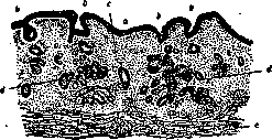
Mucous membrane of the stomach in malignant anemia
Anemia [figure caption]: a, b - surface epithelium, c - proliferated stroma; d - remnants of glandular tissue; e - muscularis mucosae. Changes in the digestive tract in malignant anemia are quite constant and characteristic. A frequent and very early symptom is the so-called Hunterian glossitis, expressed in the smoothing, hyperemia, and a sort of excoriation of the papillae ('papillitis') against a background of general atrophy of the epithelium, especially of the root of the tongue, with simultaneous atrophy of its lymphadenoid tissue; shallow cracks may also be present here, as well as mild inflammatory changes (infiltrates). The clinical reflection of this is the symptoms of burning or pricking at the root of the tongue and in the pharynx during meals and conversation. This symptom, even in the absence of others, has a certain diagnostic significance; however, it is also observed in other blood diseases, for example, in leukemias; apparently, it does not occur in Bothriocephalus anemia. To a lesser extent, but essentially the same processes are described in the pharynx and esophagus. In the stomach, with very rare exceptions, a picture of atrophy of the mucous glands (anadenia) and of the entire mucosa in toto is also observed; it becomes low, almost devoid of folds (see figure), the surface epithelium is low, and the glandular [epithelium] is atypical, being represented almost exclusively by chief cells; the interglandular spaces are wide, filled with lymphoid cells, often with an admixture of myeloid forms. Clinically, this is accompanied by the most important symptom for malignant anemia, achylia and achlorhydria, as well as the absence of pepsin and rennin in the gastric juice. These symptoms belong to the earliest ones, developing as if preliminarily in relation to anemia itself. In the stomach, polypous growths, less often cancers, and hypertrophic changes of the pylorus have also been observed. However, the indicated anatomical-clinical symptoms are not specific for malignant anemia and, although not to the same degree and not as often, are also observed in other severe forms of general anemia. Atrophic changes also extend to the intestinal mucosa; there are indications of degenerative processes in Auerbach's and Meissner's plexuses, and in some cases of ulcerative or stenosing processes in the intestine; examination of duodenal contents, as well as feces, always reveals high numbers of bilirubin. The constant presence of significant changes in the digestive tract prompted authors to seek the pathogenetic essence of the disease in these changes; indeed, it was established (Seyderhelm) that, whether in connection with the absence of HCl in the stomach or in connection with atrophic changes in the intestine, with a disturbance of enzymatic processes in it, in malignant anemia, an ascent of intestinal flora into the upper, normally almost sterile, section of the intestine, up to and including the duodenum, is noted; this entails the abundant absorption of toxic substances into the blood and the further development of the disease. In experiments on dogs with artificial stenosis of the ileum, a picture of hyperchromic anemia with characteristic anisocytosis, megaloblasts, hemosiderosis of organs, etc., with flooding of the upper segments of the intestine with bacteria, sometimes indeed developed; in negative experiments (the majority), such flooding did not take place. Seyderhelm, on the basis of this, not only comes to the conclusion that malignant anemia is the most severe degree of intestinal autointoxication, but also proposes an operation to create an artificial anus to prevent the ascent of flora from the large intestine. This operation did not have much success. Seyderhelm's theory became widespread, mainly in Scandinavia, where cases of Bothriocephalus anemia were also cited in its favor, in which the presence of helminthic toxins in the intestine played a role in the origin of the disease. An objection to this theory is that out of thousands of carriers of the indicated worm, a pernicious anemic blood picture develops only in individual persons, that these anemias are far from covering all the symptoms of typical Biermer's malignant anemia; finally, a number of control studies by other authors did not confirm any constancy of the described changes in the flora of the gastrointestinal contents. In the central nervous system, phenomena of severe anemia are noted, and sometimes, and according to some authors in 1/3 of all cases (especially with a relatively acute course of the disease), focal degenerative changes of the white matter of the brain, mainly in the posterior columns of the cervical region of the spinal cord; clinically, various disorders of the motor and sensory spheres take place; in the brain, the indicated changes are also observed simultaneously with the degeneration of groups of ganglion cells of the cortex. The glands of internal secretion do not present special changes; in recent years, attention has been drawn to changes in the thyroid gland in the form of diffuse or nodular goiter (struma), sometimes atrophy and sclerosis of the organ. The genital organs and respiratory organs do not present anything characteristic. The kidneys were mentioned above. The urine is of low specific gravity, but of a saturated yellow color, and contains urobilin and urobilinogen. Albuminuria and cylindruria are sometimes encountered but are not characteristic. Hematuria and glucosuria are usually absent. The diazo reaction is negative. Indicanuria is observed almost always. In its course, malignant anemia has several remissions (usually two), occurring, apparently, independently of the treatment being administered. During remissions (as well as at the beginning of the disease), all the main symptoms partly soften, partly disappear altogether. The number of erythrocytes and Hb increases, rouleaux formations appear; the color index falls to 1 and below. It is pointed out, however, that even during remissions, findings of megalocytes are not rare, which makes one doubt that there is an ordinary (non-malignant) anemia or that there is a real recovery of the patient. Thrombopenia and relative lymphocytosis are more often preserved during remission, or here too the ratios approach normal. There are observations that the bone marrow in the period of remissions also returns to its original state, again turning into yellow [marrow] in the long tubular bones. Simultaneously, a number of other symptoms disappear or soften, such as: bilirubinemia, urobilinogenuria, waxy pallor of the skin, glossitis; a flush appears and well-being improves, work capacity increases, etc. Achylia always remains during the period of remission. Remissions last for months, rarely years, and inevitably lead to a new exacerbation of the disease, which, in particular, can be judged quite early by the intensification or appearance of signs of hemolytic processes (urobilinogenuria, etc.). Among the rarer clinical forms of malignant anemia, the following are distinguished: a) Pernicious anemia in children occurs more often in an aplastic and hemorrhagic form. The course of the disease is mostly acute; in children with congenital syphilis, pernicious anemia proceeds with splenomegaly, mild fever, nosebleeds, and hemorrhages in the retina; the liver is enlarged; diarrhea and gastric achylia are quite frequent. b) Anemia with sickle-shaped erythrocytes (sickle-cell anemia) is usually a hereditary and familial disease; it occurs only in Negroes; picture of the disease: fever, yellowish-green coloration of the sclera, often enlargement of the liver; the spleen is normal; intense pain in muscles and joints. The course is in attacks with temperature up to 38°, duration 2-3 weeks; in the blood - a decrease in Hb and erythrocytes, the color index is within normal limits; anisochromia; poikilocytosis and anisocytosis; sickle-shaped erythrocytes, stained more intensely than others, are characteristic; some of them have nuclei; leukocytes - from 15 to 60 thousand; the remaining symptoms are the same as in ordinary malignant anemia. c) A. phagocytaire of French authors - a syndrome observed by only three authors (Malins, Barhett, Rowley) and characterized by sharply expressed phenomena of anemia and splenomegaly (which may be absent), usually ends in death; characteristic is the presence in the blood of a large number of polynuclear cells and giant monocytes, reaching 30-50µ and densely packed with ingested erythrocytes (sometimes more than 20); in the phagocytes, in addition, deposits of blood pigment are found. Statistics, Russian and foreign, show that malignant anemia has been increasing in number over the last decades; in particular, in Moscow, according to data from pathological-anatomical offices, in the single year 1925, 25 patients were autopsied against 10-15 in previous years. IV. Chlorosis. Chlorosis (from Greek chloros - green), or green sickness, is a special form of chronic anemia almost exclusively in young women, characterized by a number of clinical-hematological signs distinguishing it from other forms of anemia. Chlorosis is not only a disease of the blood and hematopoietic organs; the clinical picture and pathology of the disease are far from being exhausted by blood pictures. The disease is usually clearly outlined by the period of puberty of girls, and sometimes earlier; individual cases are noted at an older age (chlorose tardive Hayem's - late chlorosis) and even in the climacteric period; in individual cases, chlorosis is observed in young men having a somewhat feminine type of build.
However, in older women and in men, the disease does not proceed quite typically (Noorden's chlorosoid). The disease lasts for a number of years and usually ends in recovery. Mortality is zero—a circumstance that partly explains the poor familiarity with the pathological anatomy of the disease. The causes of the development of chlorosis are not yet known with precision. They point to the significance of a number of external living conditions related to profession, type of diet, upbringing, housing conditions, etc.; in particular, they point to the insufficiency of time spent in the fresh air and in the sun, which is associated with physical activity. A prominent place must also be given to congenital constitutional factors, such as anomalies of an anatomical and physiological order, in particular, on the part of the skeletal and cardiovascular systems and the blood-forming function, and especially anomalies on the part of the reproductive sphere, namely the ovaries. Hereditary predisposition also plays a role, mainly on the mother's side; Tandler pointed to cases of eunuchoidism in brothers, etc. On the basis of these predisposing factors, the influence of external factors acquires great significance. Chlorosis is generally not a frequent disease, and in recent decades it has become increasingly rare. There is no explanation for this phenomenon, but it is suggested that the disappearance of a number of external factors contributes to this; they point, for example, to the cessation of lacing in girls, which entailed a disturbance in the function of the liver and spleen—these primary organs of iron metabolism, since ultimately a direct or indirect disturbance of this metabolism plays a most important role in chlorosis; changes in the lifestyle of adolescents and young women have had an influence, as they do not hide from the sun, which enlivens the processes of decay and regeneration, but cultivate sports in their environment alongside boys. Among the poor classes of the population, chlorosis is apparently no more frequent than among the affluent groups of the population, which speaks in favor of the idea of Martius that if a young girl is free from a predisposition to chlorosis, then under unfavorable external conditions she may become anemic, weak, or tuberculous, but not chlorotic. Much of what was previously considered a cause of chlorosis is now viewed as its consequence (e.g., menstrual disorders, enteroptosis, dislike of movement or physical exercise in connection with rapid fatigue, etc.). The skin and mucous membranes of chlorotic women are extremely pale, with the face taking on a somewhat greenish hue, especially in blondes. Paleness is combined with a general decrease in skin pigment, and some depigmentation of the hair is also possible; a weak tendency to tan is well known. In rare cases, patients look rosy (chlorosis rubra), which is explained by the thinness of the skin, in particular the epidermis, as well as the dilation of the vascular network, and perhaps by a constant rush of blood to the face, which is sometimes associated with improper ovarian function. The subcutaneous fat layer is usually well developed, sometimes excessively so, and this is so characteristic that the opposite picture is one of the arguments against chlorosis. The face is puffy, sometimes edematous; however, edema of the ankles and eyelids is usually insignificant and of a transient nature (e.g., appearing during the day and disappearing at night). The pathogenesis of this edema has not been studied, but it likely belongs to phenomena analogous to the so-called edematous disease (see); a certain role is apparently also played by a decrease in proteins in the serum of chlorotic women. In older patients, there are abundant fat deposits in the internal organs, especially in the epicardium and in the mesentery. The height of chlorotic women is usually above average, the body type is coarse, with masculine features, especially the skeleton. Tandler points to an infantile pelvis, short legs, and a certain prematurity in the development of primary and secondary sexual characteristics. Regarding the circulatory organs, since the time of Virchow, it has been customary to point to hypertrophy of the left ventricle and a narrow, delicate, unusually distensible aorta; at present, the pathognomonic nature of these symptoms is denied: cardiac hypertrophy in chlorosis is not at all a frequent phenomenon, and the question of a narrow aorta (aorta angusta) has been clarified in the sense that this is by no means a regular finding in chlorosis, but is observed in various blood diseases. During clinical examination, an increase in the transverse diameter of the heart is often found, but this is explained not by its hypertrophy, but by a certain displacement of the longitudinal axis of the heart due to the high position of the diaphragm; the latter is caused by a peculiar infantile positioning of the ribs and is considered by some as a degenerative sign. Hypotonia of the vascular wall, a certain decrease in blood pressure, and in connection with this, a large, soft dicrotic pulse, sometimes a capillary pulse, and a double tone on the thigh complete the symptomatology. Among subjective sensations, it is necessary to note: absence and perversion of appetite—the latter appears, mainly, for acidic food; taste perversions are not rare—a desire to eat chalk, toothpaste, coal, etc.; sleep is good, but does not refresh in the morning; frequent headaches, lack of capacity for work. The mass of blood is within normal limits, sometimes increased. The specific gravity fluctuates depending on the Hb content; the water content is increased, and that of iron and proteins is decreased. The blood is paler than usual, liquid, watery. Microscopically: erythrocytes form small rouleaux or do not form them at all; they are noticeably paler than normal, and along with intensely colored ones, pale (large) ones are noted. Among them are many pessary-shaped forms. Anisocytosis and poikilocytosis are not rare, and the stronger the drop in Hb and the number of erythrocytes, the more pronounced these phenomena are (see table for article 287-288, fig. 7). The amount of Hb is always lowered, more significantly than the decrease in the number of erythrocytes; sometimes erythrocytes remain within normal limits, but the amount of Hb is lowered even in such cases. Such a reduction of Hb (oligochromemia) leads to a significant decrease in the color index, down to 0.6-0.4. Its decrease remains even in those comparatively rare cases when chlorosis is combined with erythrocytosis, e.g., in the period of recovery during appropriate therapy. Polychromatophils and basophilic stippling are noted, mainly, during improvement and treatment with iron; normoblasts are present in small numbers, hemolytic processes are absent. The number of white blood cells is within normal limits; a decrease in lymphocytes is often encountered (however, there are also opposite observations). Blood platelets always remain at high figures; hence, apparently, the tendency of chlorotic women to thrombosis, especially of the veins of the lower extremities and cerebral sinuses; thrombosis of the extremities (more often on the left) is usually preceded by muscle tension. Thrombosis of the arteries is rarely observed. The spleen is of normal size or slightly enlarged; the lymph nodes are poorly developed, flat. There is always shortness of breath, even with insignificant movements. This symptom is noticeably intensified in the presence of an sometimes encountered intrauterine or congenital (as a result of endocarditis) narrowing of the mitral valve (Duroziez's disease). An important place among the symptoms of chlorosis is occupied by atonic or hypotonic phenomena on the part of organs with smooth and striated musculature and the ligamentous apparatus, depending on which one observes a tendency to constipation, dilation, ptosis, and a decrease in the motility of the stomach, irregularities in the position of the uterus, prolapse, wandering kidneys, weakness of the skeletal musculature, etc.; the hypotonia of the vessels also belongs here. The secretion of gastric juice is without special changes, but cases of hyperchlorhydria, or (more rarely) hypochlorhydria, achylia are not rare. Atonic phenomena may be absent or even replaced by spastic ones, e.g., on the part of the pylorus, the large intestines (spastic constipation, sometimes of the colica mucosa type, with pains in the stomach region, etc.). Atonic and spastic phenomena are not in direct connection with chlorosis, being a partial manifestation of general constitutional anomalies, especially on the part of the autonomic nervous system. The urinary organs are usually not changed. They often point to polyuria with a decrease in the specific gravity of the urine, and sometimes to mild albuminuria, usually without renal elements; since albuminuria almost always disappears with bed rest, it is customary to classify it as so-called orthostatic albuminuria. Nephritic phenomena are absent. A very prominent place is occupied by developmental anomalies and disorders in the reproductive sphere—small ovaries, infantile pelvis, disturbances of the menstrual and ovulatory cycles (namely, their suspension) or scarcity and painfulness of menstruation; menorrhagia is rare. Leukorrhea is observed especially during the period of the onset of amenorrhea. The mammary glands are usually well developed. Important significance is attributed to ovulation disorders and, in general, to the disturbance of ovarian internal secretion in the pathogenesis of chlorosis. By what path these disturbances cause the chlorotic symptom complex, in particular, the question of the connection of these disorders with blood changes, remains unclear; there exist, however, experimental observations concerning the reproduction of the chlorotic blood picture by castration (of rabbits), as well as observations on the strong slowing down of blood regeneration after castration.
They point to an enlargement of the thyroid gland, sometimes they even speak of chlorotic goiter with a whole series of symptoms of Basedow's disease. On the part of the nervous system, manifestations of hyperfunction of the adrenal system are typical. Changes are observed in the fundus of the eye, e.g., pulsation of the retinal arteries, venous pulse; rarely—neuritis optica, neuroretinitis, atrophy of the optic nerve head, hemorrhages. The course of the disease is, as a rule, afebrile, although exceptions here are not rare and rises in temperature up to 38-38.5° without special causes have been described (Dvukraev). Fever most often has a constant character (febris continua). Deteriorations (relapses) and improvements (remissions) in the course of the disease are not rare; relapses can be associated with certain times of the year (winter, summer chlorosis, etc.). Among the complications in the course of chlorosis, one should keep in mind, as the most frequent, tuberculosis, gastric ulcer; thrombosis and heart defects were mentioned above. Statistical data on chlorosis are difficult to provide; in recent decades, a clear tendency toward a widespread and strong decrease in morbidity has emerged, the possible reasons for which have already been discussed. Clinical forms of chlorosis: 1) Chloranemia of infants (oligosideremia of French authors); in this case, it concerns infants 12-18 months old; this disease is encountered significantly less frequently before 12 and after 30 months from birth. In patients, a waxy or greenish-tinted skin color is noted; it is striking that at the same time, the patients have no emaciation; children are always sad and apathetic; on the part of the digestive organs, there is either anorexia or bulimia and inconsistent gastrointestinal disorders; temperature is subfebrile; on the jugular veins—a humming-top murmur; on the pulmonary artery also a systolic murmur; the spleen is not enlarged; there is neither disease of the bronchial glands nor signs of rickets. In the blood, significant oligosideremia is found, sometimes up to 30 and 20%; the number of erythrocytes is either normal or subnormal; moderate anisopoikilocytosis, polychromasia, nuclear forms do not occur. There are no deviations from the norm on the part of leukocytes and platelets. The course of the disease is favorable. Heredity, too prolonged feeding with milk, and premature birth play a role in the etiology. 2) Chlorosis with severe anemia. This includes cases that clinically have all the signs of true chlorosis, and hematologically resemble pernicious anemia. In the blood, sharp oligocythemia (up to 1,500,000 erythrocytes), anisocytosis, polychromasia, and nuclear forms (normoblasts and less often megaloblasts) are found; the color index is normal or slightly lowered. The course is favorable. After appropriate energetic treatment, complete recovery occurs. 3) Chlorosis without chlorotic blood changes. Cases of chlorosis with 90% Hb and 4-5 million erythrocytes in the presence of all other clinical symptoms of true chlorosis have been published (Morawitz, Rombley, Rolly). Rolly calls these cases 'pseudochlorosis'. Clinical varieties of chlorosis also include chlorosis without menstrual disorders, encountered among young women of the wealthy classes (Hayem, Luzet, Aubertin), with sharp oligosideremia (30-40%) and oligocythemia (2-3 million red blood cells), with characteristic pallor and asthenia. I. Davydovsky. U. Anemia in childhood. Anemia in childhood is encountered extremely frequently; according to data from the Moscow prophylactic children's outpatient clinic, based on an examination of more than 20,000 children, there were from 49 to 71% anemic children in 1924-1925. The etiology of childhood anemias is very diverse and for very many forms is not yet clarified; in many cases, not one cause acts, but a whole complex of them; the majority of causes inducing anemia in children can be combined into the following groups: 1) factors of a constitutional order, 2) nutritional defects, 3) infections and intoxications, and 4) unhygienic external conditions. Constitutional anemia in children includes primary anemia observed in the first months of life in almost all premature children and in twins. The blood of a premature newborn child usually contains the same increased number of erythrocytes (6-7 million per 1 cubic mm) as the blood of a full-term one; the amount of Hb is also correspondingly increased, so that the color index = 1; only immature elements (erythroblasts, myelocytes) are encountered more often in the blood of a premature infant. During the first weeks of life, the number of erythrocytes and Hb decreases, but in a premature infant it does not stop at the normal level, but goes further (as Gundobin first pointed out), dropping to 3.5-3 million erythrocytes and to 60-50% Hb; children become sharply pale and very apathetic; anemia reaches a maximum at 3-4 months, after which in the majority of cases a slow improvement begins ('physiological anemia of premature infants'). In some children, improvement does not come for a long time; anemia may even intensify and drag on until the 2nd or 3rd year of life ('pathological anemia of premature infants' according to Finkelstein). The same anemia almost always develops in twins, even if they were born on time. The cause of this disease was previously seen in an insufficient reserve of iron in the body of the premature child. As Bunge showed, mammalian animals, which for a significant time after birth cannot feed on any food other than milk, are born into the world with a reserve of iron in the liver, which is gradually spent on building Hb until the animal switches to food richer in iron. Since the greater part of iron is deposited in the body of the fetus in the last months of pregnancy, a premature child must have less iron than a full-term one. But this hypothesis does not agree with the fact that in the majority of cases, anemia of premature infants passes after the 3rd or 4th month without treatment and without a change in diet, and that treatment with iron does not always help against this form of anemia. It is more probable that the cause here lies in the not yet sufficiently developed function of the hematopoietic organs in a premature or congenitally weak child (Finkelstein). Constitutional anemias also include congenital hemolytic anemia, which has so far been described in children in a small number of cases. Constitutional predisposition to anemia is an incidental etiological factor in many cases with a different etiology. Alimentary anemia is most often encountered toward the end of the first year or in the second year of life in children who are fed for too long on milk alone or who receive supplementary feeding that is insufficiently varied and in insufficient quantity. Children who are fed artificially fall ill more often; alimentary anemia is observed when feeding with goat's milk. This form of anemia develops very slowly; children can remain plump for a long time, but they become dull, paler and paler; muscle tone decreases, a tendency to constipation develops, the stool often takes on a 'soapy' character, sometimes already at the beginning of the disease an enlarged, rather firm spleen can be detected. In the blood, at first, only a reduced amount of Hb is found with a normal number of erythrocytes. French doctors speak in these cases of the 'chlorotic type of anemia in children'. In the further course, the number of erythrocytes also decreases, anisocytosis, poikilocytosis, normoblasts and megaloblasts appear, the number of leukocytes often increases; children become increasingly weak and pale, hemorrhages may appear on the skin; systolic murmurs are heard on the heart and neck veins; the enlarged spleen can reach the midline of the body; the liver also enlarges. If proper change in the children's diet does not occur, they usually die from some intercurrent infection, as the resistance of these patients to infections is extremely lowered. The pathogenesis of this form of anemia is not yet fully clarified. In view of the fact that breast milk contains very little iron (1-2 mg Fe2O3 per liter), cow's milk even less, and when feeding with goat's milk, iron is particularly poorly retained by the organism (Krasnogorsky), and since with a lack of iron in the food, anemia develops in experimental animals (Hosslin), it was natural to consider that the cause of anemia lies here only in the poverty of the food in iron. However, this anemia does not pass from the mere addition of iron to milk. Czerny and Keller see the cause of the disease in the milk itself; the relatively large amount of fat in this food leads, in their opinion, to the fact that fatty acids, by binding alkalis in the intestine during the formation of soaps, cause a metabolic disorder with alkalopenia. Stoltzner and Glanzmann and others see the cause in the hemolytic action of high-molecular fatty acids, of the capric, caproic, etc. type, of which there is especially much in goat's milk. But, on the other hand, signs of increased breakdown of erythrocytes in the form of an increased amount of urobilin in the intestine or bilirubin in the blood are not always detected, which should have taken place if this hypothesis were correct. New data suggest that the cause of alimentary anemia may be an insufficient supply with food of certain organic compounds, in particular, amino acids necessary for building the Hb molecule (Aron, Gyorgy). According to experimental studies (Simmond, Becker and McCollum), the assimilation of iron is connected with the presence of vitamin E in the food.
According to these data, nutritional anemia should be classified in the group of avitaminoses. Under the influence of prolonged, monotonous carbohydrate nutrition, without a sufficient amount of fats and proteins, and, in general, with quantitatively or qualitatively insufficient nutrition, nutritional anemia can also develop, both in young children and in older ones. Childhood anemias caused by infections can be observed as early as the first weeks of life (for example, in congenital syphilis or malaria), but they are more often observed after infancy. In some cases, only a decrease in the number of erythrocytes and hemoglobin is found in the blood; in others, more severe cases, there is also anisocytosis, poikilocytosis, polychromasia, basophilic stippling, normoblasts, megaloblasts, microblasts, and myelocytes. The number of leukocytes varies; it can be significantly increased, up to 50,000 and more. The spleen is usually enlarged and firm. A systolic murmur is often heard over the heart. In severe cases, dyspnea, general weakness, edema, and hemorrhages into the skin are added to the indicated phenomena. The outcome of these forms depends on the underlying affliction. Unhygienic external conditions, such as, for example, prolonged stays in poorly ventilated rooms, insufficient sleep, irregular meals, mental overwork with a lack of physical exercise, etc., are often encountered as the primary cause of anemia in older children. The number of erythrocytes and hemoglobin is not strongly reduced in this case; general weakness, unwillingness to study or participate in games are observed, and poor appetite, constipation, palpitations, murmurs at the apex of the heart, at its base, and in the cervical veins, headaches, dizziness, and poor sleep appear. This condition often appears six months or a year after starting school (so-called "school anemia"), can last for years, and usually passes with age or under the influence of an improved regimen. (Chloranemia of infants-see above, p. 719.) The pathological-anatomical changes in pronounced childhood anemias are as follows: pallor of all organs, fatty degeneration of the heart muscle, sometimes dilation of the heart; siderosis, especially in the liver; in the majority of cases, a hyperplastic and firm spleen. Sometimes it is difficult to say to what extent the listed changes depend on the anemia and to what extent on another affliction underlying the anemia. A characteristic pathological-anatomical feature of childhood anemias is the formation of additional, usually microscopic, foci of hematopoiesis in various organs-especially in the liver, spleen, lymph nodes, and kidneys-that is, outside the bone marrow, as occurs in the period of intrauterine life of an infant; this return to an embryonic state is observed more frequently the younger the child. In early childhood, sharp changes in the blood are observed under the influence of such insignificant irritants that do not cause almost any reaction in older children; therefore, in the blood of small children, normoblasts, megaloblasts, myelocytes, and other immature and pathological formed elements are found much more often than in older ones. Significant enlargement of the spleen also develops much faster in early age than in older children. A. pseudoleukaemica infantum (Jaksch, 1889), or A. splenica inf., A. pseudo-perniciosa inf., is a severe form of anemia occurring in early childhood and accompanied by splenomegaly, leukocytosis, and a significant number of megalocytes and erythroblasts in the blood. Etiologically, this form lacks any unity and is observed in various underlying afflictions-rickets (mainly), nutritional disorders, syphilis, etc. The essential features of this anemia are apparently conditioned, first of all, by the peculiarities of childhood itself; in particular, splenomegaly in anemic states is very characteristic of this age, especially in the first 3 years of life. The onset of the disease is usually before the end of the first year; girls and boys are affected equally; no definite hereditary predisposition is noted. In artificially fed children, under unfavorable conditions, the disease occurs more often; brothers, sisters, and twins are frequently affected. The first symptoms of the disease usually date back to the first weeks of life, manifesting in a disturbance of general nutrition, appetite, in a number of nutritional disorders, and progressive pallor; soon, enlargement of the abdomen due to enlargement of the liver and spleen and more or less distinct rachitic symptoms are added. Blood examination notes a sharp decrease in erythrocytes (down to 500,000), hemoglobin down to 20% and lower; the color index is usually less than one. Leukocytes, with rare exceptions, are increased in number, sometimes resembling a leukemic blood picture; among the leukocytes, lymphocytes prevail with a significant admixture of medium and large forms; myelocytes are almost always noted, sometimes myeloblasts; an almost complete absence of eosinophils and basophils is characteristic. Anisocytosis, poikilocytosis, an abundance of erythroblasts, megaloblasts with division figures, and thrombocytopenia complete the picture. The abundance of erythroblasts is particularly striking (cases of up to 20,000 per cubic mm have been described, and in individual cases, this symptom is not accompanied by the primary one, i.e., anemia proper). The spleen is enlarged, reaching the navel and lower, smooth, firm, mobile, and painless. The lymph glands are always somewhat enlarged; their microscopic examination reveals a picture of diffuse myeloid metaplasia, observed in typical cases of myeloid leukemia in adults, with the difference that in the childhood form, mast cells and eosinophils are absent. The formation of myeloid tissue, as in typical leukemias, can occur in various tissues and organs, such as: in the connective tissue, kidneys, liver, often accompanied by hemosiderosis of the parenchyma of the latter. Such changes gave many authors the right to speak of a childhood form of myeloid leukemia, and, indeed, the similarity here is, in essence, quite significant. However, in atypical cases, the similarity is lost or more similarity arises with malignant anemia; thus, for example, there may be splenomegaly and anemia without leukocytes and myelocytes and even with leukopenia, sometimes anemia with erythroblasts. Taking into account that rickets, syphilis, and other infections or metabolic disorders themselves can be accompanied by enlargement of the spleen, simple anemia, etc., one can conclude that A. pseudoleukaemica infantum is a symptom complex with an abundance of transitional forms and variants; at the base of this complex lies the peculiarity of the reaction of the child's organism, its hematopoietic organs, and the spleen, expressed in a more lively and extensive myeloid reaction and a greater tendency toward the embryonic type of hematopoiesis. In the words of Naegeli, this is a biological variant of any secondary anemia of childhood, and, moreover, of the most varied etiology. Among lesions of other organs, frequent catarrhal processes in the respiratory and digestive tracts and pneumonia are noted. Moderate fever often accompanies the disease. In very severe cases, a picture of hemorrhagic diathesis develops. The disease lasts for months, sometimes (with remissions) stretches for a longer period; individual cases end lethally in a few weeks. In half of the cases of ordinary severity, recovery occurs, with the enlargement of the spleen lasting the longest, sometimes remaining for many years as a witness to the endured affliction. More or less sharply pronounced anemia with an enlarged spleen is often encountered in rickets; whether this is merely the result of the coincidence of two very common pathological states in childhood or whether there is any etiological connection here (inadequacy of food, changes in the bone marrow) remains open for now. Diagnostics. When diagnosing anemia, one should keep in mind the "imaginary anemia" or pseudoanemia of Sahli, which is encountered in both children and adults. In it, despite the striking pallor of the skin, the content of hemoglobin and erythrocytes in a unit volume of blood remains normal. Pseudoanemia is observed either in nervous individuals with a spastic state of the skin vessels, often in combination with a narrow aorta and a small heart, or in obese subjects in whom, due to the thickness of the subcutaneous fat layer, the vessels do not show through much or at all, or when the skin vessels are generally weakly developed. Consequently, for a correct diagnosis of anemia in all cases, an examination of the number of erythrocytes and hemoglobin is necessary. As for the diagnostics of various anemias in general, it does not present special difficulties in the majority of cases; it should only be emphasized once again that the diagnosis must be based not only on one or another blood picture but also on the clinical symptomatology itself. Prevention. The prevention of anemia relates almost exclusively to anemias of the nutritional and occupational types and, in essence, coincides with the requirements of the hygiene of nutrition, upbringing, and labor. In particular, in relation to childhood anemia, the following are especially important: proper organization of nutrition in infancy (e.g., prohibition of feeding with goat's milk), timely transition from milk food to a more varied diet, prevention of acute and chronic infections, etc.
Thus, a role in the prevention of childhood anemia can be played by social-hygienic institutions: consultations for women, for children, teaching mothers proper feeding, playgrounds, summer colonies, etc.
I. Davydovsky, E. Lepsky.
VI. Therapy of anemia. Rational therapy for anemia must be directed first and foremost against the underlying disease, of which the given anemia was a consequence. However, it is not uncommon for the anemia to remain in the same state despite the elimination of the primary cause, due to insufficient regeneration of the hematopoietic organs. In practice, it is very often impossible to radically eliminate the underlying disease; in such cases, one must primarily treat the hematopoietic organs, having tested their regenerative function and stimulated it to increased activity. Thus, the therapist must be guided by the following principle: treat the underlying disease, and "irritate" the hematopoietic organs only in case of their insufficiency. In acute post-hemorrhagic anemias, the first requirement of therapy is the cessation of bleeding, i.e., the detection of the bleeding vessel and its ligation (see Bleeding, cessation of). If, due to certain conditions, for example, in internal hemorrhages (gastric ulcer, erosion of esophageal varicose veins, etc.), surgical intervention is impossible, the following measures must be taken: 1. Place the body in a recumbent position, raising the foot of the bed; thus, by gravity, a greater amount of blood will tend toward the brain and heart. 2. One can also bandage the lower extremities sufficiently tightly with an elastic bandage to displace the greatest amount of blood toward the heart (autotransfusion). 3. To increase blood pressure, introduce fluid into the bloodstream. Here, the best means is a sterile physiological solution of sodium chloride at a concentration of 7:1,000 or a more complex Ringer's solution; inject into the subcutaneous tissue or directly into a vein. The addition of 5-7 drops (per liter) of adrenaline (1:1,000) enhances the effect of such an infusion. The same goal is achieved by drinking hot liquids or by rectal infusion. This latter method is based on the ability of the mucous membrane of the rectum to absorb water. It is best to use the drip method: a thin Nélaton catheter is inserted into the rectum, connected by means of a long rubber tube to a reservoir in the form of an irrigation mug, standing nearby and slightly higher than the bed (50 cm). The reservoir contains physiological solution in a warm state (about 40°). An ordinary glass device for measuring falling drops or an accurate tap is included in the middle part of the rubber tube. Thus, during the day, it is possible to introduce a large amount of fluid into the body (up to 1-2 liters). Bayliss, based on the experience of the imperialist war, recommends infusions of a solution (3-6%) of gum arabic in Ringer's solution. 4. Recently, blood transfusions have again become widespread, taken from a suitable donor, in terms of group agglutination, from his radial artery connected to the vein of the recipient's elbow (see Blood transfusion). Robertson and Bock consider transfusion indicated only if the amount of hemoglobin falls below 25%; with a larger amount of hemoglobin, in their opinion, one can limit oneself to saline infusions. French authors consider a drop in maximum blood pressure below 9 (according to Pachon) and minimum below 3 as one of the indications for transfusion. Recently, there has been talk of blood reimplantation, i.e., the reverse infusion of escaped blood; for this, the effused blood (e.g., into the peritoneal cavity, in ectopic pregnancy) is collected sterilely, filtering it through gauze into a solution of sodium citrate, and the citrated blood is infused back into the patient's vein. 5. A good remedy against progressive symptoms of asphyxia is oxygen inhalation. 6. As for agents that increase blood clotting (calcium chloride, injection of dry extract from lung tissue, coagulen), they should not be neglected. 7. Stimulants are also prescribed: caffeine, camphor. In cases of bleeding from the gastrointestinal tract, it is recommended to use 10% gelatin or an aqueous extract of Hydrastis canadensis internally. For postpartum hemorrhages, Ergotin is prescribed. To accelerate the restoration of blood, mainly hemoglobin, the administration of significant doses of iron for a long time is useful, especially in persistent states of anemia in connection with repeated hemorrhages. As a stimulating therapy for sluggish erythropoiesis, it is recommended (Dufour and Le Hello) to inject anti-hemorrhagic serum obtained from a rabbit during anaphylaxis; Aubertin considers this serum very active and recommends it in particularly severe cases of hemorrhagic anemia. Good results in terms of erythro-regeneration from an irritating dose of thorium-X have been published. The principle of treatment for chronic post-hemorrhagic anemia is the same as for acute. Upon cessation of bleeding, stimulating therapy should be prescribed. Until now, most authors in this regard still place iron and arsenic in the first place. With normal blood and hematopoietic organs, iron preparations either do not act at all or act very insignificantly; but with damaged hematopoietic organs, iron has an energetic stimulating effect. The mechanism of action of iron is not yet known exactly. It is thought that iron accelerates the synthesis of hemoglobin and prompts the bone marrow to strengthen its hematopoietic functions (Naegeli). The fact of iron absorption was established by Hofmann; Abderhalden, when feeding young animals food with the addition of iron, found a higher iron content in their blood than in the control group. At present, most authors believe that organic iron preparations do not have any stimulating effect. On the contrary, inorganic preparations cause an increase in hemoglobin. The best iron preparation is Ferrum hydrogenio reductum, prescribed in cachets 3-4 times a day at 0.5. It must be noted, however, that some American authors (Whipple, Robscheit-Robbins) are skeptical about iron treatment and use it little. The attitude toward arsenic as a therapeutic agent has become more critical and cautious at present. Considering arsenic a hemolytic poison, Shustrov points out that an irritating effect on the bone marrow is possible only from small doses. Shustrov considers it contraindicated to use arsenic as a stimulating agent in relation to chronic post-hemorrhagic anemias. Modern American doctors are beginning to advocate exclusively dietary therapy for anemia. Whipple found that certain food products act successfully on blood regeneration. Among them, raw meat, boiled veal or beef liver, and cow butter fats are in the first place; next are spinach, cereal porridges, and milk; animal fats—fish oil, lard, as well as fish, oysters, and onions—act negatively on hematopoiesis. The mechanism of action of the mentioned diet is still completely unknown. Therapy for malignant anemia is currently still powerless in terms of its radical cure. All the variety of proposed therapeutic procedures can be reduced to three main groups: agents against intoxication, slowing down the increased breakdown of erythrocytes, and agents stimulating the hematopoietic function of the bone marrow. The fight against intoxication comes down, firstly, to establishing a general hygienic regimen (oral hygiene, fresh air, sun, etc.) for the patient and to a number of agents directed against intestinal intoxication (high enemas, collargol, large doses of HCl per os, kefir, sour milk, milk). In addition to this, Winterfeld proposed treatment with coli-vaccine, prepared from the flora of the duodenum. To reduce the breakdown of erythrocytes, splenectomy has been proposed, which in some cases has yielded good results in terms of prolonging remission; for increasing the resistance of erythrocytes, treatment with cholesterol has been proposed. Of the methods cited, none has received general recognition. A great many agents stimulating the activity of the bone marrow have been proposed. From the chemical arsenal, some authors assign the first place to arsenic, administered per os or subcutaneously; in the first case, according to many, the hematopoietic effect is better than with the second method. The dosage of arsenical preparations is either standard or very high. Neisser initially gives 10 mg per day and over the course of a week brings it up to 100-150 mg; in addition to subcutaneous injections, arsenic is introduced into the vein in the same doses. Byron Brawelle proposed salvarsan therapy with small doses. The opinions of various authors on the use of arsenobenzene preparations differ. Opotherapy. For the treatment of patients, red bone marrow of young animals is used with preliminary microscopic control. An abundance of nucleated erythrocytes indicates the suitability of the marrow for treatment. It is given internally in a pureed form with the addition of either sugar or jam; it can also be prescribed with bread or broth; dose: from 75 to 500 g daily. Marrow jelly is more pleasant and easier to digest; it is prepared from gelatin and glycerin. Dietary therapy. Americans Gibson and Howard have recently, based on the experimental trials of Whipple and Robscheit (feeding dogs with post-hemorrhagic anemia boiled beef liver), performed similar experiments on patients with pernicious anemia, prescribing them daily, in addition to liver, fresh vegetables, fruits, and egg yolks, and reducing fats to the absolute minimum.
Minot and Murphy repeated the mentioned experiments on 45 patients with malignant anemia, prescribing the following dietary regimen: 120-240 g of boiled calf or beef liver, with its replacement by an equivalent amount of lamb kidney; 120 g of lamb or beef; 300 g of lettuce and spinach; 250 g of milk and 40 g of butter. The results were good: after a month-long course of treatment, erythrocytes increased from 1.47 million to 3.4 million (on average); after two months of therapy, their number increased, on average, to 4.16 million, and after 4-6 months to 4.5 million. Ettinger modified the Minot and Murphy regimen, prescribing 200 g of raw calf liver to patients along with a normal clinical diet; the results were very good. To all severely ill patients, after 1-3 months of treatment, strength and working capacity returned; hemoglobin and erythrocytes in many reached almost normal figures. Treatment of pernicious anemia with raw liver is being conducted in Moscow clinics with a very good result. The Belgian Lemain described 6 cases of Biermer's disease treated with the Minot and Murphy diet, also with good results. Serotherapy. Based on the experiments of Roger, who showed that antidiphtheritic serum causes rapid multiplication of nucleated erythrocytes in the bone marrow of rabbits, Renon and Tixier applied this method for the treatment of patients with pernicious anemia. Practically, this method did not yield great results. Treatment with goat hemolytic serum (Gourmont, Andre) also produced an increase in the total number of erythrocytes and eosinophils, but clinically this method also proved of little use. Treatment with hematopoietic serum obtained from a horse during the period of blood regeneration after artificial bloodletting has also been proposed; such serum contains a stimulating substance for hematopoietic organs; but pernicious anemia yields less clear results than hemorrhagic anemias. Hemotherapy of pernicious anemia is quite common. Either intravenous infusion of large amounts of blood or intramuscular injections of small portions of citrated blood (20-40 cc each) are performed, with repetition every 2-3 days. According to French authors, treatment of the disease with small doses of blood is in no way inferior to large transfusions. The main goal of therapy is to induce irritation of the bone marrow; after the first blood transfusion, a more vigorous regeneration is always obtained than after the second and third. It must be agreed that hemotherapy yields only temporary success, since the erythrocytes introduced into the bloodstream are destroyed after a very short time. Radio- and thoriotherapy. X-rays, which destroy bone marrow in large doses, according to the observations of Aubertin, in moderate dosage cause its irritation and hyperplasia, which is clinically manifested by leukocytosis. A number of cases of diseases treated exclusively with radiotherapy have been published, which yielded an increase in erythrocytes to 4 million. Aubertin administered a dose of 6 H to various epiphyses and the sternum, as a result of which such an improvement occurred that patients were discharged from the hospital. After irradiation, deep pains are felt in the bones. Renon and Tixier note an increase in nucleated erythrocytes, myelocytes, and eosinophils after each session. In its effect on hematopoiesis, radiotherapy is analogous to arsenic therapy. Thorium-X is used in the form of subcutaneous injections; the dosage technique is very simple, as ready-made ampoules with thorium with a dosage from 20 to 500 mg are available for sale. Thorium injections are given weekly, starting from 50-60 mg and reaching up to 100. The course of treatment is 5-8 injections in total. A dose of 300-500 mg is used for the treatment of leukemias, but it is not permitted for pernicious anemia. A whole number of authors have noted good results with this treatment. Removal of part of the bone marrow was performed by Kofer and Schraum on 23 patients. They perform either scraping of the bone marrow from the long bones or irrigation of the bone marrow canal. After the indicated operation, active leuko- and erythropoietic reactions were noted at autopsy, but without the presence of megaloblasts and megalocytes. Treatment of chlorosis. For prophylactic purposes, physical culture exercises are recommended, wearing a corset is forbidden; in the developed stage of the disease, complete rest, life in the countryside or in the mountains are prescribed. Initially, warm, and then cool hydrotherapy and light rubbing of the whole body are useful. Food should be easily digestible (milk and milk dishes) and rich in iron—spinach, egg yolks, lentils, peas. A specific medication is iron, mainly its inorganic preparations. In typical chlorosis, iron does not act to increase the number of erythrocytes, but it quickly raises the color index. In chlorosis with strong breakdown of erythrocytes and a low color index, iron increases both hemoglobin and the number of erythrocytes. Arsenic is of little use in typical chlorosis, but in chlorosis with oligocythemia it quickly increases the number of red blood cells; however, no increase in hemoglobin is observed. In chlorosis complicated by amenorrhea, ovarian opotherapy is indicated; treatment with hematopoietic serum and medullary opotherapy have only auxiliary significance in chlorosis. Therapy of childhood anemias consists of the following methods: 1) treatment directed against the underlying affliction that caused the anemia; 2) general therapy in the form of the use of fresh air, sunlight, establishment of a proper regimen, etc.; 3) dietotherapy—for children under 1 year, the amount of milk should not exceed 100 cc per 1 kg of weight, food should be varied as much as the patient's age allows; food substances rich in vitamins are given—fruit juices, vegetable purees; for older children—fresh fruits and vegetables; boiled liver is useful (for small children—in the form of puree) from 30 to 100 g per day, depending on age; 4) drug therapy is not very reliable.
Related articles
Mentioned in
Cite this page
“Anemia.” Soviet Medical Encyclopedia. English translation of Bolshaya Meditsinskaya Entsiklopediya, 1st ed. (Moscow, 1928–1936), ed. N. A. Semashko. https://sovietmedicalencyclopedia.pages.dev/article/anemia/