Blood Transfusion
Historical document, translated for reference. It reflects medical knowledge of the 1920s–30s and is not medical advice.
Summary
This part of the 1930s Soviet medical encyclopedia article outlines the contents for sections covering the physiological action of transfused blood, contraindications, errors and dangers, and additional data on blood groups.
Encyclopedia article (1928–1936)
687 Physiological action of transfused blood ...
690 Contraindications to blood transfusion ....
707 Errors and dangers in blood transfusion . .
709 Additional data on blood groups . .
Blood transfusion (transfusio sanguinis) has become extremely widespread, especially in recent times, and the importance of this issue is constantly increasing. History. According to old legends, blood transfusion was already performed among the ancient Egyptians and Romans, but blood transfusion was understood as sucking it out with the mouth from opened blood vessels, which in antiquity and the Middle Ages was associated with the notion of "vampires." The "blood transfusion" that was performed on Pope Innocent VIII and is cited in all textbooks must also be classified in the realm of sucking blood out with the mouth. Blood transfusion became possible only after Harvey's discovery of the circulation of the blood (1628), and since then the question of blood transfusion began to assume scientific forms. Already in 1638, Potter, in connection with Harvey's discovery, raised the question of the possibility of blood transfusion, but it was only in 1666 that Lower undertook the first scientific experiments. The first direct blood transfusion from a ram to a human was performed by Denis and Emmerez in 1667, but the unsuccessful outcome of one such transfusion led to the fact that in 1668 blood transfusion was permitted in France only with the permission of the medical faculty. In the 18th century, a stagnation is noted in the question of blood transfusion, and only from 1821 is blood transfusion advanced again in connection with the proposal of Prevost and Dumas on the use of defibrinated blood. In 1838, Dieffenbach and Müller proposed warming the defibrinated blood of animals. Only in 1860 did Panum propose using human defibrinated blood, but even during the Franco-Prussian War (1870-71) a total of only 37 successful blood transfusions were performed. In 1875, Landois first described and analyzed the phenomena of hemolysis after blood transfusion. The further development of the question of blood transfusion became possible after A. Schmidt discovered the essence of the blood clotting process. In 1901, Landsteiner published his isohemagglutination groups, but only much later did this discovery acquire practical significance for blood transfusion. In 1906, Crile and in 1908 Carrel proposed direct blood transfusion via arteriovenous anastomosis. In 1914, sodium citrate began to be used in blood transfusion. Since the World War, blood transfusion has begun to spread rapidly. In our country, during the 19th century, there was the same infatuation with the transfusion of defibrinated blood as in Western Europe (Maksimenko, 1934). During the imperialist war, blood transfusion in the tsarist army found no application. The development of blood transfusion in the USSR began in 1918 thanks to the works of Shamov and Elansky. The first reverse blood transfusion in the USSR was performed by Hesse in 1918. The first proposal on the organization of blood transfusion work in the USSR was made in 1926 by Hesse. The first Institute of Blood Transfusion was organized by Bogdanov in Moscow. Since then, the USSR has been one of the most advanced countries in blood transfusion and has a widely distributed network of institutes and stations throughout the Union. Physiological action of the transfused blood. Blood transfusion should be considered as the transplantation of a liquid tissue—blood. There is no doubt that the transplant finds the most favorable conditions possible for itself in the recipient. Despite this, a part of the transfused blood dies after its transplantation into another organism, even when all the rules of the modern doctrine of isohemagglutination are observed. The question of the time of death of the transfused blood has not yet been finally resolved, and various authors give the most diverse periods (Foerster and Müller, Morawitz, Crile, Coenen, Oehlecker, Ashby and Jervell, Wearn, etc.). In recent times, thanks to an improved method of investigation and differentiation of the red blood cells of the transfused blood in the new host, it has been possible to clarify this controversial issue more precisely. For this purpose, the most expedient method turned out to be the transfusion of polycythemic blood to anemics, since the red blood cells of the donor and recipient can be differentiated within two weeks after the transfusion (Wildegans). Ashby used a serum that agglutinated the red blood cells of the recipient and did not agglutinate the erythrocytes of the donor to determine the duration of erythrocyte survival, and in the Thoma-Zeiss chamber could distinguish the donor's erythrocytes from the recipient's erythrocytes. It is clear that when using the Ashby test, it is necessary to use a universal donor of group O. According to Ashby's studies, erythrocytes died after 30-100 days (Ashby, Hotz, Wearn). Schiff combined the Ashby method with the determination of the M and N factors and was able to add further valuable observations. When transfused into the recipient's organism, a significant amount of protein is introduced. Despite this, Wildegans proved that nitrogen excretion before and after transfusion does not change significantly. In any case, it was not possible to establish signs of increased protein breakdown, which also indicates the preservation of the transfused blood. Summarizing, it must be considered established that the erythrocytes of the transfused blood are preserved in the new host for several weeks, provided compatible blood is used. This is also evidenced by the increase in the number of erythrocytes and Hb after blood transfusion and the clinical effect that is observed after transfusion. It is clear, of course, that the erythrocytes of the transfused blood participate in gas exchange. The substitutional (substituting) role of blood transfusion is based on these facts. Therefore, blood transfusion cannot be compared with the intravenous infusion of saline solution, Ringer-Locke liquid, tutofusin, ukrfusin, and other solutions that are rapidly eliminated from the bloodstream. In second place should be named the immunobiological role of the transfused blood. The antibodies of the serum undoubtedly retain their effect after blood transfusion (Kallius). Wright proposed using blood transfusion for immunotransfusion. For this purpose, there is the possibility of using immunized donors vaccinated with one or another type of disease-causing agents. The method of polyvalent immunization is also frequently used. In Leningrad and Paris, central donor organizations possess immunized donors. In third place, the stimulating role of the transfused blood can be noted. Each blood transfusion should be considered as a irritant of the hematopoietic apparatus, thanks to which the number of red blood cells increases significantly. According to the studies of Bogdanov, Belyaeva, and Mayants (from Hesse's clinic in Leningrad), using vital staining methods, it was possible to establish a significant increase in reticulocytes during the first 2-5 days after blood transfusion. The increase in the number of reticulocytes is the clearest and earliest sign of the reactive activity of the bone marrow and hematopoietic apparatus (Naegeli, Seyfarth, Moldavsky, Istomanova, etc.). In fourth place, it is necessary to point out the increase in coagulability after blood transfusion. The hemostatic effect of transfusion is still explained differently. In the case of a deficiency of fibrin ferment in the recipient, it is possible to significantly increase the thrombin content by blood transfusion from a healthy person. But it is undoubtedly necessary to reckon with the direct effect of the transfused blood on the contractile capacity of the vascular wall (Magnus, Stephan, Tannenberg, Wildegans). Substances that promote increased coagulability are contained in the plasma, which is why the latter was used by the Leningrad Institute of Blood Transfusion (Filatov and Kartashevsky) to stop bleeding. According to Wildegans, the increase in blood coagulability persists after blood transfusion for 10-12 days. The peripheral vasoconstrictor effect has been established both for defibrinated blood (Freund) and for citrate blood, wherein old citrate solutions possess a stronger vasoconstrictor effect than fresher solutions (Trendelenburg). The question of the nature of the substances that exert the above-mentioned direct effect on the vascular wall has still not been sufficiently elucidated. There are indications of the significance of breakdown hormones ("Zerfallshormone") and platelets. Next, one must also reckon with the detoxifying effect of the transfused blood, thanks to which blood transfusion can be used with success when the organism is flooded with protein, bacterial, and chemical poisons. Finally, in last place, it is necessary to mention the hypothesis of the founder of the Moscow Institute of Blood Transfusion, Bogdanov, who proposed using blood transfusion on the basis of the "rejuvenating" action of blood. This hypothesis of blood exchange between two persons, which is a propaganda of physiological collectivism, is not new. Elsholtz already writes in 1667 in his "Clysmatica nova" about "transfusio mutua", and Guerin speaks of the same physiological collectivism in 1873 ("Communaute du sang"). Such ideas are ideological delusions and inadmissible reductions of the higher forms of organized matter to the lower ones.
Therefore, Bogdanov's mechanistic and vitalistic mode of thinking, bordering on elements of mysticism, must be rejected. Indications for blood transfusion. Indications for blood transfusion must be based on the aforementioned premises. Blood transfusion is most strongly indicated and yields the best results in acute blood loss. It is known that the loss of more than 2 liters of blood, i.e., half of a person's total blood volume, places the organism on the boundary between life and death. In these cases, blood transfusion is of decisive importance. The administration of saline or colloidal solutions cannot make up for the deficiency in oxygen, whereas transfused erythrocytes, replacing the lost blood, perform its functions and serve as carriers of O2 throughout the body. When blood transfusion is performed for acute blood loss for the purpose of replacement, preliminary care must be taken to stop the bleeding. The effect of blood transfusion in acute blood loss is striking. Patients who have bled out, lost consciousness, and have dilated pupils can be brought back to life (Wederhake, Vaughan, Oehlecker). In the most severe cases, blood transfusion must be repeated and administered in large doses (1 liter or more). In certain cases of severe blood loss (bleeding gastric ulcer, uterine, and hemorrhoidal bleeding), a radical operation becomes possible only after copious blood transfusion. The effect of blood transfusion is especially dramatic in bleeding gastric ulcer (v. Haberer, Dzhanelidze, Hesse, and others). Filatov, Mayants, Kartashevsky, and Depp report on 218 cases of blood transfusion with 107 cases of acute and 111 chronic blood loss. In acute blood loss, 87% good results were obtained from blood transfusion. In chronic and subacute blood loss as well, blood transfusion renders excellent service. The same authors in 91 cases out of 111, i.e., in 82%, obtained unquestionably favorable results. Absolute indications for blood transfusion exist when the hemoglobin content is below 50%. Blood transfusion is of great importance in traumatic shock. In the British, American, and French armies during the World War, the method of blood transfusion for shock became widespread. It became clear that blood transfusion is of the greatest importance in traumatic shock caused by hemorrhage. In so-called reflex primary shock, the results of blood transfusion are poorer (Butler, Wildegans). According to the data of the British Shock Commission, blood transfusion in shock causes not only the replacement of lost blood, an increase in blood pressure, and an improvement in gas exchange, but also the elimination of capillary stasis. Weil and Isch-Wall also emphasize that blood transfusion exerts a beneficial effect in shock due to the elimination of capillary stasis. From these data stems the immense importance of blood transfusion for military surgery and in combating shock in peacetime traumas. Experience of the World War showed that blood transfusion proved its worth even in frontline sanitary units (Wederhake, Coenen, Haberlandt, Constantini-Vigo, Fonio, Kocher, and others). Based on the material of the 1st Surgical Clinic of the 2nd Leningrad Medical Institute, blood transfusion was tested in 72 cases of shock. Out of 43 cases of traumatic shock, there were 20 good, 10 relative, and 13 poor results. Out of 29 cases of postoperative shock, there were 9 good, 12 relative, and 8 poor results (Filatov, Mayants, Kartashevsky, and Depp). The figures of 40% good results and 60% relative and poor results do not prove anything by themselves, but upon analysis of the group of hopeless patients, the usefulness of blood transfusion in these patients is obvious. When blood pressure drops below 70–80 mm, it is necessary to proceed to an urgent blood transfusion. It is impermissible to proceed with an operation if blood pressure is below these figures. After an operation, it is unacceptable to leave a patient without a blood transfusion when blood pressure is below 75 mm. All this dictates the necessity of repeated blood transfusions. The average dose in shock is 500 cm3. The earlier blood transfusion is performed in shock, the better the result (Paunet). Blood transfusion is most effective in the sense of preventing shock. From this follows the significance of blood transfusion as a preparatory measure before major surgical interventions. The importance of preparing patients with blood transfusion before operations is striking. Out of 253 cases of blood transfusion before and after operations, Hesse had a favorable effect in 180 cases (i.e., in 75%). All weak and exhausted patients, especially cancer patients undergoing major surgical interventions, must be prepared in the preoperative period by blood transfusion. Crile points out that since his introduction of the method of blood transfusion, he has managed to reduce postoperative mortality from 12% to 1%Cushing used blood transfusion during brain operations, chiefly in operations in the posterior cranial fossa region, and reports good results. Hesse also used permanent blood transfusion in operations on the brain and spinal cord. When blood pressure drops below 70 mm, 100–200 cm3 are infused. During major operations, such infusions sometimes have to be repeated 3–4 times. In agreement with Foerster, Shields, and others, Hesse notes that this method makes it possible to successfully complete the largest interventions without fearing shock and blood loss. To increase blood clotting, blood transfusion is used in chronic recurrent bleedings, as mentioned above. These include bleeding from a gastric ulcer, intestinal bleeding in typhoid fever and dysentery, pulmonary bleeding in tuberculosis, and bleeding from esophageal veins in liver cirrhosis, and others. With equal success, plasma transfusion can also be performed in these diseases (Filatov and Kartashevsky). Excellent results were produced by blood transfusion in melaena neonatorum. In this dismal disease of infancy, it was possible to reduce mortality from 75% to 5%. In such cases, some authors prefer to perform blood transfusion into the sinus longitudinalis. The dose is 30 cm3 per 1 kg of weight. The most convincing data in terms of increasing blood clotting are achieved in the prevention and treatment of cholemic hemorrhages. Blood transfusion in cholemia was first proposed by Pendl and currently enjoys general recognition (Breitner, Denk, Oehlecker, Wildegans, Hesse, Filatov, Mayants, Depp, Elansky, S. P. Fedorov, and others). According to compiled statistics, 10% of mortality in operations for cholemia is accounted for by cholemic hemorrhages. Kehr cites 5%, Korte 10%, and Fedorov 15% mortality. Hesse had 5.4% mortality in 37 cases with systematic blood transfusion. Once bleeding has begun, blood transfusion is not as effective as when used for the prevention of hemorrhage. Preparation of patients before operation with repeated transfusions of 200–300 cm3 of blood is necessary. In acholic hemorrhages, the results are poorer than in cholemic ones, but blood transfusion is fully indicated here as well. Berczeller and Schonbauer recommend small doses of incompatible blood and report good results. Blood transfusion for the purpose of increasing clotting was also used in malaria biliosa haemoglobinurica—one of the most hopeless forms of malaria. The greatest hopes were pinned on blood transfusion in hemophilia. Blood transfusion is certainly absolutely indicated in hemophilic bleedings and plays a major role as a prophylactic agent before operations in hemophiliacs. Unfortunately, failures have also been described (Bürkle de la Camp, Spitzmüller, Lewisohn, and others). Affecting the hemophilic constitution has not yet succeeded. Blood transfusion is indicated in all hemophilic diatheses. Good results have been obtained in essential thrombocytopenia and Werlhof's disease (Anschutz, Morawitz, Oehlecker, and others). The same results were obtained in purpura haemorrhagica. Blood transfusion must also be used as a preparatory measure before splenectomy (Schaak, Haselhorst). Blood transfusion is also indicated after splenectomy in the event of bleeding. Blood transfusion in primary pernicious anemia for the purpose of a stimulating effect is currently receding more and more into the background, but before the introduction of liver and gastric therapy, it played a fairly large role in the treatment of this disease (Morawitz, Vlados, Boiler); however, this requires repeated transfusions and eventually the bone marrow becomes exhausted. Sometimes remissions are quite long. The best results were obtained in the Biermer form of pernicious anemia (Vlados). Blood transfusion is of great importance in all forms of secondary severe anemia, e.g., in Bothriocephalus latus. Blood transfusion is also indicated in aplastic anemia, whereby the fatal outcome can be delayed for some time. Modest results have been obtained in agranulocytosis (Schultz, Friedemann). Blood transfusion is of considerably less importance in leukemia. Usually, one does not see any improvement from blood transfusion.
In chronic myeloid leukemia, even deteriorations have been described (Jagic). On the other hand, improvements have been observed in chronic lymphatic leukemia. Blood transfusion in leukemia is indicated only in the case of a significant decrease in red blood cells. In myelopathic anemias, lymphomas, and pseudoleukemia, blood transfusion makes no sense. In lymphogranulomatosis, fairly prolonged remissions have been described. In severe cases of chlorosis, an effect from blood transfusion has sometimes been observed. Data on the effect of blood transfusion in severe forms of scurvy are contradictory. For immunobiological purposes, blood transfusion has been used in a number of acute infectious diseases. Blood transfusion has become most widespread in general purulent infection. The opinions of authors on the value of this method vary significantly. The extremely optimistic views of Spasokukotsky and Bürkle-de la Camp are far from shared by everyone (Hesse). Blood transfusion from immunized donors apparently has some advantage over the transfusion of non-immunized blood. The best results seem to be obtained from small doses of incompatible blood (Levin). In chronic general purulent infection, the results are better; especially in the recovery period, the latter can be accelerated by blood transfusion. In endocarditis lenta, blood transfusion gave no result. In acute infectious diseases, blood transfusion was used in typhoid fever and scarlet fever. In septic forms of the latter, good results were obtained by Promptova, who used the blood of convalescents. Benedict recommended treating malignant diphtheria with blood transfusion and obtained good results. In diphtheritic myocarditis, however, blood transfusion is strictly contraindicated. In anemia on the basis of Malta fever (Schottmüller) and in pellagra (Oehlecker), fairly good results were obtained. In pulmonary tuberculosis, blood transfusion has been used repeatedly, but the data are contradictory and at present a definitive conclusion cannot be given (Leitner, Gamble, and others). Good results have recently been obtained in ulcerative colitis (v. Bergmann, Strauss, and others); Ryss, Stroikova, and Vvedensky report good results in heterogeneous blood transfusion. Haberlandt and others recommend blood transfusion in gas phlegmon. Recently, there have been indications of a good effect of blood transfusion in psittacosis. From the point of view of detoxification, blood transfusion has been used in the most diverse types of poisoning. Success can also be expected with that group of poisons which convert hemoglobin into methemoglobin (potassium chlorate, aniline, phenetidine, and others). Mayants reports successful cases in the treatment of aniline poisoning. The effect of blood transfusion in potassium chlorate poisoning is considerably worse. Individual cases with variable success have been described in poisoning with hedonal, chloroform, and other poisons. In case of poisoning, it is recommended to remove the poisoned, altered blood by bloodletting and replace it with fresh blood. The earlier this therapy is initiated and the more persistently it is carried out, the better the result that can be expected. It is clear that repeated blood transfusions are required. In poisoning with carbon monoxide, illuminating gas, and other gases, good results have been described. In some forms of chemical warfare agent poisoning, blood transfusion may be successful, but in poisonings accompanied by pulmonary edema, blood transfusion is contraindicated. In diphosgene poisoning, no result from blood transfusion was obtained (Elansky). For the purpose of detoxification, Spasokukotsky and Seltsovsky, based on experimental and clinical data, persistently recommend blood transfusion in intestinal obstruction. Hesse, based on 20 observations, holds a less optimistic point of view (Filatov, Mayants, Kartashevsky, and Depp). High hopes were placed on blood transfusion in burns. Undoubtedly very good results were obtained with blood transfusion for shock after major burns. Unfortunately, hopes were not justified in burn toxinemia. In severe cases, a fatal outcome cannot be prevented, but thanks to blood transfusion, it is possible to prolong life. The results of Riehl, Imazu, Bogomolova, and Lindenbaum are very modest. In granulating extensive surfaces after burns, blood transfusion, by improving the general condition, promotes more rapid healing of wounds and shortens the recovery period (Bagdasarov and others). Bogomolets persistently recommends blood transfusion in cancer. According to this author, repeated transfusions after a radical operation can prevent recurrences. Blood transfusion has some significance in the fight against cancer cachexia. Sometimes cases that seemed inoperable due to a severe general condition become operable after blood transfusion. There can be no question of a curative effect of blood transfusion on cancer. Blood transfusion has been used in a number of skin diseases (mycosis, pemphigus, lupus, and others), but the results are doubtful. Blood transfusion has been tried in some eye diseases. Arkhangelsky observed a good result after blood transfusion for cataract. Significant achievements have been made in blood transfusion in urology, mainly in various kinds of hemorrhage (Elansky). The effect of blood transfusion in uremia is doubtful (Martin, Lespinasse, Pauchet, and others). Many authors consider blood transfusion in uremia dangerous, because the damaged kidney can be permanently put out of action by the action of the transfused blood (Kuttner, Oehlecker). In gynecology and obstetrics, blood transfusion is indicated for hemorrhages of various origins. In eclampsia, results can be expected only with bloodletting followed by blood transfusion (Seitz, Hempel, Scholl, and others), but opinions on the value of this method differ and failures have been described. Haselhorst, Schmid, and others report good results in hyperemesis gravidarum. In pediatrics, blood transfusion is used besides melaena neonatorum (see above) and in nutritional anemias (Opitz). Contraindications to blood transfusion. There is no doubt that indications for blood transfusion are often set too broadly. Blood transfusion has nowadays become a fashionable method of treatment and is sometimes used for the purpose of "ut aliquid fiat". Blood transfusion is permissible only when the effect is within the bounds of possibility. Unfortunately, there are still no clearly developed contraindications. Recently, the following absolute contraindications to blood transfusion have become clear: 1) All diseases with congestion in the lesser circulation (pneumonia, severe bronchitis). In such cases, blood transfusion can lead to pulmonary edema and death. 2) Organic diseases of the myocardium and decompensated heart defects. Blood transfusion is especially dangerous in diphtheritic myocarditis and acute endocarditis. 3) Purulent thrombophlebitis and thrombosis with phenomena of softening. 4) Embolism with clots and fat. Relative contraindications: 1) Arteriosclerosis and elevated blood pressure. In such cases, bloodletting should be performed preliminarily and functional tests of the heart should be used. 2) Kidney diseases proceeding with oliguria and anuria, chronic nephritis and nephrosis. In doubtful cases, urine examination and the Volhard functional test are required. 3) Vascular thrombosis. 4) In the treatment of leukemia, great caution is required. Serious complications have been described with the infusion of 80–150 cm3. 5) Diseases in which activation of the process is possible (pulmonary tuberculosis). 6) Cachexia. 7) Liver insufficiency. Contraindications for large doses exist in uncontrolled hemorrhages, in particular in bleeding gastric ulcer and in typhoid and dysenteric hemorrhages. Technique of blood transfusion. To this day, questions of technique do not leave the pages of the medical press, and new proposals concerning the technique of transfusion appear incessantly. Filatov and Kartashevsky counted more than 150 different designs and apparatuses for blood transfusion. Such a large number of proposed methods shows that there is still no ideal method that would satisfy in all respects. The classification of blood transfusion methods is still not uniform, and there are major disagreements here. The most widespread is the division of all methods into two groups—direct and indirect. However, while some authors understand transfusion without the addition of stabilizers to it as a direct method (Laqua and Liebig, Kubanyi, Wildegans), others consider a direct transfusion to be one in which there is a direct connection of the donor and recipient vessels using cannulas or apparatus (Beck, Unger). To avoid these disagreements, a proposal has been made to classify blood transfusion methods into the transfusion of whole and altered blood. But it is also difficult to agree with this classification. Finally, recently the division into mediated and immediate methods has become widespread. Filatov (1934) proposed the following detailed classification based on these premises: I. Immediate blood transfusion. 1.
Direct connection of blood vessels (vascular suture, cannulas). 2. Connection using apparatus (Oehlecker, Beck, Tzanck, Landsberg, and others). II. Indirect blood transfusion. 1. Paraffin and non-thrombotic apparatus (Kimpton-Percy, Lampert-Neubauer, Bürkle de la Camp, Merke, and others). (using syringes, 2. Fresh citrated blood with the Landsberg three-way stopcock, 3. Preserved blood; funnels, cylinders, stands, 4. Cadaveric blood
standard jar, Philatov's siphon, and others. III. Reverse blood transfusion (reinfusion). To this classification, one could add on the fourth place heterogeneous blood transfusion, which has recently gained some distribution as a stimulating factor and a potent irritant of the reticuloendothelial apparatus (Cruchet, Bier, Hesse and Philatov, Ryss and Stroykova, Halpern). At the Leningrad Institute of Blood Transfusion, the infusion of heterogeneous blood was used 566 times for gastric ulcer and enterocolitis. The immediate effect of this shock therapy is good. It is too early to speak of long-term results. A 14-month observation period has so far yielded 17% relapses (Ryss, Stroykova, Vvedensky, and Bogdanov). Very recently, Philatov's investigations at the Leningrad Institute have established that the transfusion of defibrinated blood is possible using settled and preserved defibrinated blood. The advantage of this blood lies in its use without stabilizers, due to which complications are observed less frequently. Direct blood transfusion using vascular suture and cannulas has historical significance and is no longer used. Among the apparatuses for direct blood transfusion, the most widespread are those of Oehlecker, Beck, Tzanck, and, in our Union, the Landsberg apparatus. The principle of the Oehlecker apparatus consists in the simple pumping of blood from the donor's vein into the recipient's vein using a tap with two openings and glass syringes. The connection of the donor's and recipient's veins is achieved by an arched metal tube having in the middle a triple tap and an opening for glass syringes. Both ends of the metal tube are connected by means of short rubber tubes with glass cannulas (Figs. 1, 2). Oehlecker originally conceived blood transfusion itself with the help of venesection, and in this one must also see the main shortcoming of the old model. Currently, there are new Oehlecker apparatuses adapted for venipuncture. The experience of the Leningrad Institute of Blood Transfusion with the Oehlecker apparatus encompasses 192 blood transfusions. For the first 50 cases, there were about 10-15% failures due to blood clotting in the cannulas and due to sticking of the syringe piston. When the technique of this transfusion was mastered, failures dropped to 1-2% (Philatov and Kartashevsky). The shortcomings of the device consist in the fragility of the glass cylinders. Experience is needed for the transfusion to go smoothly. The Oehlecker apparatus can be used while performing venipuncture. Transfusion of large doses (500-600 cm3) with needles is difficult, because due to the intermittent blood flow and the large size of the syringe, thrombosis in the system often occurs (Philatov and Kartashevsky). The necessity of using venesection when using old models of the Oehlecker syringe prompted the search for other methods by which blood transfusion could be performed via venipuncture. This principle is observed by Beck, who proposed the Satrans apparatus of a more complex design. Blood transfusion with this apparatus occurs by milking blood from the donor's vein by a system of rollers sliding along rubber tubes passing through the apparatus and being in connection with the recipient's vein. With this method, exposure of veins in the donor and recipient is not required, but venipuncture through the skin is performed. The advantage of the apparatus lies in this and in the fact that blood is outside the vessels for no more than 1 second. Figs. 3 and 7 show the technique of blood transfusion according to Beck. Beck also proposed very practical cannulas for venipuncture, which can be successfully used with other methods of blood transfusion (Fig. 5). The Beck apparatus was tested by the Leningrad Institute of Blood Transfusion in 125 cases. Failures were encountered in 1-2%. The latter occurred mainly with poor donor veins. The shortcomings of the apparatus are cumbersomeness, the necessity of having very well-expressed veins in the donor, and a certain traumatization of erythrocytes due to the milking process (Philatov and Kartashevsky). Based on the same principle of milking is the extremely primitive device of Khrustalyov, used by the author in a number of cases with success. In 1931, Landsberg proposed a new apparatus for blood transfusion. The originality of this apparatus lies in the fact that blood flows directly from the donor's vein into the recipient's vein through an opening made in the piston of a simple Record syringe. To prevent sticking of the piston, there is a special washing system. The Landsberg apparatus was tested by the Leningrad Institute of Blood Transfusion in 97 cases. In 10%, failure was obtained due to thrombosis in the tubes and sticking of the piston. The shortcomings of this device are the absence of a continuous blood flow and the need for great skill, because if certain minor details are not observed, misfires are possible. Finally, when transfusing large doses, thrombosis in the tubes is possible. The Landsberg apparatus is suitable for transfusion of small doses of blood (200-300 cm3). After the publication of the Landsberg apparatus in the foreign press, a report appeared by Küper, who proposed some additions to the Landsberg apparatus. Essentially, he combined two apparatuses—Landsberg's and Oehlecker's—taking from the first the two-way tap and from the second the original syringe with an opening in the piston and a washing system for the piston. All this indicates that Landsberg's ideas, despite some shortcomings in their execution, are undoubtedly fruitful. One of the most perfect apparatuses for blood transfusion by the direct route is Tzanck's apparatus, the advantage of which consists in its simplicity, portability, convenience of maneuvering, absence of fragile parts, and small size of the syringe (Figs. 10, 11). Among Soviet authors, Braytsev and Tsimkhes proposed modifications of this apparatus. Summarizing the analysis of the main apparatuses for direct blood transfusion widespread in the USSR, it must be said that there is still no perfect device and further work in this direction is necessary. The advantages of direct methods of blood transfusion are of course very great, since the blood spends a short time outside the blood vessels and its biological properties are preserved most perfectly. Nevertheless, one should not close one's eyes to the shortcomings of the direct method: the complexity of the equipment and the need to place the donor next to the recipient. In addition, direct methods are contraindicated in acute and chronic infectious diseases of the recipient (general pyogenic infection, syphilis, malaria), because there is a threat of infecting the donor, which can become real in case of equipment malfunction and backflow of blood into the donor's veins. In addition, methods of direct blood transfusion are not always applicable and are definitely inapplicable in advanced institutions under wartime conditions. Therefore, indirect blood transfusion in a number of cases should be given preference. Here the principle of paraffinized vessels is used (Kimpton, Brown, Percy, Davis). The Percy apparatus has become the most widespread. When using it, a rapid transfusion is required, since clotting is delayed for only 10-15 minutes. In addition, venesection is required (Figs. 4, 6, 8). In 1930, Neubauer and Lampert proposed a new apparatus made of a special composition of artificial amber, which they named athrombit. Experimental studies have shown that athrombit is characterized by low wettability in relation to blood and therefore delays its clotting. The most perfect of the athrombit apparatuses is the Bürkle de la Camp apparatus. Philatov and Kartashevsky used this apparatus in 9 cases, and it turned out that preliminary paraffinization of the athrombit apparatus is required. The necessity of paraffinization is also confirmed by Unger. Athrombit apparatuses are expensive, cumbersome, require very careful maintenance and special dexterity in technique. In 1929, Merke proposed an apparatus that deserves attention. Proceeding from the assumption that clotting is delayed when cooling of the blood is prevented, Merke began to use a paraffinized vessel placed in a special heating device—the Merke thermos. Flessa, Philatov, and Kartashevsky could confirm that blood clotting in a paraffinized vessel kept in warm water (38-39°) is delayed for 30-40 minutes. In the USSR, blood transfusion with the help of paraffinized apparatuses is not used; there are only isolated reports (Barinshtein); meanwhile, this method deserves great attention, because it allows blood transfusion to be performed even in the absence of stabilizers. All devices for transfusing citrate blood are divided into 2 groups. To the first belong pressure devices, where blood is injected into the patient's vein under pressure applied to it. These include syringes, the Salibi-Bobrov apparatus (Fig. 13), and others. To the second belong gravity methods, where blood is poured thanks to the pressure difference in two communicating vessels; these include funnels, cylinders, and others. A middle position is occupied by the standard jar and the IM apparatus.
ROKK, in which the infusion can be performed both by gravity and under increased pressure by pumping air with a Richardson bulb. In 1932, Landsberg designed a simple device for transfusing citrated blood. The Landsberg tee consists of a simple Record syringe. Filatov and Kartashevsky describe 378 blood transfusions by this method. Hesse considers the Landsberg tee to be the best apparatus among all available pressure devices. It is simple, portable, cheap, and makes it possible to perform a blood transfusion from any vessel regardless of size and shape (Filatov and Kartashevsky).-Among gravity apparatuses, blood infusion using a funnel or apparatuses working on the principle of the Esmarch mug is most widespread. Disadvantages include too long a path for the blood to travel. In addition, it is impossible to warm the blood and it is necessary to have a separate person to hold these devices in a raised position. When using a funnel, there is also the possibility of infection; this disadvantage is absent when working with a closed standard jar and the ROKK apparatus. The latter apparatus consists of a bottle with a side opening near the bottom. The disadvantage of this method is the difficulty of warming the blood, the necessity of having precisely fitted stoppers, and special devices for holding the stoppers (Figs. 9 and 14) (Filatov and Kartashevsky). The standard jar occupies one of the leading places among methods of blood transfusion by the citrate method due to its simplicity (Fig. 15).-In 1932, Filatov proposed transfusing blood by the siphon method. This set consists of a glass tube 30-35 cm long, bent at an acute angle at one end, onto which a rubber tube 60-70 cm long is placed. The free end of the rubber tube is connected to a needle inserted into the vein (Fig. 12). If this entire system is preliminarily filled with physiological saline solution and the glass tube is lowered into a vessel with blood located at some elevation relative to the level of the recipient's body, then according to the law of the siphon, the blood spontaneously flows into the vein. The Leningrad Institute of Blood Transfusion tested the siphon method in 125 cases, and it proved itself to be on the best side. The device is extremely simple and there is no need to have special glassware. Blood can be collected in any glass or bottle and poured out of it according to the law of the siphon. Furthermore, there is no need to have stoppers specially fitted to the bottles. The vessel with blood can be placed in another vessel with warm water. Extra hands to hold the vessel with blood are not required, and the entire transfusion can be performed by a single person. The method is so simple that it has received widespread adoption (Filatov, Shatunov). Taking into account the possibility of clots getting into the glass tube, Kartashevsky proposed a modification consisting in the fact that the lower end of the glass tube immersed in the blood is bent upwards and thanks to this...
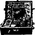
Figure 15. Field kit for blood transfusion.
The tube opening is located 1–1.5 cm above the bottom. A standard jar can be used in combination with the siphon method. In such cases, there is no need to heat this jar during the infusion. Filatov and Kartashevsky showed that this combination unites all the advantages of the two methods, namely: the blood flows by gravity, the possibility of blood clots entering is eliminated, there is no need to hold the jar in the hands, blood can be heated during the transfusion, and if necessary, increased pressure can be created through the short tube of the Richardson bulb. Separately, the method of backward blood transfusion (rein-fusio sanguinis) of blood poured into serous cavities must be considered. The first suggestion to use blood poured into one of the cavities of the human body belongs to Highmore (1874), but in practice this method was first applied by the gynecologist This (1914). The method consists in the fact that the blood poured into the pleura or into the peritoneal cavity is collected, filtered through citrated gauze, mixed with a solution of sodium citrate (the proportions are the same as in blood transfusion, see below) and, in combination with a physiological salt solution, is injected back into the patient's vein. The amount of blood transfused back in some cases was quite significant. For example, Sokolov transfused 1,700 cm3 in a case of tubal rupture in ectopic pregnancy and had a good result. This method is applicable in wounds of the chest cavity (wounds of the lung, heart, chest vessels) and parenchymatous organs of the abdominal cavity (liver, spleen, pancreas) and in general in hemorrhages in the abdominal cavity (ectopic pregnancy, hemorrhages from abdominal vessels, etc.). Backward blood transfusion is contraindicated: 1) with simultaneous wounds of hollow organs, when infection of the poured blood is possible, 2) in very late cases in which decomposition of the blood is possible (after 24 hours, the poured blood is unsuitable), 3) in acute inflammatory processes in the corresponding serous cavities (e.g., pyosalpinx and in inflammatory kidney diseases). Filatov collected a cumulative casuistry of 570 cases. Most often, this method was used for the rupture of a pregnant tube (534 cases). In the vast majority of cases, an excellent result is obtained. In 4.8% of cases, a transient reaction to the infusion in the form of an increase in temperature, chills, and sometimes hematuria and jaundice was noted by Filatov. In 14 cases out of 250 (2.7%), a fatal outcome from reinfusion resulted after the backward infusion. In each of these cases, errors can be found either in the indications or in the technique. Thus, for example, in Wolf's case, knowingly infected blood was transfused in a wound of the stomach and liver. The patient died of a general infection. In Grossmann's case, blood that was too old was transfused—hemolysis, anuria, and death on the 6th day occurred. In Schweitzer's case, the blood was whipped and apparently the erythrocytes were destroyed by this. The patient died of hemolysis on the 8th day. In Schaefer's case, the transfusion was performed with an excessively large amount of sodium citrate—hemolysis and death ensued. If correct indications are set and the correct technique is adhered to, the method of backward blood transfusion deserves wide application (Hesse, Filatov). To more precisely clarify the indications and technique, a number of experimental studies were undertaken by Filatov. It was possible to clarify that the complications and harmful effects observed after backward blood transfusion depend on the backward introduction of hemolyzed blood, but with spontaneous defibrination of blood (fibrinolysis) poured into the abdominal cavity, the blood is not toxic. The hemolysis of the spilled blood is explained by its decomposition in the abdominal cavity. During the first 24 hours after hemorrhage, hemolysis usually does not occur. Hemolysis is explained in some cases by mechanical damage to the blood during the very act of transfusion (rough handling of blood, squeezing tampons, whipping, and defibrination of blood). For the same reasons, dilution of blood with a hypotonic sodium citrate solution is unacceptable. In cases where the hemorrhage occurred more than 24 hours ago or the bleeding took place from the liver, and the spilled blood mixed with bile acids, blood transfusion can be performed only with a negative hemolysis test. The hemolysis test is very simple and is performed by centrifugation. By the color of the plasma, one can judge the hemolysis that has occurred. If the plasma is colored pink, partial hemolysis is unquestionable. With normal plasma coloration (a slightly yellowish tint) or with a barely noticeable pink coloration, hemolysis is absent. Into the group of blood transfusions, some authors mistakenly also refer the infusion of blood into the abdominal cavity, its introduction into the subcutaneous tissue, and into the rectum. These manipulations have no relation to the question of blood transfusion, since here the blood is absorbed by the lymphatic system and the elements of the foreign introduced blood enter the bloodstream through the entire filter of the lymphatic system, so that the favorable success in such cases is explained by the introduction of nutritional protein substance and the stimulation of hematopoiesis. The method of leaving blood in the abdominal cavity or infusing it there should be rejected for other reasons as well, since the introduction of blood into the abdominal cavity causes a reaction of the organism to a foreign body, which can ultimately lead to the formation of intraperitoneal adhesions. In acute oxygen starvation, this method cannot save the patient's life, since absorption occurs too slowly and help arrives too late. In addition, orientation in the peritoneal cavity with unremoved blood encounters serious difficulties. General rules for all methods of blood transfusion. After determining the group affiliation of the donor's and recipient's blood, the number of erythrocytes and hemoglobin in the recipient is examined for subsequent comparison after the transfusion. The rubber bandage on the donor's shoulder is tightened until the pulse disappears, and then slowly loosened until it appears. This is how the correct dosage of stasis is obtained (Yelansky). Strict asepsis. Needles should lie in a 3% solution of paraffin in ether, the tube system boiled and moistened in a 4% solution. In all methods of blood transfusion, the choice of a vein in the donor must be coordinated with local anatomical conditions. The median cubital vein communicates via a wide anastomosis, which in most cases has no valves, with the deep elbow veins. This valveless anastomosis is the main source of inflow during blood suction, which is of great practical importance. It is recommended to choose a well-palpated, swollen superficial vein of the elbow bend, which soon disappears in the distal direction, i.e., goes from the depth. The superficial veins of the forearm, which can be traced over a long distance along the course of the forearm, are not suitable for suction. The donor and recipient are placed side by side and necessarily both in a lying position. Using the blood of a donor who is in a sitting position is incorrect, since he may lose consciousness and thereby disrupt the operation. All restrictive parts of clothing must be removed. The donor's arm must lie opposite the operator's left hand. Disinfection of the operational field according to all the rules. If venesection is contraindicated for the donor under all conditions, then for the recipient in the case of poorly expressed subcutaneous veins, venesection may be considered permissible. When puncturing vessels, it is a mistake to use novocaine with adrenaline, since the vessel may narrow and the outflow of blood stop. The recipient must be closely monitored throughout the transfusion. In the so-called citrate method of blood transfusion, one has to use sodium citrate as a stabilizer, first used for this purpose in 1914. Sodium citrate, dissolving in liquid blood, significantly slows down the clotting process. It turns out that sodium citrate binds the calcium salts of the blood without exerting, at the same time—at a certain concentration—its harmful effect. Therefore, its use is scientifically grounded. All other chemical agents that also possess the property of delaying the blood clotting process (sodium phosphate, sodium carbonate, hirudin) are too toxic. True, sodium citrate in large doses is not harmless: 11–15 g of it is a lethal dose. If we assume that during the largest transfusions, from 1 to 2, maximum up to 5 g enters the organism, then these doses, if not indifferent, are in any case not dangerous. Experimental studies (Horsley, Unger) have shown that sodium citrate 1) increases the fragility of red blood cells and thereby promotes hemolysis; 2) decreases the phagocytic properties of leukocytes and 3) decreases the opsonin-forming properties of the serum. Despite the foregoing, a special American commission, having weighed all the objections raised against the transfusion of citrated blood, came to the conclusion that the introduction of sodium citrate into the organism in the dosage that usually has to be used is practically harmless.
Recently, a number of other stabilizers have been proposed instead of sodium citrate. Heparin, germanium, and others have been tested abroad and have proven to be agents that inhibit the clotting process. The Central Institute of Blood Transfusion in Moscow deserves special credit for finding new stabilizers. Synantrin and several other stabilizers have been proposed here (Brukhonenko, Yankovsky). The stabilizing qualities of these agents have been proven, but the question of their practical application has not yet been finally resolved. When using the citrate method, complications known as the nonspecific protein reaction (see below) are observed. These phenomena are explained by the influence of foreign protein that enters the blood with distilled water or in the form of decomposed proteins from previous transfusions stuck in the apparatus. Using a special technique for preparing the apparatus (washing in a 0.1% caustic alkali solution) and triple distillation of water, Lewisohn brought these complications almost to zero. To avoid possible hemolysis, the transfusion of the first 10 cm3 of blood should be performed very slowly, carefully observing the recipient for 5 minutes. This procedure is called the biological test (Oehlecker) and is mandatory in all cases of blood transfusion. If everything goes well during this period, then in the future the danger of hemolysis is negligible. If formidable phenomena occur indicating hemolytic shock, the transfusion should be stopped. From 500 to 1,000 cm3 can be taken from one donor. If larger amounts of blood are required, it is necessary to use two donors. In acute anemia, 500-1,000 cm3 should be infused, as an exception 1,500-1,800 cm3. Too large doses cause acute cardiac dilatation, pulmonary edema, and visceral hemorrhages. In chronic anemia and diathesis, for the purpose of stimulating blood formation and increasing coagulability, small doses of 50-150 cm3 are indicated. Too rapid administration of large quantities of blood (500-700 cm3 in a few minutes) is erroneous and dangerous. Acute cardiac dilatation occurs, which can cause death on the operating table. No more than 100 cm3 of blood should be infused within three minutes. If formidable symptoms appear after too rapid administration of blood, the patient's head should be lowered and the chest compressed rhythmically. Every blood transfusion is fraught with the danger of air embolism, but this is possible only with insufficient attention; 6 cases have been described so far during blood transfusions. The entry of blood clots into the venous circulatory circle can cause phenomena of pulmonary infarction. In view of this, any forced administration of blood or saline solution to the recipient, in case of sudden manifestation of obstruction of the system, is contraindicated. If it is necessary to preserve blood, which is of particular importance in wartime and in emergency surgery, it can be stored for several weeks in a closed glass vessel. The experience of the Americans during the World War showed the complete suitability of preserved blood for transfusion (Turner, Robertson, Percy). Recently, Belenky, Bagdasarov (Moscow and Leningrad Institutes of Blood Transfusion) have developed a method for blood preservation. Belenky's mixture consists of 6 parts of blood, 1 part of citrate (3.8%), and 5 parts of glucose (5.4%). In this mixture, blood is preserved for 2-3 weeks. The vessel with preserved blood is stored in an icebox in a resting state. Repeated shaking can lead to premature hemolysis (Depp, Lindenbaum, and Stroykova). After 24-36 hours, the precipitation of erythrocytes occurs, and the upper part of the solution takes on a turbid yellowish-green color. If a reddish tinge appears (hemolysis!), this blood cannot be used. Accidental contamination of the blood quickly leads to hemolysis. The glucose and citrate solution is sterilized, according to Belenky, by pasteurization (three times for 2 hours up to 70° for 4-5 days), since glucose caramelizes upon boiling. However, as shown by the studies of Lindenbaum, Stroykova, and Depp, preserved blood can also be prepared on caramelized glucose by sterilization in an autoclave. In wartime conditions, pasteurization may not always be possible. Therefore, Elansky recommends using a citrate-salt solution (Sol. Natrii chlor. physiol. 100.0; Natrii citrici 0.6-1.0). Blood and the above solution are taken in equal amounts. The citrate-salt solution is sterilized in an autoclave and protects the blood from hemolysis for one week. Before transfusion, the blood is warmed to 40°. The Moscow Institute of Blood Transfusion recommends using an ordinary pharmacy bottle with a capacity of 750 cm3 for preserving and transfusing blood. The bottle is closed with a sterile stopper and stored in an icebox. Questions related to the technique of preparation and use of preserved blood have not yet been sufficiently developed. Blood harvested for preservation must not come into contact with the hands of the operator and the air of the room. It is necessary to establish a direct flow of blood from the donor's vein into a closed vessel, which must be sterilized in advance with a preserving stabilizer. The largest dangers are clotting and contamination of preserved blood. In the latter case, the blood hemolyzes. Transfusion of hemolyzed blood is a grossest mistake. The raspberry color of the plasma is an indicator of hemolysis. The question of the storage periods of preserved blood is under investigation. Lindenbaum and Stroykova were able to find out that hemolysis occurs earlier if blood is not stored in an icebox and not in the dark. Overheating of preserved blood is extremely dangerous (Petrov, Filatov, and Bogomolova). When warming preserved blood of longer storage periods above 42°, one has to observe its hemolysis. When transporting preserved blood, it is necessary to remember careful packaging without the presence of air in the flasks. Shaking and agitation with air lead to disintegration and hemolysis (Bryaitsev). Preserved blood of longer storage periods (more than 10-12 days) should be used only with great caution. Preserved incompatible blood, according to the experiments of Hesse and Filatov (1932), is less toxic in terms of the appearance of a drop in blood pressure and the effect on the kidney than blood taken on the same day. From a biological point of view, preserved blood is less wholesome than fresh blood, since the resistance of erythrocytes in it decreases (Depp), while the resistance of hemoglobin increases (Popova), but from a practical point of view, the method of infusing preserved blood has a number of advantages. Most recently, for blood transfusion, it has also been possible to use blood taken several hours after death from a corpse (Shamov). It is clear that corpses of people who died suddenly, for example from severe trauma, can be used for this purpose. The method has proven itself in practice and, despite the complexity of the organizational side of the issue, can be recommended (Sakayan, Yudin, Skundina). Spasokukotsky (1932) used cadaver blood from uremics and eclampsia patients for blood transfusion, but the practical evaluation of this proposal is yet to come. Sorokina and Breite tested Spasokukotsky's proposal on the use of uremic blood in an experiment on dogs, investigating residual nitrogen and aromatic substances in recipients, and were able to prove that the amount of aromatic substances often doubles; the amount of residual nitrogen also increases significantly. On the 4th-7th day, equilibrium is established. The authors believe that the erythrocytes of uremic blood in the recipient's blood vessels die in moderate quantities. In this regard, the authors urge some caution and consider the use of uremic blood contraindicated in poisonings. The problem of donor service is a very complex issue and in its organizational part has not yet been finally resolved. Initially, the blood of relatives of patients in need of blood transfusion was used everywhere, then they switched to using the blood of volunteers. In the West, there are two systems of donor organizations: the American (since 1917) of professional donors and the English (since 1921) of voluntary ones. Volunteer donors give blood for free out of altruistic motives. What these two systems have in common is that the donor service is centralized. Donors are thoroughly examined before and after blood collection and do not have the right to give their blood without the knowledge of the center. As an example of the American donor system, one can point to the organization of donors in Minnesota-Rochester (Mayo Brothers Clinic) and New York (Department of Health; Coca). The fee is 10 dollars for 100 cm3 of blood. Secret surveillance is maintained over the donor. In addition to the USA, this system has been introduced in other countries, e.g., in China. An example of the English (voluntary) system is the organization in London under the Red Cross Society (Oliver), numbering 2,000 donors.
Donors are recruited from medical personnel, youth and other organizations, and are accepted with great selectivity. For each transfusion, the donor receives a license. For 10 donations of blood, a medal featuring the image of Landsteiner is awarded. Besides England, this system has been introduced in Holland (Rotterdam, van Dijk). In Paris, donors are centralized at the Institute of Emergency Care and, just like in England, receive no remuneration. In Germany and Austria, the donor system was organized recently. In Berlin, donor service is centralized at the Virchow City Hospital (Schiff, Friedemann, Unger). In places where the donor system is insufficiently organized, instances have been observed of blood being given 2–3 times a month by the same person, the appearance of a different donor for the transfusion, and other abuses (Boiler). The current state of blood donation in the USSR has been summarized by E. P. Burtseva, who gathered material from 51 institutions. 2/3 of our institutions take blood from registered donors, 1/3 from relatives (due to the lack of special funds). Regarding the number of donors, Moscow is in first place (250), and Leningrad is in second (200). It turned out that among persons wishing to be donors, 36.2% are rejected [most frequently (28.3%) due to blood inferiority, with negative reports from venereologists (21.4%)—lues taking second place]. In southern branches, rejections frequently have to be made because of malaria. In the USSR, 40% of donors belong to medical personnel, followed by university students and workers. In military institutions, the donors are Red Army soldiers. The vast majority of donors in the USSR are between the ages of 18 and 35 (82.4%). 2/3 of all donors are women. 60% of institutions take blood in equal amounts from men and women, on average 400 cm3 each, and 40% of institutions in varying amounts from men and women, with men averaging 450 cm3 and women 350 cm3. The majority of institutions take blood once a month. Blood renewability is monitored in 40 institutions. It turned out that 32% of all institutions still widely use group O as a "universal" donor (see below). Compensation is issued to the donor in the form of monetary and ration allowances. The average monthly ration of a donor in calories is 29,345 (meat—3.5 kg, butter—1 kg, sugar—1.25 kg, eggs—25 pcs., rice—1.5 kg, and milk—15 l). The highest rations in terms of caloric content are issued by Sverdlovsk (68,452), Kaluga (55,000), and Leningrad (39,901). Only in 15 cities of the Union do donor organizations also serve other hospital institutions of the city. It turned out that a donor usually arrives at the call location in 1 hour during the day and 2–3 hours at night, which is why many institutions use conserved blood. Home examinations are systematically conducted by Leningrad and Grozny, and sporadically by Vladivostok and Taganrog. Legal registration is carried out in 18 institutions only in the form of a receipt given by the donor upon admission stating that they have no chronic diseases and that they will appear for blood transfusion without fail. Only seven institutions register the donor by issuing them a booklet (Leningrad, Grozny, Tiflis, Moscow, Tashkent). Donor recruitment in the overwhelming majority of cases occurs through the donors themselves, followed in second place by recruitment through lectures, and in 6.5% through popular literature. Blood donation in the USSR is not a profession, since all donors have jobs. Blood is taken from men in amounts not exceeding 500 cm3 and from women 350 cm3, since the latter take a longer period to restore their blood. Blood is taken on average once a month, no more than 8 times a year. The Leningrad Institute fundamentally always uses the identical group, and blood is taken exclusively by venipuncture. Venesection is categorically prohibited. For 1933, it was never necessary to grant leave after a blood transfusion. The Institute conducts systematic home examinations of donors. Upon enrollment, an investigation of seroreactions and the morphological composition of the blood is performed, along with an examination by a therapist and a venereologist. Examination of social and living conditions is very valuable for the selection of donors and transferring them from full-fledged for transfusion to non-full-fledged (serological), which to some extent helps protect the recipient from the transmission of infection. When comparing donors who demonstrated high blood renewability after giving it for transfusion with donors who demonstrated poor renewability, a definite dependence of renewability on social and living conditions, in particular nutrition, becomes clear. The better the latter, the faster blood regeneration occurs (Burtseva and Zalkind). Blood donation from a biological point of view, subject to the above rules, is harmless. This has been proven by thorough psycho-physiological studies by Khvilivitsky. Among donors, even those who gave blood many times, individuals with clearly pathological reactions were not found; however, on the other hand, blood donation from a social point of view is not indifferent for certain professions. Finely differentiated functions react sensitively to blood loss, which dictates special caution when recruiting donors from persons of highly differentiated labor (for example, precision mechanics, high-grade locksmiths and turners, mechanical transport drivers, etc.). In view of the presence of certain changes in the area of motility and autonomic changes associated with blood loss, blood donation is contraindicated for neurotics. The general criterion for determining the dose of blood extraction is the donor's weight (for primary donors 2–3.5 cm3 per 1 kg of weight, for experienced donors 4–6 cm3). The rest provided to the donor after blood loss accelerates the process of recovery from psycho-physiological changes, while physical work following bloodletting somewhat slows it down. This must be especially emphasized for primary donors. The recovery of psycho-physiological changes in primary donors proceeds faster than in repeat donors, which allows for the possibility of repeated blood extraction in primary donors after a somewhat shorter interval than is usually accepted (Khvilivitsky). The question of organizing blood donation in wartime conditions is a separate issue. The experience of the French, English, and American armies during the imperialist war showed that these armies could successfully use voluntary donors from medical personnel and the slightly wounded. The American medical service used conserved blood, and in a future war this method must be used. Conserved blood will need to be delivered to that sector of the front where the need to use this blood arises. Errors and hazards in blood transfusion constitute a special chapter developed recently. In this area, only the most important data can be provided. Among the errors that can arise in determining the group properties of blood, fluctuations in the agglutinability of erythrocytes should be mentioned. Sometimes agglutinability is so weak that it can only be proven with very active sera. Sometimes agglutination is sharp, delayed, and does not occur within the usual 5–10 minutes of waiting. Pseudo-agglutination should not be confused with agglutination. The former belongs to non-specific reactions and is related to the erythrocyte sedimentation reaction. Upon adding a weak saline solution, pseudo-agglutination does not occur (Lattes). Auto-agglutination, cold agglutination, or panagglutination are synonyms. All these concepts share the phenomenon that the serum agglutinates erythrocytes of all groups, including those of its own blood. This is rarely observed (1 : 1,500) and occurs most often in patients suffering from diseases of the hepatolienal system. Blood transfusion in such cases is completely permissible, since these "cold" agglutinins begin to act only in vitro, remaining inactive in vivo. The upper limit of temperature at which cold agglutination occurs is 25°; consequently, it cannot manifest itself at body temperature. In this connection, it must be emphasized that the determination of group properties must always be performed under known temperature conditions. At an ambient temperature above 40°, agglutination may not occur, and such blood may be mistakenly classified as group O; on the other hand, at temperatures below 10°, agglutination occurs in blood of any group affiliation, and such blood may be mistakenly classified as group AB. This fact must be taken into account in hot and cold countries, in summer and winter, and especially in field conditions. A false characterization of groups can also occur with so-called defective blood groups. Recently, the practical significance of subgroups A1 and A2 has become clear. In practice, taking these subgroups into account during transfusion is desirable, but since the danger of severe hemolysis due to slow absorption is not great, the absolute requirement for accounting cannot yet be stated. Standard sera should be prepared from A2 as being more strongly agglutinating. Finally, the significance of subgroups for forensic medicine in determining paternity must be emphasized. Complications during blood transfusion. Until relatively recently, the mortality rate during blood transfusion was relatively high.
In 1928, some authors (Clairmont, v. d. Velden, and Wolff) determined the mortality rate to be 0.03% according to collected statistics. Statistics from individual institutions are slightly worse, but closer to reality. Thus, Brines (1930) reports 0.05% for 4,000 transfusions, and Hesse for the Leningrad Institute of Blood Transfusion (1933) reports 0.07% for 1,300 transfusions. There is no unity in the classification of complications following blood transfusion. Hesse proposes to divide these complications into: 1) phenomena of nonspecific protein reaction, 2) hemolytic shock, 3) anaphylactic shock. Any transfusion of even compatible blood is an irritating factor causing changes in the organism. The strongest irritant is the administration of blood of a different group and heterogeneous blood transfusion. - N o n s p e c i f i c p r o t e i n r e a c t i o n. In this symptom complex, which occurs in 5-20% of all cases with the nitrate method, the phenomena appear 1/4 hour after blood transfusion and later, and are expressed in chills, elevated temperature, vomiting, dizziness, urticaria, and malaise. These phenomena are of no serious significance. With the citrate method, these complications are much more frequent than with direct transfusion methods. These disorders are explained by the organism's reaction to the introduction of foreign protein, due to which phenomena of nonspecific protein irritation occur. These phenomena have no relation to agglutination or hemolysis. When these disorders appear, intravenous administration of calcium is recommended. Lewison (1933) proved that foreign proteins enter the recipient not from the donor, but are introduced with insufficiently distilled water and in blood particles remaining in the apparatus from previous transfusions. Prophylactic measures have reduced these complications to negligible figures (triple distillation of water and thorough cleaning of instruments by rinsing with cold water, washing in a diluted soapy solution with the addition of a 1% lysol solution, rinsing with water, 5-minute boiling in a 0.1% sodium hydroxide solution, and rinsing in distilled water; Lewisohn). Hemolytic shock. Despite familiarity with the doctrine of isohemagglutination, this complication still occupies the very first place among all complications during blood transfusion. The symptoms of hemolysis consist primarily of the phenomena of hemolytic shock. The patient begins to become restless, complains of headache, a burning sensation throughout the body, malaise, and pain in the pit of the stomach. The pulse quickens, becomes weaker, and a drop in blood pressure is observed. Nausea appears, sometimes vomiting. The complexion changes drastically. First, redness occurs, then sharp paleness. Flickering before the eyes, buzzing in the ears, dizziness, and sweating appear. One of the most characteristic signs is pain in the lower back, which is explained by spasm of the renal arteries (Hesse and Филатов); In more severe cases, cyanosis, sharp shortness of breath, a drop in cardiac activity, and clouding of consciousness may join these phenomena. In rare cases, death may occur in a state of hemolytic shock. More often, however, patients recover after the initial severe phenomena of shock. In severe cases, after some improvement in the general condition, phenomena of renal insufficiency occur (oliguria, hemoglobinuria, anuria, uremia). In such cases, patients may die on the 5th-14th day after transfusion. Unity has not been achieved in the classification of hemolytic shock. Hesse proposes the following classification: 1) Acute form with a predominance of phenomena on the part of the cardiovascular system with a sudden drop in blood pressure. This form can quickly lead to a fatal outcome or pass into a second, more chronic form. 2) Acute form with a predominance of phenomena on the part of the kidneys without visible sharp damage to the cardiovascular system. In this form, patients die on the 5th-14th day. 3) Acute form with a predominance of mild subjective phenomena with rapid equalization. This form is observed upon the admission of an insignificant amount of incompatible blood (up to 60 cm3), with the hemolyzed blood being captured by the reticuloendothelial cells of the liver and spleen; 4) Late form, in which the phenomena of hemolysis occur several hours or even the next day. This form is observed extremely rarely. Hemolysis during blood transfusion occurs in the vast majority of cases due to blood incompatibility and failure to observe the basic laws of isohemagglutination. However, a very small group remains in which hemagglutination relationships are so complex that an error is fully understandable. The appearance of hemolysis with identical groups can be explained by the existence of defective groups (Schiff), the properties of the recipient's serum, and the failure to account for subgroups in groups A vs. AB (Ridel, Blain, Blinov). There are very significant data on the occurrence of hemolysis when using a "universal" donor. Recently, the question of the "universal" donor has been revised, and group O cannot be recognized as "universal." In total, 43 cases of hemolysis have already been described when transfusing from a universal donor, with 18 fatalities. The dilution of the donor's blood and the two agglutinins a and ̢ contained in its serum in the recipient's blood is so significant that the agglutinating and hemolyzing action of the donor's blood is of no importance and the agglutinins lose their strength. But experience shows that with large transfusions from a universal donor to a recipient with marked anemia and a limited total amount of blood, sometimes the donor's serum with high-titer agglutinins will not be sufficiently diluted in the recipient's blood. As a result, the concentration of hemolysin in the recipient's serum reaches the limit necessary for hemolysis to occur. A universal donor in the present sense of this word does not exist, and during blood transfusion it is safer to use only blood of the same group. Group O is especially strictly contraindicated for transfusion to different groups when it is necessary to transfuse more than 100-200 cm3 of blood with marked anemia of the recipient. In chronic anemia and diseases of the hematopoietic apparatus, when the recipient's erythrocyte count drops below 2 million, this danger is particularly great. The use of a group O donor with a high titer is unacceptable. The essence of hemolysis has been insufficiently studied until now. Only recently have Hesse and Filatov (1932-34) and then I. R. Petrov, Veselkin, Stroykova, Bogomolova, Lindenbaum, and Kartashevsky (1933-1934) from the Leningrad Institute of Blood Transfusion published data from experimental and clinical studies that shed some light. According to the studies of the above-mentioned authors, in hemolytic shock there is a sharp drop in blood pressure, a decrease in the stroke and diastolic volume of the heart, and a decrease in the volume of the kidney and spleen during their onchographic measurement. When washing the vessels of the mesentery and intestinal loops with preserved connections, a sharp decrease in the outflow of Ringer-Locke fluid was found. Comparing these data, one can come to the conclusion that during hemolysis, the following develop: 1) vascular spasm, which is especially sharply expressed in the splanchnic region, 2) weakening of cardiac activity due to a decrease in the filling of the heart during diastole. These vascular changes occur mainly due to the direct effect of hemolyzed blood on the vascular walls, but along with this, action through the central nervous system also takes place. Furthermore, phenomena of intoxication of the organism with hemoglobin breakdown products develop. From clinical observations, it is known that death in hemolysis in the vast majority of cases is the result of renal function disorders. Hesse and Filatov reject the mechanical theory of blockage of renal capillaries by agglutinated erythrocytes and blockage of urinary tubules by hemoglobin crystals. Changes in the kidneys during hemolysis leading to their insufficiency and expressed in oliguria and uremia are explained by the initial spasm of the renal arteries and then phenomena of intoxication (Hesse and Filatov, Ilyin, B. F. Malyshev). The precipitation of hemoglobin in the renal tubules during hemolysis is a secondary phenomenon. Prognosis in hemolysis depends on the state of the kidneys. Healthy kidneys with normal function suffer less than those impaired in their function. From this follows the practical requirement of a thorough examination of renal function before the intended blood transfusion. Pathological-anatomical examination of the organs of individuals who died from hemolysis (Lemke, Lindau, Irsigler, B. F. Malyshev) shows the following: in serous cavities, most often in the pleura, bloody fluid is observed, the heart is usually stretched, the muscle is flabby. The liver has undergone fatty degeneration. The kidneys are enlarged, brown in color, the pattern is blurred. At later periods (5-10 days after transfusion) they are paler. The cortical substance has undergone fatty degeneration. Microscopic examination reveals deep damage to the epithelium of the renal tubules, sometimes their necrosis, degenerative changes in the parenchyma, and hemorrhages in the intestinal walls.
Prevention of hemolysis consists in observing the rules of isohemagglutination and using the biological test of Eleker. - Treatment. Relatively recently (until 1932), there were no expedient measures against hemolytic shock. The usual prescription of symptomatic remedies (cardiacs, diuretics, bloodletting, saline infusion) is useless. Out of five cases of kidney decapsulation, one patient survived. This procedure should be rejected. The only expedient therapeutic method for hemolytic shock is (according to experimental and clinical observations by Hesse and Filatov) immediate transfusion of compatible blood. This method achieves immediate relaxation of the renal artery spasm. The time frame after the onset of hemolytic shock within which it makes sense to resort to this method has not yet been elucidated. Hesse and Filatov saved one patient after eight minutes, the second after 24 hours, Christiansen after an hour, Bogina after several hours, and Hess de Calve even after 24 hours had complete and immediate success. At later times, this procedure will of course prove less effective. In institutions where blood transfusions are frequently performed, it is necessary to have prepared conserved blood of various groups ready for use in case of hemolytic shock. Anaphylactic shock during repeated transfusions is a real danger. Frequently, the boundary between anaphylactic and hemolytic shock and a non-specific protein reaction is blurred, and these concepts are treated without sufficient criticism. Anaphylaxis in the true sense of the word can occur only when a small amount of completely harmless protein, introduced into an organism previously sensitized by injections of the same protein, produces the phenomena known as anaphylactic shock. Therefore, anaphylaxis must be rejected in all cases where it was supposedly observed after a single blood transfusion. These cases must be assigned to the group of non-specific protein reaction. With repeated blood transfusions, anaphylactic shock is possible and is observed most frequently with the repeated use of the same donor. The symptoms are characteristic: towards the end of the transfusion, sometimes a few hours later, flushing of the face, headache, dizziness appear, rapid pulse, sweating, coughing, and phenomena of bronchospasm with slowed breathing appear. Sometimes, after a chill, body temperature rises. Urticaria appears, which can rapidly spread throughout the entire body. Sometimes a drop in cardiac activity, cyanosis, and hemoptysis appear (Lundberg). The duration of these phenomena is from 6 to 48 hours. The prognosis is generally favorable. The causes of the appearance of anaphylaxis after blood transfusion have still not been sufficiently elucidated. The occurrence of anaphylactic shock must be explained by the emergence during previous transfusions of isoantibodies against specific antigens of the donor's blood. There is no doubt that the newly discovered factors M, N, and P by Landsteiner and Levine will play a role in deciphering anaphylaxis after blood transfusion. - Prophylaxis. When repeated transfusions are necessary, most authors advise taking a different donor, but this is completely insufficient, since the issue is not individual, but group antigens, which may accidentally also be present in another donor (Schür). Therefore, it is most correct to determine factors M and N if a secondary transfusion is necessary. If it is impossible to determine these factors, it is recommended, in case a secondary transfusion is necessary, to induce an antianaphylactic state in the recipient. For this purpose, on the eve of the expected transfusion, 4 cm3 of the recipient's blood is injected intravenously into the donor (Nather). György and Witebsky previously inject 0.1 cm3 of the expected donor's serum into various areas of the recipient's skin. In the case of an anaphylactic state, a large bluish spot appears at the injection site. Hesse recommends using desensitization of the recipient by a preliminary injection of a small dose of blood (5 cm3) from the prospective donor one hour before the transfusion. - Treatment. When an attack of anaphylactic shock has broken out, there is no specific remedy. Intravenous administration of 10–20 cm3 of a 1% calcium chloride solution every 4 hours is recommended. Twelve hours after the attack, caffeine and theobromine are prescribed. For facial edema, cyanosis, and dyspnea, adrenaline is prescribed (Hartmann). Transmission of acute and chronic diseases during blood transfusion. During each blood transfusion, the transfer of pathogenic agents present in the donor's blood is possible. All acute and chronic contagious diseases whose pathogens are in the blood can be transmitted. This is easily possible with a donor who is in the incubation period. Cases of transmission of so-called measles, smallpox, and typhus are known (Golandsky). Chronic contagious diseases that may escape detection if they are in a latent state can also be transmitted during blood transfusion. Tuberculosis is particularly widespread among the population, and the possibility of using a donor with a dormant infection is not excluded, although tuberculosis bacilli are not present in the blood in all cases. In addition, the bactericidal properties of the recipient's blood must also be taken into account. Only 3 cases of tuberculosis transmission are described in the literature. In the tropics, there is a danger of transmitting filariasis. To avoid inoculation of the aforementioned diseases, the most thorough clinical and laboratory examination of the donor and observation of him are required. The greatest practical importance is the question of the transmission of syphilis and malaria. According to modern syphilidology, it must be considered proven that the blood of syphilitics can contain virulent spirochetes in all stages of the disease. In terms of transmitting syphilis during blood transfusion, the primary and primary-secondary periods of syphilis are the most dangerous (Zalkind). The literature describes 28 cases of syphilis transmission during blood transfusion. All cases are characterized by the absence of a hard chancre, swelling of regional glands, and a long incubation period (about 70 days). The Leningrad Blood Transfusion Institute, which has had no syphilis infections out of 2,000 blood transfusion cases, takes the following precautionary measures: 1) The donor is obliged to sign a receipt stating that they have not had syphilis and undertake to report the infection if it occurs in the future. Such a receipt does not relieve the physician of responsibility, but has moral significance. 2) The most thorough examination of donors by a specialist syphilidologist. Special attention should be paid to the state of the peripheral lymph glands. Perinipple adenitis (Zalkind) and swelling of the ulnar glands must be considered particularly pathognomonic. The medical examination of the donor must take place every 20 days. 3) Performance of specific seroreactions of the blood. Modern syphilidology requires a comprehensive study using several seroreactions. A donor with a positive reaction must be rejected. A negative result of seroreactions does not exclude the possibility of syphilis; therefore, seroreactions by themselves cannot serve as a decisive criterion when concluding on the suitability of a donor. The conclusion is made based on the totality of all the aforementioned clinical and laboratory research methods. Hovhannisyan, Kudryavtseva, and Zalkind were able to ascertain very recently that spirochetes in conserved blood five days old die. The addition of quinine accelerates death. Very important practical conclusions can be drawn from this. Such a real danger is the transmission of malaria during blood transfusion, especially in malarial localities. The literature has published 19 cases of malaria transmission during blood transfusion. The first symptoms usually appear on the 9th to 30th day after transfusion. In all cases, the administration of quinine quickly cured the patients. Malaria in a latent state is diagnosed with great difficulty. The question of preventing the danger of malaria transmission has been resolved very recently. Akkerman and Filatov transfused artificially charged blood with malaria plasmodia of various conservation periods to patients suffering from progressive paralysis. It turned out that conserved malaria blood when stored in the cold (+4 to +6°) after 4 days can be considered sterile and practically harmless with respect to any possibility of infection with these forms of malaria during blood transfusion in general. In vitro experiments with conserved blood containing tropical fever plasmodia show that these parasites also die after 5 days. Based on these established facts, in malarial localities, one should use exclusively conserved blood with a storage period of more than five days, by which the possibility of malaria transmission is completely prevented. Effect of blood collection on the donor. With the correct blood transfusion technique, no dangers threaten the donor; however, if the rules are violated, complications may also arise for the donor. The most minor surgical intervention is always associated with the possibility of infection.
During venipuncture this danger is negligible. The danger of infection is especially great, however, if the blood recipient suffers from a general purulent infection. A case of a donor transmitting syphilis is known. In such cases, it is permissible to use only the citrate method. In rare cases, thrombosis of the donor vein is possible. Cases of keloid formation have been observed. The danger of anemia from blood loss in the donor is slight. A single blood withdrawal of 500 cm3 for men and 350 cm3 for women over the course of 4-5 weeks is borne without any complications and is completely safe for the donor. Donors with polycythaemia rubra and high blood pressure are especially well-suited for blood transfusion, because such blood extraction gives them a therapeutic effect.
E. Hesse. Additional data on blood groups (see Hemagglutination). Initially by Landsteiner, then by Jansky and Moss, humanity was divided into four hemagglutination groups, which currently play a very important role in matters of blood transfusion, forensic medicine, and anthropology. For about two decades, this division scheme held without any additions. But over time, a whole series of facts accumulated that forced a revision of the question of blood groups and the isolation, on the one hand, of subgroups, and on the other, of a number of new additional factors existing in erythrocytes and making it possible to establish a new additional classification of humanity. A thorough study of group A made it possible for Landsteiner, Thomson, and other researchers to prove the heterogeneity of this group. At the present moment, it is precisely established that group A breaks down into two groups that are qualitatively distinct from one another, but share common characteristics in relation to other groups. The latter circumstance does not allow these types to be isolated into special groups, like O and B, which is why it is necessary to speak of two subgroups, A1 and A2. Erythrocytes of subgroup A1 differ from A2 by stronger agglutinability and greater adsorption capacity. The inhibiting effect of pepsin and group A serum is more pronounced on A1 erythrocytes. Upon immunization of guinea pigs with A1 and A2 erythrocytes, specific antisera were obtained. The serum of subgroups, apart from the /? agglutinin, sometimes contains two types of extraagglutinins, which differ from the main agglutinins in that they give an optimum agglutination reaction only at a temperature of 15-18°; at human body temperature, agglutination with them usually does not occur. In the serum of subgroup A2, the a1 extraagglutinin is often encountered, agglutinating only A1 erythrocytes; in the serum of A1, another type of extraagglutinin, a2, is observed, giving agglutination with A2 and O erythrocytes and not agglutinating A1 erythrocytes; this extraagglutinin is encountered significantly less often. A1 and A2, as qualitatively different genes, are inherited, with A1 dominating over A2. Among the population, A1 is encountered four times more often than A2. Methods for isolating subgroups. 1) If group B serum is completely adsorbed by A2 erythrocytes, the resulting serum will agglutinate A1 erythrocytes and not agglutinate A2 erythrocytes. 2) Group B serum with the addition of an equal amount of a 1/8% pepsin solution agglutinates A1 erythrocytes within 1.5-2 minutes, and A2 erythrocytes after 10-15 minutes. The simplest method for separating subgroups was developed at the Leningrad Institute of Blood Transfusion (Blinov) and consists of the following: if one drop of standard group A serum is added to two drops of group B serum diluted in half, the resulting mixture will give agglutination only with A1 erythrocytes. One can also use extraagglutinins a1 and a2, which are obtained from the corresponding sera, to separate subgroups. Since factor A breaks down into two types, group AB will also break down into two subgroups, A1B and A2B, as a result of which at the present moment we have 6 phenotypes: O, A1, A2, B, A1B, A2B, each of which possesses certain characteristic properties. The isolation of subgroups has played a rather large role in the systematization of various exceptions to the four-group scheme; furthermore, it is an additional factor in matters of forensic medical expertise. Studying animal immune sera, Landsteiner discovered in them, besides the specific anti-A and anti-B agglutinins, also special agglutinins which proved the presence of additional factors in human erythrocytes capable of producing specific immune sera in animals. These factors were named M and N. They differ from factors A and B in that corresponding antibodies (agglutinins) are not found in human serum, therefore these factors cannot be detected by human serum; their presence in erythrocytes is revealed with the help of special sera obtained from rabbits immunized with human erythrocytes having the corresponding factor. Usually, washed erythrocytes in an amount of 2.5-3 cm3 are injected into the rabbit's abdominal cavity or vein 6-7 times at intervals of 2-3 days. The resulting serum is inactivated at 56° and adsorbed to remove heteroagglutinins by erythrocytes not containing the required factor. Factors M and N are not isoreceptors, but immune receptors, and are encountered in every person either individually or both together, forming an independent system, independent of factors A and B. According to their content in erythrocytes, humanity is divided into three groups: M, N, and MN, with the MN group occurring in approximately 50% of the population, the M group in 30%, and the N group in 20%. Factors M and N are inherited independently of one another. The transmission scheme is presented in the following table: Parents / Children: M x M (M: 100%); M x N (MN: 100%); M x MN (M: 50%, MN: 50%); N x N (N: 100%); N x MN (N: 50%, MN: 50%); MN x MN (M: 25%, N: 25%, MN: 50%). These factors have practical significance in matters of forensic medical expertise in determining paternity. In case of repeated transfusions to the same person, they must also be taken into account, because antibodies against M and N can form in the recipient's serum, as a result of which complications can arise after blood transfusion. Besides the indicated factors, Landsteiner, with the help of adsorbed horse sera, discovered an additional factor in human erythrocytes named P, and Schiff, with the help of sera from immunized sheep, isolated a factor named by him i. These factors are independent of one another and can be present or absent in human erythrocytes. By adsorbing anti-i serum with various O erythrocytes, Schiff managed to isolate factor G, which is almost always encountered in erythrocytes having agglutinogen A or B. All these factors do not yet have practical significance. Agglutinogens A and B are contained not only in human erythrocytes, but also in all cells of the body, tissue juices, and some excreta (saliva, urine), and in the majority of people they can be detected in excreta, while in 30% they cannot be detected (Schiff). Therefore, all people having any agglutinogen in their erythrocytes can be divided into two types: secretors and non-secretors (Schiff). In some (secretors), the presence of agglutinogen can be proven in various excreta, while in others (non-secretors), it cannot be detected in excreta. All this large number of different phenotypes can have significance mainly in matters of forensic medical expertise. In blood transfusion, the classical four groups with their division into six continue to play the main role. N. Blinov.
Related articles
Mentioned in
Cite this page
“Blood Transfusion.” Soviet Medical Encyclopedia. English translation of Bolshaya Meditsinskaya Entsiklopediya, 1st ed. (Moscow, 1928–1936), ed. N. A. Semashko. https://sovietmedicalencyclopedia.pages.dev/article/blood-transfusion/