Vascular Suture
Historical document, translated for reference. It reflects medical knowledge of the 1920s–30s and is not medical advice.
Summary
This article describes the historical technique of vascular suture from the 1930s, including the development of the procedure, specialized instruments, and various methods for suturing blood vessels. It covers both lateral and circular sutures, as well as anastomosis techniques.
Encyclopedia article (1928–1936)
VASCULAR SUTURE. Any ligation of a blood vessel of an organ necessary for life or for preserving a part of the body should be considered a defect in the surgeon's art, provided there is a technical possibility to close the vessel wound through a lateral or circular suture or some equivalent method and thereby restore circulation (Schmieden). At the initiative of the English surgeon Lambert (Lambert), Hallowell in 1759 first successfully sutured a lateral wound of the brachial artery. The next operation of this kind was performed only more than 100 years later (Durante, 1892). By this time, the suture (parietal) of veins had already gained acceptance. The first circular suture of an artery was performed by Murphy (Murphy) in 1897 on the femoral artery, and the first circular suture of a vein by Kümmel in 1900. Modern technique of V.s. is based on numerous experimental works (Yasinovsky, Carrel and others), which established that sutures can be passed through the entire thickness of the vessel wall without damage and that intima, when applied to each other, quickly, like peritoneum, adhere together and that the processes of healing originate from the same intima. Vessels are sensitive to drying, therefore they cannot be exposed for a long length and for a long time, and when exposed, they should be moistened with saline solution or vaseline oil. For applying a vascular suture, special instruments are required: Moore's vascular clamps (see Carotis arteria, fig. 30) or Hepfner's (see Blood vessels, fig. 24 b), sharply curved non-cutting needles with an average diameter of 1 cm and straight needles; a needle holder with a rather sharp point so that curved needles do not break; fine anatomical forceps and the finest No. 000 silk, specially treated (after ordinary sterilization with alcohol and boiling, the silk is impregnated with sterile vaseline). The silk is so thin and all instruments are so delicate in size that for this case Carrel introduced certain special conditions facilitating orientation in such fine work: he introduced and recommends black threads and black-colored gauze and napkins. Some details of preparation for the operation are not without a certain practical expediency: it is very difficult to thread the eye of a needle, and with a wet one it is quite impossible, therefore it is necessary to thread the needle before sterilization, stick the needle into a small piece of paper, and wrap the thread around it. Carrel's method. The artery or vein is exposed and separated from neighboring veins and nerves, but not from connective tissue; only the ends of the vessel segments are carefully cleaned from it with forceps and scissors; then clamps are applied to the vessel, and if they are not available, the vessel should be compressed with a thin drainage tube, a strip of gauze, or a doubled thick catgut thread. The edge of the vessel wound is grasped with a forceps, the needle is inserted perpendicular to the vessel wall, passed completely through into the lumen of the vessel, the other edge of the wound is pierced with the needle in the opposite direction—from the intima to the adventitia; the insertion and exit of the needle are located farther from the edge of the vessel wound than the holding edge of the forceps. The ends of the thread are clamped with a hemostatic forceps. Such guiding (situational) sutures are placed 3-4 around the vessel at equal distances from each other (fig. 1). The ends of the ligatures, both or one of them, are left long and when pulled, the lumen of the vessel—in arteries round, in veins collapsed flat—takes the form of an isosceles triangle or quadrilateral; in this position the operator has before him the edges of the wound lying flat against each other, which greatly facilitates suturing. The threads are tied so that the edges of the wound are everted outward and
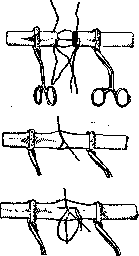
FIG. 1.
lie against each other with the intima. Between the first (situational) sutures, continuous sutures are placed—with a straight needle, starting from the posterior semicircle of the vessel (see Carotis arteria, fig. 30). If dealing with a large vessel, it is recommended to cover the suture site with a strip of fascia or, in appropriate cases, peritoneum. A lateral V.s. in lateral ruptures of vessels can be applied, like a transverse (circular) one, if, by applying a clamp at some distance on both sides of the wound, first one suture is placed on each corner of the latter, and by pulling on them, the wound is turned into a linear one, which is sutured with a continuous suture as described above. A flap wound by placing a Y-shaped suture on the apex of the flap is turned into two linear wounds and sutured according to general rules.--For connecting completely ruptured vessels of considerable caliber, at present the invagination method is preferred, since the circular method is considered unreliable. Murphy invaginates the central segment into the peripheral one and connects them with sutures, but in this case there is no apposition of the intima, which is achieved by Payr with his method: with the help of three strong threads passed through one end of the artery, a special magnesium tube, rather a ring, is placed over the artery, the latter is wrapped around the tube with the intima outward and fixed with a circular catgut ligature; the other end of the vessel with the help of similar threads is placed over the first and there is again fixed by a ligature. The magnesium tube (prosthesis) dissolves after some time. The negative side of the method is the rigidity of the vessel wall until the tube dissolves.-To avoid narrowing of the lumen when suturing vessels of not very large caliber, Dobrovolskaya proposed several methods for treating the ends of vessels: 1) the ends are cut obliquely, so that the incision takes the form of an oval (fig. 2); 2) two tongue-shaped flaps are cut out, when suturing, one end of the vessel is rotated 90° along its long axis so that the ends come together at their apices (fig. 2); 3) after transverse section of the ends, each is additionally incised along the ends of the diameter of the lumen, the edge of the incision becomes quadrangular; these flap-like ends are sutured (fig. 2); 4) for connecting vessels of different calibers, two tongue-shaped flaps are cut out in the smaller one; by stretching them, this vessel can be sutured with slight tension to a larger vessel (fig. 2).-Anastomoses of vessels are performed side by side (side to side) or end to side. The technique is in principle the same as in surgery of the biliary-intestinal canal, with the only difference that the walls of the vessels are not inverted, but everted. The suture of the common carotid artery is indicated for partial injury of the vessel not only under aseptic conditions, but an essential condition for the possibility of applying a suture must be the short time elapsed since the moment of injury (preferably within the first 6 hours after injury), with mandatory 'primary' treatment of the wound tissues. The suture of the common carotid artery is contraindicated in infection, in complete transection of the vessel, in atheromatous degeneration of the vessel, and in the general poor condition of the wounded. Exposure of the artery—see Carotis arteria, clinic; the artery is isolated above and below the vessel wound from nerves and veins while preserving the connective tissue on it; vascular clamps are applied to these areas; the vessels should not be compressed too strongly to avoid damage to the intima, which may be a source of thrombosis. The wall of the vessel in the area of the wound is carefully isolated from connective tissue, but not circularly, in order to preserve part of the vasa vasorum. The edges of the vessel wound are smeared with Oleum paraffini to prevent drying of the intima. The technique of suturing is described above. The results of suturing the common carotid artery—see Carotis arteria, clinic.
FIG. 2.
Related articles
Mentioned in
Cite this page
“Vascular Suture.” Soviet Medical Encyclopedia. English translation of Bolshaya Meditsinskaya Entsiklopediya, 1st ed. (Moscow, 1928–1936), ed. N. A. Semashko. https://sovietmedicalencyclopedia.pages.dev/article/vascular-suture/