Aneurysm
Historical document, translated for reference. It reflects medical knowledge of the 1920s–30s and is not medical advice.
Summary
Aneurysm is a term used to denote the dilation of an artery's lumen. The article distinguishes between true aneurysms, involving all layers of the arterial wall, and false aneurysms, while also differentiating from arteriectasias and angiomas.
Encyclopedia article (1928–1936)
ANEURYSM (from Greek aneuryno - I expand), a term used to denote the dilation of an artery's lumen. The concept of A. is distinguished from arteriectasias, which represent a uniform dilation of an arterial system with its branches, without particular changes in their walls, as well as angiomas (see), tumors of blood vessels. A. in the narrow sense is the dilation of an artery developing as a result of pathological changes in the vessel wall. True and false A.s are distinguished.-Under a true A. is meant the dilation of an artery's lumen in which all layers of the vascular wall participate (see Figure 1 a),
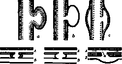
Figure 1. Aneurysms (their types): a - true aneurysm (saccular); b - false aneurysm; c - dissecting aneurysm (spindle-shaped); d - varicose aneurysm (true); e - varicose aneurysm (false); f - aneurysmal varix; in figures a, b, c the inner membrane is indicated by a dotted line, the middle by cross-hatching, the outer by black; A - artery, V - vein.
whereas a false aneurysm is essentially a hematoma, i.e., a collection of extravasated blood, located near a vessel and communicating with its lumen through an opening in the vessel wall (see figure 1 b). This division has relative significance and cannot always be practically implemented, since, on the one hand, in a true aneurysm there is usually such a marked change in the vessel wall that the continuity of its layers is to some degree disrupted, on the other hand, in a long-existing false aneurysm, a dense connective tissue capsule forms around the hematoma, which may serve as a continuation of the vessel wall; furthermore, to a true aneurysm (in origin), a false aneurysm may be added when its wall ruptures. Finally, there exists an intermediate form between true and false aneurysms, called dissecting aneurysm (A. dissecans); the latter is the result of a rupture of the inner or also the middle coat of the artery and the formation of an intramural hematoma (haematoma intramurale), spreading along the vessel in the space between the inner and middle or middle and outer coats of the wall, dissecting them apart (see figure 1 c).- According to shape and extent, diffuse and limited aneurysms are distinguished (A. diffusum, A. circumscriptum). In the first cases, it is a matter of expansion of the vascular trunk, circularly extending over a certain length of it, and such expansion may be cylindrical or spindle-shaped; in those cases where, along with diffuse expansion of the artery, there is also elongation of it, the dilated vessel is arranged in a tortuous course, which gives a tortuous or serpentine aneurysm (A. cirsoideum s. serpentinum), most frequently observed in the splenic artery. A limited aneurysm is an expansion originating only from a limited place in the vessel wall and having the appearance of an appendage to the main vascular trunk; sac-like (see fig. 1a), boat-shaped, and funnel-shaped forms of limited aneurysms are distinguished.- A hernial aneurysm (A. herniosum) is the rare form of limited aneurysm in which there is a breach in the integrity of the outer and middle coats of the vessel and a hernial protrusion of the inner coat through the defect in the outer layers. In some cases, the cavity of the aneurysm communicates with the cavity of an adjacent vein, which may be the result of the aneurysm being attached to the vein with a rupture into it, or more often is the result of simultaneous damage, for example, trauma to the walls of the artery and vein. Under these conditions, an arteriovenous or varicose aneurysm is formed, in which the lumen of the aneurysm opens into the lumen of the vein (see figure 1 d), the latter being somewhat dilated at the level of the communication. - From this form, a false varicose aneurysm must be distinguished (see figure 1 e), when there is a hematoma between the artery and vein, the cavity of which communicates with the lumens of both vessels, as well as the so-called aneurysmal varix (varix aneurysmaticus), in which there is no aneurysm at all, but only a communication between the artery and vein with dilatation of the latter (see figure 1 f).- The size of aneurysms can be extremely varied; in general, the size of an aneurysm depends on the caliber of the original artery, as well as on the duration of the disease. In such large vessels as the aorta, an aneurysm sometimes can reach the size of an adult's head; on the other hand, in small arteries, for example, of the brain, aneurysms can develop that are discoverable only with the aid of a microscope (miliary aneurysms).- The structure of the aneurysm wall corresponds only at the very beginning to those layers of the artery wall that participate in the formation of the dilatation; subsequently, however, there occurs a gradual separation and disappearance of the elements of the wall with their replacement by coarse, calloused connective tissue. In a long-existing true aneurysm, the wall usually consists entirely (without the possibility of distinguishing individual layers) of such connective tissue, among which the microscope sometimes finds only isolated bundles of elastic fibers; closer to the main vascular trunk in the aneurysm wall, one can sometimes find remnants of the muscular tissue of the middle coat, as well as signs of that process, b. m. e. t. I. which may underlie the formation of an aneurysm (e.g., the picture of syphilitic mesoaortitis in an aneurysm of the aorta). In some cases, the vessel wall in the aneurysm apparently completely disappears, being replaced by dense connective tissue proliferated from a neighboring area. The inner surface of the aneurysm is rarely smooth and retaining an endothelial covering; it is usually uneven due to the uneven growth of connective tissue and the manifestation of arteriosclerosis (see). In those places lacking endothelium, mural thrombi (see) very often form, which, arranged in the form of sometimes very voluminous layered plates, can frequently reduce the actual cavity of the aneurysm; long-standing thrombi undergo replacement (organization) by connective tissue growing from the aneurysm wall. The frequency of aneurysm formation in various parts of the arterial network is more or less regular. The most common site for the development of aneurysms is the thoracic aorta, in second place are the popliteal arteries, followed in descending order by the abdominal aorta, femoral, carotid, subclavian, innominate, cerebral, splenic, pulmonary arteries; aneurysms of the mesenteric arteries and coronary arteries of the heart are rarely encountered. Aneurysms most often develop at the age of 30-40 years; in men, aneurysms occur almost twice as often as in women. The course of an aneurysm is almost always progressive; once having arisen, an aneurysm has a tendency to increase under the pressure of the blood circulating in it. Only in very rare cases, especially if it is a small sac-like aneurysm communicating with the vessel through a narrow opening, can the cavity of the aneurysm become completely filled with thrombotic masses, which subsequently organize; under such conditions, the growth of the aneurysm may stop and even a relative cure may occur.- The effect of an aneurysm on the organism is varied. An aneurysm of the ascending part of the thoracic aorta, especially if it involves the valve ring, affects the function of the heart and is the cause of its hypertrophy, which may have a certain general significance for the organism. In other cases, an aneurysm can affect neighboring tissues and that part which this artery supplies with blood. Neighboring tissues, under the pressure of the increasing aneurysm, undergo atrophy, which results in the formation of loss of substance, defects, erosions in these tissues; this is especially common in aneurysms of the thoracic aorta, which can cause destruction of the adjacent wall of the bronchus, trachea, or esophagus or a significant loss of substance, erosion of the sternum or bodies of the vertebrae; sometimes in this way the vertebral canal can be opened and the spinal cord compressed. The pressure of an aortic aneurysm on the recurrent nerve (n. recurrens) causes paralysis of the vocal cords of the larynx. An aneurysm of any peripheral artery can be the basis for a decrease in blood flow to the area supplied by this artery and a series of changes in such an area, associated with a deficiency of blood in it, anemia (see); if, however, a thrombosis develops in such an aneurysm, which also involves the lumen of the artery, then the matter can end in necrosis of the area supplied by this artery (e.g., gangrene of the lower extremity in an aneurysm of the popliteal artery). The most common outcome of an aneurysm is its rupture, perforation with subsequent hemorrhage. The basis of rupture is the gradual thinning of the aneurysm wall, facing one or another cavity or in contact with some soft, yielding tissue. Such ruptures most often occur in aneurysms of the thoracic aorta, with hemorrhage, usually fatal, into the pleural cavity, into the trachea, bronchus, esophagus; less frequently into the pericardial cavity or outward through the skin; very rarely into the atrium, into the superior vena cava, into the pulmonary artery. Perforation of an abdominal aortic aneurysm results in hemorrhage into the retroperitoneal tissue or into the abdominal cavity. Rupture of an aneurysm of a branch of the pulmonary artery, e.g., in pulmonary tuberculosis, usually occurs into the cavity of a cavern or into a bronchus, which often gives fatal hemorrhage. Perforation of an aneurysm of a cerebral artery results in hemorrhage into the meninges or into the substance of the brain. Rupture of aneurysms of peripheral arteries leads to the formation of a hematoma with this or that consequence depending on the size of the hemorrhage.- In terms of pathogenesis, an aneurysm is based on some change in the artery wall, which disrupts its elasticity and thereby weakens the wall's resistance to blood pressure, the main importance belonging to the disruption of the normal elasticity of the middle coat of the vessel. Such a change in the wall can begin from the inner, outer coat or develop directly in the middle coat of the vessel; it can relate to chronic processes or be an acute disruption of the normal state of the wall. For this reason, the specific causes or etiology of aneurysms are very diverse. In first place among the causes of aneurysms of the thoracic aorta is syphilitic damage to it in the form of specific mesoaortitis (see Aorta). Second place among the causes of aneurysms of the thoracic aorta is occupied by arteriosclerosis (see), which plays a large role also in relation to other.
A.; if the arteriosclerotic changes in the inner lining of the vessel extend to its middle layer, then areas of discontinuity of its muscular-elastic tissue arise in the latter, which weakens its elasticity and creates conditions for the formation of a bulging under the influence of blood pressure, and subsequently - an aneurysm (arteriosclerotic A.); moreover, the ulceration of the inner lining caused by arteriosclerosis can sometimes serve as a site for blood penetration between the inner and middle layers, which gives a picture of dissecting A.; the same is observed in ruptures of the inner lining of other origins (trauma, increased blood pressure). Furthermore, various inflammatory processes in the artery wall caused by microorganisms, whether they begin with the outer layer (for example, in phlegmon or in tuberculosis of the surrounding tissue), or with the inner layer (in mycotic embolism, see), or arise directly in the middle layer (for example, streptococcal, as well as rheumatic mesoarteritis) - can subsequently lead to the formation of A. due to impaired elasticity of the middle layer; in those cases where the inflammatory process disrupts the integrity of the outer and middle layers, the inner lining can form a bulging through the defect (hernia-like A.). A. caused by acute infection (streptococcus, staphylococcus) of the inner lining, which most often occurs in septicemias, particularly in sepsis lenta, are traditionally distinguished under the name mycotic or mycotic-embolic A.-Simple embolic A. are those that develop on the basis of trauma to the inner layers of the vessel by a dense embolus, for example, a piece of heart valve or thrombus impregnated with lime. Close to those caused by inflammatory processes are those that develop as a result of chemical effects on the artery wall; for example, A. forming in the base of a gastric ulcer from the action of gastric juice on the exposed artery. Finally, various kinds of external trauma can be the basis for traumatic aneurysms.
A. Abrikosov.
Traumatic aneurysms are the result of traumatic damage to the arterial wall. In relatively rare cases, traumatic A. are true A., for example, when a bruise-type trauma disrupts the integrity of the middle vessel membrane, and in this place an acute bulging of the wall develops (acute traumatic A.), or when a scar of the arterial wall subsequently develops at the site of injury, which under the influence of blood pressure begins to bulge, leading to the formation of an A. However, in most cases, traumatic A. is a false A., resulting from a complete through-and-through disruption of the arterial wall, with the damage being of two types: subcutaneous or open, penetrating. In the first case, there is a bruise or partial subcutaneous rupture of the arterial walls. Such cases are rarely observed, for example, in shoulder dislocation and attempts at reduction, or in closed fractures; in the latter case, the sharp end of a fragment injures the vessel, which leads to the later formation of a traumatic A. Open external injury, in the vast majority of cases, is the result of gunshot wounds, or cut or stab wounds. A bullet knocks out a section of the vessel wall, a projectile fragment irregularly tears it. A knife or dagger produces a cut wound in the wall of the vessel in any direction. The mechanism of formation of the aneurysmal sac becomes completely clear if one imagines that the wound channel formed during the penetration of the piercing instrument into the body subsequently loses its straightness due to the varying degree of elasticity or contractility of different layers of tissues; muscles and behind them fasciae, due to changes in the position of the body and limb, change their relationships with each other and, thus, close the lumen of the wound channel. Blood that has escaped from the damaged vessel penetrates into the interstitial spaces of the wound area by the force of arterial blood pressure; in the peripheral parts of the hematoma, blood clots under the influence of tissue thrombokinase and binds together various tissues: torn muscles, fasciae, tendons, bone fragments, etc.; the pressure and pulsation of blood gradually flatten this powerful, though of varying thickness, plate, while the inner part of the hematoma consists of the remaining liquid blood. The inner surface of the cavity, initially uneven, covered with fibrinous and plate-like deposits, gradually becomes more even and smooth. The liquid part of the hematoma blood is in communication with the blood filling the vessel bed through the vessel wall defect. As a result, the blood content of the hematoma cavity is subject to the influence of the arterial pressure changing during systole or diastole; in other words, a hematoma formed as a result of injury to an arterial trunk pulsates, and therefore is called a pulsating hematoma. After several days from the appearance of the pulsating hematoma, signs of the beginning organization of clots are noted. At first, after opening the hematoma, clots can be easily separated from the neighboring blood-infiltrated tissues. After 15-20 days, this is already difficult to do because the wall of the hematoma is already firmly connected with the underlying organs by newly formed fibrous tissue. In this way, as a result of the organization of thrombotic deposits of the primary hematoma and the fibrous transformation of the damaged tissues, a dense wall of the aneurysmal sac is formed. The inner surface of the wall is not in fact covered with a uniform layer of endothelium, as is observed on the wall of a normal artery; the characteristic intima does not exist, the typical elastic membrane of the vessel wall is absent, although in well-formed aneurysmal sacs elastic fibers can be found in the thickness. Some authors find in proper sacs an endothelial layer on a greater or lesser extent of their inner surface, near the arterial opening. Many authors speak of A. when, in essence, there is still a pulsating hematoma, which is incorrect; without an organized wall, one cannot speak of an aneurysm. For the latter to form, 6-8 weeks are required from the moment of injury and hemorrhage; Clinical signs in traumatic A. will be different for the period of occurrence and further evolution of the pulsating hematoma until it transitions into a false A. The first period lasts from 1-2 weeks to a month. In the first period, one has to deal with a pulsating hematoma, in the second-with a false A. At the site of injury, a tumor forms, sometimes insignificant, sometimes, on the contrary, very large. In a fresh case with a pulsating hematoma, the tumor appears as a more or less diffuse swelling, without sharp boundaries; the size of the swelling can be very significant, for example, over the entire inner half of the thigh, and over the entire back of the arm, over the entire supraclavicular and neck regions. The covers are edematous and tense, often there are signs of an inflammatory nature in the area of injury; also often increases in temperature as a result of the absorption of decay products and fibrin-ferment. This sometimes gives reason to think of an abscess. Confusion of a pulsating hematoma with an abscess is possible in cases where suppuration develops along the course of the bullet channel, and under the skin, i.e., above the pulsating hematoma, an abscess forms. In such cases, extraordinary caution is required; the decisive diagnostic sign is the presence of pulsation (the classic sign). Pulsation varies in intensity. Sometimes almost the entire area of swelling expansively (not by transmission) pulsates; these are mild cases for diagnosis. On the contrary, there are cases when only with very careful examination can pulsation be detected and then only on a very limited area of the swollen region. Between these extremes, all degrees are encountered. The absence of pulsation or its insignificant degree in such a hematoma is explained by the temporary blockage of the opening leading to the artery by a clot or by the insignificance of the size of the pulsating hematoma cavity with liquid blood in relation to the very thick layer of clotted blood; pulsation is poorly perceived also under the condition of strong blood pressure inside the pulsating hematoma cavity, and finally also in cases with generally very low blood pressure. For diagnosis, the still rarely occurring sign - aneurysmal noise - is also used. This noise should not be confused with arterial noise, which can appear in individual cases as a result of compression of the artery and narrowing of its lumen by displaced bone, tumor, etc. Aneurysmal noise presents to the listening ear as a blowing or sharper, rustling noise. The noise is intermittent in cases of arterial A. and then synchronous with the arterial pulse, or continuous with a well-marked systolic intensification in cases of arteriovenous A. The origin of the noise has been explained differently; the most satisfactory explanation is the following: noise arises when rapidly flowing, strongly pressurized arterial blood collides with slowly moving, weakly pressurized blood located in a space much wider than the arterial bed. From the instantaneous decrease in pressure and from the eddies when the two currents collide, friction and vibration occur both in the blood and in the wall of the pulsating hematoma and A., perceived from the outside as noise. The indicated conditions are obtained both when blood passes from the arterial bed into the cavity of A., and when the same blood passes into the expanded central part of the vein in arteriovenous fistula and A. Other symptoms are rather sequential; the main ones are weakness of the pulse wave on the periphery due to a decrease in blood pressure below the site of arterial injury and a certain compression of the main arterial trunk and collaterals by the hematoma. Of course, veins are compressed to an even greater extent; this last moment undoubtedly plays a certain role in maintaining blood circulation up to a certain height on the periphery of the injured limb. Cooling, edema, venous stasis, initial stages of gangrene are further phenomena of the development of impaired blood circulation. Pain, however, indicates compression of nerves by the hematoma. In the imperialist war, attention was drawn to contracture of joints in aneurysm due to mechanical damage to muscles and nerves. Signs of the formed A. The tumor from the hematoma during the 1st and 2nd months significantly decreases due to organization and the subsequent dissolution of the components of the hematoma. In connection with this, the aneurysmal tumor will always be much more limited than the hematoma tumor. In addition, due to the constant influence of arterial pressure, the tumor of A. takes a more or less rounded shape. Expansive pulsation in A. is a much more constant symptom than in hematoma. Noise is noted in A. also invariably, but sometimes it is difficult to find; the causes of origin are the same as in hematoma. The noise spreads in the direction of blood flow and is often heard on the branches of the given trunk.
For precise localization of the noise in a specific arterial trunk, compression of this trunk with a finger proximal to the A. is performed, then the noise disappears, simultaneously the pulse peripherically also disappears, and upon removal of the compressing finger, the noise and peripheral pulse reappear. With A., the pulse peripherically is always weaker than on the opposite normal side of the body, blood pressure peripherically from the A. is lowered, although in this respect there are significant fluctuations. With a formed aneurysm, the phenomena of nerve compression appear much more vividly than with a hematoma; the consequences of this compression are expressed in painful sensations, muscular atrophy, skin dystrophy, etc. Arteriovenous A. A projectile penetrating the body often produces damage to the artery and accompanying vein. These vascular injuries occur in the form of lateral injuries to the walls or in the form of a bullet piercing through both vessels, with all possible varieties (see figure 2-9). They occur in frequency in 35% (Gertsen), 36% (Punin), 61% (Savvin) of all observed A. A pulsating hematoma in such a case develops in the same way as with injury to one artery, but can be significantly smaller in size due to the fact that injury to a vein greatly facilitates outflow of blood. Therefore, as a result of injury to both vessels, only a fistulous opening connecting the artery with the vein may remain, with both vessels sometimes being fused together over a large extent (see figure 4). In other cases, the hematoma develops either from the side of the artery or from the outer side of the vein or between both vessels (see figure 5). Characteristic for arteriovenous A. are changes on the part of the vein; its lumen, mainly in the central direction, expands, while the walls of the vein thicken and become arterial. - Signs of arteriovenous A. Expansive pulsation in these A. is significantly less than in arterial A., because the sac is smaller, and with a fistula it is absent altogether. Instead, a strong pulsation of the veins is characteristic, which occurs because the arterial pulse wave is transmitted directly through the aneurysmal opening from the artery into the vein. Venous pulsation is also transmitted peripherically due to difficulty in outflow of blood during systole and diastole of the arterial pulse. A characteristic symptom of arteriovenous A. is considered to be the continuous aneurysmal noise with systolic intensification, taking on the character of a murmur or purring. This noise is perfectly perceived by the hand applied to the area of the A. In these A., the larger part of the arter

Figure 2. Aneurysma varicosum.

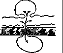
Figure 6. Aneurysm with arterial and venous sac.
Figure 3. Aneurysmal node.
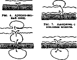
Figure 5. Aneurysm with arterial, venous and intermediate sac.
Figure 8. Aneurysm with arterial sac. between both vessels (see figure 5). Characteristic for arteriovenous A. are changes on the part of the vein; its lumen, mainly in the central direction, expands, while the walls of the vein thicken and become arterial. - Signs of arteriovenous A. Expansive pulsation in these A. is significantly less than in arterial A., because the sac is smaller, and with a fistula it is absent altogether. Instead, a strong pulsation of the veins is characteristic, which occurs because the arterial pulse wave is transmitted directly through the aneurysmal opening from the artery into the vein. Venous pulsation is also transmitted peripherically due to difficulty in outflow of blood during systole and diastole of the arterial pulse. A characteristic symptom of arteriovenous A. is considered to be the continuous aneurysmal noise with systolic intensification, taking on the character of a murmur or purring. This noise is perfectly perceived by the hand applied to the area of the A. In these A., the larger part of the arter

Figure 9. Indirect arteriovenous aneurysm (semi-schematic drawing).
of blood passes into the vein, and only a smaller part reaches the periphery of the limb completely; as a result, atrophic phenomena at the periphery are particularly pronounced. In addition, the communication between the artery and vein creates a great difficulty for the heart, part of the propulsive force of which is lost due to the presence of the arteriovenous communication. Thus, as a consequence of arteriovenous aneurysms, significant dilation of the heart is noted. Course. Pulsating hematomas initially increase in size until the pressure in the hematoma reaches a level equal to arterial pressure; then they contract, and an aneurysmal sac is formed. During this complex process of restructuring, dangerous complications may appear. The first is the rupture of the outer layer of clots and fibrin with secondary hemorrhage into the tissues or with external bleeding. Such a complication arises from two main causes: 1) mechanical, due to the thinning of the sac and its rupture as a result of the strong pressure inside the sac, 2) infection of the sac in one or another of its parts and, as a result, the softening of the otherwise firm wall of the sac. In both cases, the rupture occurs, predominantly, at the moment of accidental increase in blood pressure (from standing up, sharp movements, coughing, excitement, etc.). Secondary, sometimes repeated, ruptures of the pulsating hematoma are very dangerous; for the limb they also give a poor prognosis due to the weakening of the body and, mainly, the heart, on the one hand, and on the other hand, the compression of the collaterals by the suddenly enlarged hematoma. The same danger continues to exist even after the final formation of the aneurysm, but it is observed much less frequently. Here something else happens, namely: a) gradual increase and expansion of the sac, locally with thinning of its walls; b) atrophy of the tissues lying above the sac down to the skin (or internal cavities-pleura, peritoneum) inclusive, and c) a fatally ending rupture of the sac outward, resp. into the cavity. The next complication consists of peripheral embolization, which occurs in such a way that a free clot inside the sac cavity or one formed at its neck is thrown along the blood flow to the periphery and blocks some artery of this or that caliber, as a result of which this part of the limb is in danger of gangrene. Such embolization acquires special significance in aneurysms of the carotid arteries (embolism of the brain) and, mainly, in large arteriovenous aneurysms, in which the embolism is directed not to the periphery but along the draining vein to the heart and lungs. From the above, the danger of developing gangrene of the limb in false aneurysms is clear. Gangrene results from insufficient blood flow through the arterial trunk, from compression of the collaterals, and from embolization. It is true that the danger of gangrene after vascular injury gradually decreases due to the progressive expansion of the collaterals, which is facilitated by an increase in arterial pressure above the site of arterial narrowing; however, individually, the strength of collateral circulation can be extremely variable, and the complications mentioned above can significantly disrupt the restored circulation in the area below the site of injury. Therefore, in all false aneurysms and pulsating hematomas, gangrene is a real danger that forces the surgeon to always be ready to intervene. Self-healing. The blood in the cavity of a pulsating hematoma is in conditions in which its clotting is easily possible. In aneurysms, however, this possibility, although hindered, is also not excluded, so it is possible for all the blood that has flowed out of the vessel to clot, followed by its organization and, thus, the emptying and destruction of both the pulsating hematoma and sometimes the aneurysm. The process mentioned is by no means an exceptional rarity, and in practice one must take it into account very seriously: according to Dobrovolskaya, it is observed in no less than 12% of cases, and according to Savvin - about 2%. In such cases, there were typical signs of aneurysms, which then disappeared, and everything returned to normal. - The prognosis of false aneurysms depends entirely on the form of the wound, on infection, on the caliber and importance of the injured vessel, and on all other conditions that have been discussed above. - Indications for surgical treatment exist: always and absolutely, when the pulsating hematoma increases continuously instead of decreasing; in cases of infection; in cases giving secondary bleeding, and finally, when gangrene of the limb is threatening. - The treatment of false aneurysms is a difficult task both in relation to the whole organism and in relation to the injured limb. From the side of the organism, it is necessary to achieve relief for the burdened heart, and from the side of the limb - improvement of blood circulation and nutrition of peripheral tissues, as well as elimination of the danger of fatal bleeding. To achieve this purpose, there are two paths: conservative and operative. --The conservative method of treatment should all the more attract attention because a certain percentage of pulsating hematomas or aneurysms heal spontaneously when they are located on small arteries. Here it is appropriate to use systematic, regular, daily compression of the main feeding arterial trunk for 10 to 30 minutes 1-2 times a day (the daily duration of compression is very difficult to carry out in practice). This procedure not only promotes the process of blood clotting in the sac but at the same time prepares the collaterals, which expand each time with this procedure (gymnastics of the collaterals). On this basis, this procedure is also used to prepare the patient for radical surgery of the aneurysm. In the literature, successful cases of such therapy are described, but they are few in number. The administration of calcium orally does not significantly improve the modest results (Savvin). - Surgical treatment is indicated for the vast majority of cases. In addition to what has been said above, an indication for surgery is the increase in volume of the pulsating hematoma or aneurysm, worsening of trophic symptoms, intensification of painful phenomena. The question is only when to operate on a false aneurysm - in the stage of pulsating hematoma or in a later stage. There are ardent supporters of both views. The decision of the question often depends on the combination of pathological phenomena in both cases; often the surgeon, regardless of his theoretical views and experience, is forced to operate on the pulsating hematoma, despite the fact that during the formation of the aneurysmal sac, as is well known, the limb gradually gets used to the reduced blood flow, and at the same time the collaterals begin to develop and function. On the other hand, the surgeon, a convinced advocate of early intervention, must wait for the final formation of the aneurysm, i.e., will operate under the best conditions for preserving the limb and when the general condition is already good, because the operation is then performed more easily, the vessels are found more quickly, and the sac, consisting of more or less organized clots, is dissected out more gently and better than in the earlier stage. An experienced clinical surgeon, who knows how to weigh the value of indications for surgery, will encounter cases of both kinds. It was mentioned above that pulsating hematomas, at a certain time in their growth, may take the appearance of an abscess; sometimes, by mistake, the cavity of the hematoma is opened by an incision; to stop the threatening bleeding that has opened, it is necessary to complete the operation and tie off the injured artery in the wound (one cannot rely on tight tamponade and bandaging). In planned operations, both for pulsating hematoma and for aneurysm, one must first protect oneself from strong bleeding. This is achieved by taking the artery and vein on a strong thread over a length, above and below the aneurysm; the threads are not tied in a knot, but serve only for temporary tightening and bending of the vessels in case of unexpected bleeding; entering into the cavity of the hematoma, resp. aneurysm, the clots and the soft wall of the pulsating hematoma are removed or the dense fibrous wall of the aneurysm is carefully separated from the nerves and vessels and it is completely or partially excised. Thus, the site of damage to the arterial wall, with edges protruding in white, is naturally exposed. Usually, a ligature is then placed under the artery, directly above and below the site of its injury, and the damaged segment is excised. In this case, concerns arise - whether gangrenous phenomena will develop on the periphery or subsequent phenomena of insufficient nutrition of the tissues of the limb (pain, atrophy, etc.). The experience of recent wars (Japanese, imperialist) has shown that intrasac ligation is well tolerated, and only in rare cases do mild gangrenous phenomena develop on the fingers; the final results are excellent, sometimes including the restoration of the pulse on the periphery of the tied artery. Everything depends on the development of the collaterals. Even before the operation, to some extent, the strength of collateral circulation can be determined by the method of Korotkoff or Moshkovich.
These methods are based on studying the ability of blood vessels to react to anemia after artificial bloodletting using an elastic bandage and tourniquet for several minutes (3-5) in the form of active hyperemia (redness). Depending on the width of the collateral pathways, under conditions of continued isolated compression of the main arterial trunk, active hyperemia, due to severe damage to the skin coverings, occurs either quickly over a large area of the limb or slowly over a smaller area. For orientation, it is recommended to perform a similar experiment on a healthy limb. Further, under the same conditions, the blood pressure, which had fallen almost to 0, rises to a higher or lower degree. A minimum pressure height of 30 mm Hg according to Riva-Rocci is considered a favorable moment for determining whether the strength of the collaterals is sufficient to maintain nutrition of the limb after ligation of the arterial vessel supplying the limb. These methods are inaccurate and can be misleading, mainly because it is almost impossible to isolate one artery during compression and a vein is always more or less compressed simultaneously; and this moment can cause, in addition to blood retention in the limb, an increase in blood pressure. The filling of skin vessels in the Moskovich test, despite its convincing demonstrability, does not allow one to conclude about the filling of vessels in the depth of the muscle tissue - necrosis of the muscle tissue was often observed with good skin nutrition (Stich and Fromme). During the operation, the strength of collateral circulation is determined very approximately by the filling of the vein with blood during prolonged compression of the arterial trunk ad oculos, as well as by the abundance of back-bleeding from the peripheral segment of the artery when the central segment of the artery is compressed above the aneurysmal opening. In some cases of arterial aneurysm, blood pressure on the periphery falls too low after ligation of the artery; then simultaneous ligation of the corresponding vein raises the pressure on the periphery somewhat higher (reduced circulation of Oppel). Due to frequent simultaneous injury of arteries and veins during the imperialist war or inclusion of a vein in the fibrous wall of the sac with compression of the lumen, ligation of the vein was usually performed without any adverse consequences, and sometimes with a visible positive effect. The operation of pulsating hematoma and A. undoubtedly improves the existing preoperative collateral circulation: it eliminates the pressure of the hematoma, resp. sac, on the collaterals and eliminates tension in the tissues; in addition, it eliminates pain sensations depending on compression and irritation of nerves, which often leads to spasm of collaterals and vessels on the periphery. The operation in cases of arteriovenous A. aims at complete separation of the arterial and venous pathways and elimination of A. This is achieved by exposing the site of vascular injury and applying a quadruple ligature to the artery and vein above and below the injury with subsequent excision of the affected segment. This difficult operation gives good results, but here, as with other A., the question arises - whether the ideal method, providing for the restoration of the arterial and venous pathways, i.e., restoration of normal blood circulation conditions, can be applied here. Vascular suture (see) is used in A. infrequently. It has been successfully applied in fresh incised wounds of arteries (Herzen), as well as in accidental injuries of vessels during operations; but in fresh gunshot wounds of vessels, it is applied under certain and limited indications (Sencert), although there are references in the literature of a more radical nature: Stich and Fromme expand the indication for vascular suture in fresh wounds and insist on its early application. Many objections have been raised against this opinion; the most significant of them is the fear of infection in the surgical wound. It is more rational to apply the suture later, when the bullet channel does not show inflammatory phenomena and when bullet injuries have smoothed out in the form of a smooth opening. This happens, mainly, in cases of arteriovenous fistula: then, after separation of the pathways, the opening, both in the artery and in the vein, is closed by a lateral vascular suture. In A. that deform the arterial wall, it is better to abandon the suture, in this case necessarily circular. Artery suture, in any case, does not guarantee at all the disappearance of the pulse on the periphery, secondary bleeding due to insufficiency of the suture, or gangrene of peripheral parts due to local thrombosis of the artery with thromboembolism of arteries on the periphery. Other methods of operation for false A. are not currently used or are used only under special indications. These methods - ligation on the central segment of the artery, ligation on the peripheral segment of the artery, double ligation, central and peripheral, or finally, the Anschluß method with double ligation and opening of the sac with subsequent tamponade; the latter method of operation is reliable.
Related articles
Mentioned in
Cite this page
“Aneurysm.” Soviet Medical Encyclopedia. English translation of Bolshaya Meditsinskaya Entsiklopediya, 1st ed. (Moscow, 1928–1936), ed. N. A. Semashko. https://sovietmedicalencyclopedia.pages.dev/article/aneurysm/