Brachium
Historical document, translated for reference. It reflects medical knowledge of the 1920s–30s and is not medical advice.
Summary
The brachium is the anatomical region of the upper limb between the lower border of the pectoralis major muscle and a line two transverse fingers above the condyles of the humerus. The article details the skeletal structure, muscular attachments, and developmental aspects of the humerus.
Encyclopedia article (1928–1936)
Brachium, brachium, part of the upper limb within the boundaries between a transverse line drawn along the lower border of the pectoralis major muscle, the latissimus dorsi muscle, and the teres major muscle (above), and a similar line drawn two transverse fingers above the condyles of the humerus (below). The shape of the brachium depends on the degree of development of its musculature and its nutritional state. In women and children, the subcutaneous fat layer smooths out all the relief of the musculature and gives the brachium the form of almost a regular cylinder, while in a muscular man, the brachium in cross-section appears elongated from front to back due to the developed musculature of the anterior and posterior parts of the arm; at the top, the volume of the brachium is greater than at the border with the elbow region. -The skeleton of the brachium is the humerus (humerus, os humeri) (fig. 1)-a long, tubular bone; articulating4 above with the scapula, below-with the bones of the forearm.- The upper epiphysis of the humerus is thickened, carries on itself a cartilage-covered head (caput humeri), which has the shape of a hemisphere, is directed upward and inward; the axis of the head is located at an angle of 130 - 145° to the longitudinal axis of the bone; immediately behind the head there is a constriction of the bone, the anatomical neck (collum anatomicum); below it are two tubercles; the forward-facing lesser tubercle (tuberculum minus), to which the subscapularis muscle (m. subscapularis) attaches, and the lateral and more voluminous greater tubercle (tuberculum majus); posteriorly - upper
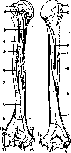
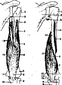
fig. 1. Humerus with sites of muscle attachment. . Front view (a): 1-m. supraspinatus; 2-m. latissimus dorsi; 3-m. teres major; 4-m. pectoralis major; 5-m. deltoideus; 6-m. coracobrachialis; 7-m. brachialis; 8-m. extensor carpi radialis longus; 9-m. extensor carpi radialis brevis; 10-m. extensor digitorum communis; 11-m. extensor digiti minimi; 12-m. extensor carpi ulnaris; 13-m. flexor carpi radialis; 14-m. flexor digitorum sublimis; 15-m. flexor carpi ulnaris. Posterior view (b): 1-m. deltoideus; 2-m. brachialis; 3-m. brachioradialis; 4-m. anconaeus; 5-m. extensor digitorum communis; 6-m. extensor digiti minimi; 7-m. extensor carpi ulnaris; 8-m. flexor carpi radialis; 9-m. flexor digitorum sublimis; 10-m. flexor carpi ulnaris. The lateral surface carries on itself three areas: the upper area is designated for the attachment of the supraspinatus muscle (m. supraspinatus), the middle area for the latissimus dorsi muscle (m. latissimus dorsi), the teres major muscle (m. teres major), and the pectoralis major muscle (m. pectoralis major); the lower area is for the deltoid muscle (m. deltoideus). From each tubercle downward extends a crest (crista tuberculi minoris et majoris); to the first attach the latissimus dorsi muscle and the teres major muscle, to the second-the pectoralis major muscle; between the tubercles and their crests there is a groove 6-8 cm long, lined with cartilage at the top (intertubercular groove-sulcus intertubercularis); in this groove lies the tendon of the long head of the biceps brachii muscle. The part of the humerus immediately below both tubercles, of smaller diameter, is called the surgical neck (collum chirurgicum). The middle part of the humerus (diaphysis, body, corpus humeri), almost straight, twisted along its axis, is cylindrical in shape above, triangular below. On it are distinguished the anteromedial surface (facies ant. medialis), the anterolateral, lateral (facies ant. lateralis) and the posterior surface (facies post.); the latter is clearly delimited from the first two by a well-expressed, especially in the lower half of the bone, medial edge (margo med.) and lateral edge (margo lat.). In the upper part of the middle third of the humerus on the anterolateral surface there is a V-shaped, downward-pointing deltoid tuberosity (tuberositas deltoidea), serving for the attachment of the muscle of the same name. Behind it from top to bottom spirally runs the groove for the radial nerve (sulcus n. radialis)-the location of the radial nerve and the deep brachial artery with its two veins. At the middle of the medial edge of the humerus is the nutrient foramen (for. nutricium), through which begins the nutrient canal (canalis nutricius) going to the lower end of the bone. The diaphysis of the humerus serves as the site of attachment of several muscles (see below). The lower end of the humerus, flat and wide, is bent forward at an obtuse angle, open forward, to the long axis of the body of the humerus; on the sides of the lower end of the humerus on the continuation of the medial and lateral edges lie the rough tubercles, the medial and lateral condyles (condylus medialis et lateralis). The medial condyle is larger, on its posterior surface it has a flat groove for the ulnar nerve (sulcus n. ulnaris). The lower
end of the humerus has cartilage-covered articular surfaces: the medially located part, the trochlea, articulates with the ulna, and the lateral part, the capitulum (capitulum, the cap-like elevation, eminentia capitate), has the shape of a part of a sphere and articulates with the radius; above the trochlea in front is located the shallow coronoid fossa (fossa coronoida), into which during flexion at the elbow is placed the coronoid process of the ulna; above the trochlea posteriorly is located the deep olecranon fossa (fossa olecrani), into it during extension is immersed the olecranon process of the ulna. "The bone between these fossae is very thin and sometimes perforated. Above the cap-like elevation
Fig. 2.
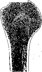
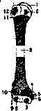
in front lies the radial fossa (fossa radialis). The three named fossae lack cartilage. Above the articular parts of the condyles of the humerus on the sides there are bony projections, intended for the fixation of muscles and ligaments on them; the medial epicondyle (epicondylus medialis) is more developed, where are attached: the medial collateral ligament of the elbow (lig. collaterale ulnare) and five flexor muscles: mm. pronator teres, flexor carpi radialis, palmaris longus, flexor digitorum sublimis and flexor carpi ulnaris and occasionally an accessory head of the deep flexor of the fingers; the lateral epicondyle (epicondylus lateralis) is less developed and serves as the site of attachment of the lateral collateral ligament (lig. collaterale radiale) and 6 extensor muscles: mm. extensor carpi radialis longus, supinator (brevis), extensor digitorum, extensor indicis, extensor carpi ulnaris, anconaeus. Internal structure of the humerus. Both ends consist of a relatively thin compact cortical layer4 and the spongy substance of bone, occupying all the remaining mass, consisting of thin bone plates, trabeculae; these plates are a continuation of the cortical layer of the bone (fig. 2), pass in various intersecting directions along the lines that mathematicians draw in graphical statics when constructing bodies having the same shape as bones and serving to counteract the same forces that act on the bone (Lesgaft). The cells of the spongy substance are filled with bone marrow. The medullary canal of the diaphysis occupies its entire height, having a mean transverse diameter of 8 mm in the lower third, 10 mm in the middle third and 12 mm in the upper third. The walls of the bony cylinder thin from bottom to top. The main mass of the lower epiphysis consists of spongy substance with denser plates, mostly vertically arranged in relation to the articular surfaces. The cortical layer on the lower epiphysis is thicker here than on the upper epiphysis. The development of the humerus occurs from 8 ossification points (fig. 3)-one primary and 7 secondary. The primary point appears around the 45th day of intrauterine life in the middle of the length of the diaphysis, growing upward and downward. Of the 7 secondary, 3 are in the upper epiphysis and 4 in the lower; the first point of the upper epiphysis appears on the head of the bone in the 2-4th month after birth; the points of the greater and lesser tubercles appear in the 2-3rd year. In the lower epiphysis, the first point appears on the cap-like elevation at the beginning of the 3rd year. In the 5th year appears the point of the medial epicondyle. In the 12th year-3rd and 4th points of the trochlea and lateral epicondyle. The lower epiphyseal line disappears around the 16-18th year; the same between the medial epicondyle and the epiphysis-a year or two later, but cases are not rare when it exists even at 25 years. The upper epiphyseal line persists until 25-26 years. The brachium is anatomically and functionally divided into two regions: the anterior-flexor bed (regio brachii ant.) and the posterior-extensor bed (regio brachii post.).-The anterior part has the form of a convex forward oval, more pronounced with developed musculature. On the lateral and medial sides of the brachium
run shallow grooves (sulci bicipitales lat. et med.), delimiting the anterior part of the brachium from the posterior; both grooves converge at an acute angle in the cubital fossa; the medial groove is more sharply expressed, the lateral is shorter than the medial; at the top, at the lower end of the deltoid muscle, it divides into two branches, of which the anterior passes into the deltopectoral groove (sulcus deltoideo-pectoralis), and the posterior runs along the posterior edge of the deltoid muscle.- The skin of the brachium is loosely connected with the underlying tissues and is thicker on the lateral surface; it is devoid of hair on the anterior and medial surfaces of the brachium. The subcutaneous tissue consists of two layers, between which
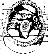
Fig. 3. Points of ossification: 1-head; 2-lesser tubercle; 3-body; 5-medial condyle; 7-trochlea; 8-capitulum; 9-lateral condyle; 12-greater tubercle (4 and 6-epiphyseal lines of the medial condyle; 10 and 11-lower and upper epiphyseal lines).
are located
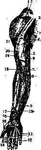
Figure 4. Superficial nerves of the arm: 1 and 2-branches from the cervical plexus; 3,4, 5-cutaneous branches of the median nerve; 6-9-n. cutaneus brachii med.; 10-branch of the n. medianus; 11-15 and 22-cutaneous branches of the nn. mediani et ulnaris; 16, 17 and 19-branches of the n. radialis; 18-cutaneous branch of the musculocutaneous nerve; 20 and 21-cutaneous branches of the axillary nerve. The superficial vessels and nerves are located here. From the forearm extend two large venous trunks - v. basilica and v. cephalica. V. basilica runs in the subcutaneous tissue along the sulcus bicipitalis med., but only in the lower half of the groove, above it pierces the brachial aponeurosis and runs in the canal of this aponeurosis, often to the axillary fossa or else on the middle of the arm it empties into the brachial vein. V. cephalica passes in the subcutaneous tissue along the sulcus bicipitalis lat., then upward in the deltopectoral groove to its emptying into the axillary or subclavian vein. Superficial nerves of the anterior part of the arm (fig. 4): below-lateral cutaneous nerve of the arm (n. cutaneus brachii lateralis from the axillary nerve; on the anteromedial surface of the Brachium above-intercostobrachial nerve (n. intercosto-brachialis) and medial internal cutaneous nerve of the Brachium (n. cutaneus brachii medialis); in the lower half of the Brachium-medial cutaneous nerve of the forearm (n. cutaneus antibrachii medialis). Aponeurosis (aponeurosis, fascia brachii). It envelops the Brachium like a sheath and passes into the aponeurosis of the areas adjacent to the Brachium. From the inner surface of the aponeurosis extend thin septa for the muscles. Between the aponeurosis and the humerus in the frontal plane are located the intermuscular septa of the Brachium-internal and external (septum intermusculare mediale et laterale) (fig. 5). The aponeurotic sheath is divided by them into two compartments: anterior-compartment of the flexors and posterior-compartment of the extensors. Both compartments communicate with each other by vessels and nerves passing from one compartment to the other. The anterior compartment is divided into two-compartment of the biceps muscle and compartment of the brachial muscle (fig1. 5). The aponeurosis of the Brachium is thicker behind. The main mass of fibers of the aponeurosis consists of circular bundles with a slight oblique inclination, the longitudinal bundles (there are fewer of them than circular ones) run perpendicular to the first ones. The pectoralis major and
Figure 5. Transverse section of the Brachium in its lower third (diagram): 1-m. biceps; 2-n. cutaneus brachii med.; 3-v. basilica; 4-n. medianus; 5-n. cutaneus antibrachii med.; 6-a. brachialis; 7-internal tendinous septum; 8-n. ulnaris; 9-a. collateralis ulnaris sup.; 10-humerus; 11-m. triceps; 12 and 20-aponeurosis; 13-sulcus n. radialis; 14-a. profunda brachii; 15-n. radialis; 16-external tendinous septum; 17-m. brachialis int.; 18-n. musculo-cutaneus; 19-v. cephalica.
latissimus dorsi send wide aponeurotic expansions to the aponeurosis of the Brachium (Sappey). The subaponeurotic layer is occupied by 5 muscles arranged in two layers: in the first, the deltoid and biceps muscles (see Biceps and Deltoideus m?«-culus); the following three muscles-coracobrachialis (m. coraco-brachialis), brachialis (m. brachialis) and brachioradialis (m. brachio-radialis)-are located under the first two (see Muscles of man). In the axillary fossa, the coracobrachialis muscle forms part of its lateral wall, in front of the muscle lie the deltoid and pectoralis major muscles, behind-tendons of the subscapularis, latissimus dorsi and teres major muscles, inside-the vascular-nerve bundle. The brachialis muscle (m. brachialis)-see Muscles of man. Relation to neighboring organs: on the anterior surface of the brachialis muscle lies the biceps muscle; between them is an intermuscular septum, through which passes the musculocutaneous nerve (fig. 5) and inside it from it, the vascular-nerve bundle of the Brachium. The posterior surface of the muscle above lies on the humerus, below-on the elbow joint. The inner edge above borders with the triceps muscle, below-with the round pronator, between them is formed a groove, in which pass the brachial artery with veins and the median nerve; the upper part of the lateral edge borders with the triceps muscle, and the lower part-with the brachioradialis muscle, in the groove between which pass the radial nerve and the collateral radial artery (a. collateralis radialis).-Brachioradialis muscle (t. brachio-radialis) (see Muscles of man) passes lateral to the brachialis muscle on the forearm, borders on the dorsal side with both radial extensors of the wrist, on the palmar side-with the round pronator and radial flexor of the wrist; this muscle attaches
Figure 6. Arteries of the arm from the front: /-a. profunda brachii; 2-a. brachialis; 3-a. collateralis ulnaris sup.; 4-a. collateralis ulnaris inf.; 5-a. ulnaris; 6-a. radialis; 7 and 8-muscular branches.
by a thin flat tendon to the radius above the styloid process; the tendon of the brachioradialis muscle is often pierced by the radial nerve. Most of the vessels and nerves of the anterior part of the Brachium go from the axillary region to the forearm and hand. The main arterial trunk of the Brachium is the brachial artery (a. brachialis, fig. 6), which is the continuation of the axillary artery (a. axillaris). The artery runs along the median side of the Brachium, along the edge of the biceps muscle, along the projection line from the apex of the axillary fossa to the middle of the cubital fossa. The two accompanying veins (v. brachiales) run on both sides of the artery behind: 1 and 2-aprofunda brachii; 3 and 9-network of anastomoses; 4-a. subscapularis; 5-a. transversa scapulae; 6-a. circumflexa humeri; 7-an
Figure 7. Anastomoses of the axillary artery from the front: 1-a. axillaris; 2-a thoraco-acromialis; 3-anastomoses between a. thoraco-acromialis and circumflexa humeri; 4-anastomoses between a. circumflexa humeri and a. brachialis; 5-a. brachialis; 6-a. circumflexa humeri; 7-a. circumflexa scapulae; 8, 10 and 13-network of anastomoses; 9 and 12-a. transversa colli; 13-a. subclavia.
astomosis between aa. circumflexa and a. profunda brachii; 10-a. thoraco-acromialis; 11-a. trans
versa scapulae. In the upper third of the Brachium, outside 1 and profunda brachii; s-from the artery, lies
...... the median nerve (fig. -see Nerves of man), crossing it on the middle of the Brachium from the front and in 10-0-from behind; after the crossing the nerve is located on the inner side of the artery. The artery, veins and nerve pass along the anterior surface of the median intermuscular septum, behind which passes the ulnar nerve and the upper and lower collateral ulnar arteries. From the upper part of the brachial artery emerges the deep artery of the Brachium (a. profunda brachii), which immediately goes backward and downward between the lateral and medial heads of the triceps muscle ' I and with the radial nerve "v it curves around the humerus from behind. The deep artery of the Brachium e--'' anastomoses with the branches of the anterior and posterior circumflex arteries of the Brachium, through them with a. tho-raco-acromialis on fig. 8. Anastomoses of the axillary artery from the front: 1-a. axillaris; 2-a thoraco-acromialis; 3-anastomoses between a. thoraco-acromialis and circumflexa humeri; 4-anastomoses between a. circumflexa humeri and a. brachialis; 5-a. brachialis; 6-a. circumflexa humeri; 7-a. circumflexa scapulae; 8, 10 and 13-network of anastomoses; 9 and 12-a. transversa colli; 13-a. subclavia.
from above and with the arterial network of the posterior
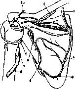
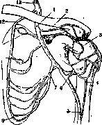
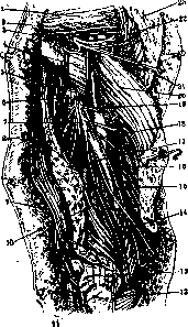
of the scapula, as a result a collateral pathway is formed from the subclavian artery to the brachial artery (figs. 7 and 8). Directly from the brachial artery or from one of its muscular branches originates the nutrient artery of the humerus (a. nutricia humeri), which penetrates the bone through the nutrient canal. Next4, somewhat lower than the previous one, departs the ulnar collateral superior artery (a. collateralis ulnaris sup.); it goes with the ulnar nerve to the elbow joint; it branches in the neighboring muscles. 2-3 transverse fingers above the medial epiconyle departs the ulnar collateral inferior artery (a. collateralis ulnaris inferior), going forward of the epiconyle downward and participating together with the previous one in the formation of the arterial network of the elbow joint. Variations of the brachial artery. Often instead of a single trunk of the brachial artery there are on the arm several parallel arterial trunks, departing either from the axillary or from the brachial artery at its very beginning and then replacing it; such arteries are called vasa aberrantia. Very rarely they fuse into one trunk or give continuation again in the form of a single brachial artery, more often each passes into one of the arteries of the forearm, and six times more often into the radial artery (Giacomini). A case is known of the transition of vasa aberrantia into the artery of the median nerve at its detailed end and a case of transition into the superficial palmar arch. Division of the brachial artery very rarely occurs below the olecranon fossa (low or late division) and more often (10-12%) above the elbow, such high or early division occurs most often in the upper third of the arm and very rarely in its middle and lower thirds. The place of division of the brachial artery into branches can rise to the axillary fossa and even to the neck. High division of the artery can occur in one of the following five ways: the brachial artery breaks down into 1) a. radialis and truncus interosseo-ulnaris, 2) a. ulnaris and truncus interosseo-radialis, 3) a. interossea or mediana and truncus radioulnaris, 4) a. radialis, a. ulnaris, a. interossea, 5) a. aberrans and a. brachialis. This anomaly of division of the brachial artery (according to Quain) out of 61 cases in 43 cases it is unilateral and in 18 cases bilateral. The volume, length, course and branching of such arteries are very diverse. Coming to the forearm, they often become superficial, i.e. they go between the muscles and aponeurosis or between the latter and the skin. Often the radial artery on the arm passes close to the ulnar and, in order to become lateral on the forearm, it crosses the ulnar. Deep veins accompany the arteries in pairs, the external brachial vein goes to the middle of the axillary fossa and there, crossing the artery from the front, merges with the previous vein. Lymph. system of the arm-see Lymphatic system. Deep nerves of the anterior part of the arm: musculocutaneous nerve (n. musculo-cutaneus) and median (n. medianus)-see Nerves of man, Medianus nervus. Posterior part of the arm. The skin is thicker than in front, movable, often covered with hair. Superficial arteries and veins are insignificant, they represent branches of the deep artery of the arm and a network of veins flowing into vv. basilica and cephalica. Superficial nerves-the lateral cutaneous nerve of the arm from the axillary nerve, the posterior cutaneous nerve of the arm (n. cutaneus brachii posterior) and in the lower part of the department the dorsal cutaneous nerve of the forearm (n. cutaneus antibrachii dorsalis); both last from the radial nerve. Aponeurosis-part of the common aponeurosis of the arm. The main content of the posterior part, if we do not count the lower part of the deltoid muscle, is the triceps brachii muscle (m. triceps brachii, see Muscles of man). This muscle is a strong extensor of the forearm-consists of 3 portions (heads), one of which, the long (caput longum), goes with a flat tendon from the infraglenoid tubercle of the scapula (tuberositas infraglenoidalis scapulae) between the minor and major round muscles, partly covered by the deltoid muscle (figs. 9 and 10), and lies behind and medial to the medial head (caput mediale); the latter originates from the posterior surface of the humerus, below and medial to the radial nerve groove, and from the medial and lateral intermuscular septa; in the form of a wide muscular belly they go downward, covering the ulnar muscle (t. anconaeus); here the fibers pass into a flat tendon, with which the tendon bundles of the long and lateral heads merge. The lateral head (caput laterale) originates from the posterior surface of the humerus, above and lateral to the radial nerve groove and from the lateral muscular septum; its fibers go downward and pass over the radial nerve to the tendon of the medial head. The triceps muscle attaches to the olecranon of the ulna; besides extension, at the elbow the long head of the muscle adducts the arm. The triceps muscle is innervated by the radial nerve, with branches for the long head separating from the main trunk immediately below the lower edge of the major round muscle; for the lateral head several twigs depart lower than the previous ones; the nerve of the medial head enters its upper part, in the thickness of the muscle it reaches the ulnar muscle and innervates it (fig. 11). On the arm also begin the brachioradialis muscle and both radial extensors of the hand. On the posterior wall of the axillary region two openings are formed: 1) for. quadrilaterum, bounded above by m. subscapularis, below by m. latissimus dorsi and m. teres major, in front by collum humerale chirarg., behind by cap. long. m. tricipitis and 2) for. trilaterum, located medially from cap. long. m. tricip. and bounded above by m. subscapularis, below by m. teres major, in front and laterally by cap. long. m. tricip. Through the first opening pass vasa circumflexa hum. et n. axillaris, through the second- a. circumflexa scapulae. The main artery of the posterior bed of the arm is the deep artery of the arm (a. profunda brachii). On its entire path it gives off small branches for the coverings of this area and more significant ones for the triceps muscle; its most significant branches are the deltoid artery (a. musculus deltoideus), lateral middle (a. colla
teral superior), radial collateral (a. collateralis radialis), and middle collateral (a. collateralis media). The deep artery of the arm (a. profunda brachii) is a large branch of the brachial artery, departing from it at the lower edge of the major round muscle. It goes backward between the medial and lateral intermuscular septa, accompanying the radial nerve. In the middle of the arm it gives off the radial collateral artery, which goes backward and downward, accompanying the radial collateral nerve, and anastomoses with the ulnar collateral artery. The middle collateral artery departs somewhat lower, goes backward and downward, accompanying the middle collateral nerve, and anastomoses with the ulnar collateral artery. The deep artery of the arm continues as the radial collateral artery, which goes downward between the lateral intermuscular septum and the humerus, accompanying the radial nerve, and at the elbow divides into the radial recurrent artery and the interosseous artery. The radial recurrent artery goes upward and laterally, anastomoses with the radial artery. The interosseous artery goes downward between the radius and ulna, gives off the anterior and posterior interosseous arteries, and anastomoses with the anterior and posterior ulnar recurrent arteries. The anterior interosseous artery goes downward between the radius and ulna, accompanies the anterior interosseous nerve, and anastomoses with the deep palmar arch. The posterior interosseous artery goes downward between the radius and ulna, accompanies the posterior interosseous nerve, and anastomoses with the dorsal carpal arch. The ulnar collateral artery (a. collateralis ulnaris) is a branch of the brachial artery, departing from it at the level of the insertion of the coracobrachialis muscle. It goes backward and downward, accompanying the ulnar nerve, and anastomoses with the radial collateral artery and the ulnar recurrent artery. The ulnar recurrent artery is a branch of the brachial artery or of the deep artery of the arm, departing from it at the level of the elbow. It goes upward and medially, anastomoses with the ulnar artery and the ulnar collateral artery. The anterior and posterior ulnar recurrent arteries are branches of the ulnar artery, departing from it at the level of the elbow. The anterior ulnar recurrent artery goes upward and medially, anastomoses with the anterior interosseous artery. The posterior ulnar recurrent artery goes upward and medially, anastomoses with the posterior interosseous artery. The nutrient artery of the humerus (a. nutricia humeri) is a branch of the brachial artery or of one of its muscular branches. It enters the humerus through the nutrient canal, which is located on the medial surface of the bone, below the insertion of the deltoid muscle. The deltoid artery (a. musculus deltoideus) is a branch of the deep artery of the arm or of the brachial artery. It goes to the deltoid muscle, which it supplies. The lateral middle artery (a. collateralis ulnaris superior) is a branch of the brachial artery, departing from it at the level of the insertion of the coracobrachialis muscle. It goes backward and downward, accompanying the ulnar nerve, and anastomoses with the radial collateral artery and the ulnar recurrent artery. The radial collateral artery (a. collateralis radialis) is a branch of the deep artery of the arm, departing from it in the middle of the arm. It goes backward and downward, accompanying the radial collateral nerve, and anastomoses with the ulnar collateral artery. The middle collateral artery (a. collateralis media) is a branch of the deep artery of the arm, departing from it somewhat lower than the radial collateral artery. It goes backward and downward, accompanying the middle collateral nerve, and anastomoses with the ulnar collateral artery. The anterior and posterior interosseous arteries are branches of the interosseous artery. The anterior interosseous artery goes downward between the radius and ulna, accompanies the anterior interosseous nerve, and anastomoses with the deep palmar arch. The posterior interosseous artery goes downward between the radius and ulna, accompanies the posterior interosseous nerve, and anastomoses with the dorsal carpal arch. The radial recurrent artery is a branch of the deep artery of the arm or of the interosseous artery. It goes upward and laterally, anastomoses with the radial artery. The ulnar recurrent artery is a branch of the brachial artery or of the deep artery of the arm. It goes upward and medially, anastomoses with the ulnar artery and the ulnar collateral artery. The anterior and posterior ulnar recurrent arteries are branches of the ulnar artery. The anterior ulnar recurrent artery goes upward and medially, anastomoses with the anterior interosseous artery. The posterior ulnar recurrent artery goes upward and medially, anastomoses with the posterior interosseous artery. The nutrient artery of the humerus enters the bone through the nutrient canal, which is located on the medial surface of the bone, below the insertion of the deltoid muscle. The deltoid artery supplies the deltoid muscle. The lateral middle artery accompanies the ulnar nerve and anastomoses with the radial collateral artery and the ulnar recurrent artery. The radial collateral artery accompanies the radial collateral nerve and anastomoses with the ulnar collateral artery. The middle collateral artery accompanies the middle collateral nerve and anastomoses with the ulnar collateral artery. The anterior and posterior interosseous arteries are branches of the interosseous artery. The anterior interosseous artery accompanies the anterior interosseous nerve and anastomoses with the deep palmar arch. The posterior interosseous artery accompanies the posterior interosseous nerve and anastomoses with the dorsal carpal arch. The radial recurrent artery goes upward and laterally, anastomoses with the radial artery. The ulnar recurrent artery goes upward and medially, anastomoses with the ulnar artery and the ulnar collateral artery. The anterior and posterior ulnar recurrent arteries are branches of the ulnar artery. The anterior ulnar recurrent artery goes upward and medially, anastomoses with the anterior interosseous artery. The posterior ulnar recurrent artery goes upward and medially, anastomoses with the posterior interosseous artery.
The skin of the posterior part of the arm is thicker than that of the anterior part, movable, and often covered with hair. The superficial arteries and veins are insignificant, representing branches of the deep artery of the arm and a network of veins flowing into the basilic and cephalic veins. The superficial nerves are the lateral cutaneous nerve of the arm from the axillary nerve, the posterior cutaneous nerve of the arm (n. cutaneus brachii posterior), and in the lower part of the department the dorsal cutaneous nerve of the forearm (n. cutaneus antibrachii dorsalis); both last from the radial nerve. The aponeurosis is part of the common aponeurosis of the arm. The main content of the posterior part, if we do not count the lower part of the deltoid muscle, is the triceps brachii muscle (m. triceps brachii). This muscle is a strong extensor of the forearm and consists of 3 portions (heads). The long head (caput longum) originates from the infraglenoid tubercle of the scapula, between the minor and major round muscles, partly covered by the deltoid muscle. It lies behind and medial to the medial head (caput mediale), which originates from the posterior surface of the humerus, below and medial to the radial nerve groove, and from the medial and lateral intermuscular septa. The fibers of the medial head form a wide muscular belly that goes downward, covering the ulnar muscle (t. anconaeus). Here the fibers pass into a flat tendon, with which the tendon bundles of the long and lateral heads merge. The lateral head (caput laterale) originates from the posterior surface of the humerus, above and lateral to the radial nerve groove and from the lateral muscular septum. Its fibers go downward and pass over the radial nerve to the tendon of the medial head. The triceps muscle attaches to the olecranon of the ulna. Besides extension at the elbow, the long head of the muscle adducts the arm. The triceps muscle is innervated by the radial nerve. Branches for the long head separate from the main trunk immediately below the lower edge of the major round muscle. For the lateral head, several twigs depart lower than the previous ones. The nerve of the medial head enters its upper part, and in the thickness of the muscle it reaches the ulnar muscle and innervates it. On the arm also begin the brachioradialis muscle and both radial extensors of the hand. On the posterior wall of the axillary region two openings are formed: the quadrilateral foramen (for. quadrilaterum) and the triangular foramen (for. trilaterum). The quadrilateral foramen is bounded above by the subscapularis muscle, below by the latissimus dorsi and teres major muscles, in front by the surgical neck of the humerus, and behind by the long head of the triceps muscle. Through this opening pass the circumflex humeral vessels and the axillary nerve. The triangular foramen is located medially from the long head of the triceps and is bounded above by the subscapularis muscle, below by the teres major muscle, and in front and laterally by the long head of the triceps. Through this opening passes the circumflex scapular artery. The main artery of the posterior bed of the arm is the deep artery of the arm (a. profunda brachii), which is a large branch of the brachial artery departing at the lower edge of the major round muscle. It goes backward between the medial and lateral intermuscular septa, accompanying the radial nerve. In the middle of the arm it gives off the radial collateral artery, which goes backward and downward, accompanying the radial collateral nerve, and anastomoses with the ulnar collateral artery. The middle collateral artery departs somewhat lower, goes backward and downward, accompanying the middle collateral nerve, and anastomoses with the ulnar collateral artery. The deep artery of the arm continues as the radial collateral artery, which goes downward between the lateral intermuscular septum and the humerus, accompanying the radial nerve, and at the elbow divides into the radial recurrent artery and the interosseous artery. The radial recurrent artery goes upward and laterally, anastomoses with the radial artery. The interosseous artery goes downward between the radius and ulna, gives off the anterior and posterior interosseous arteries, and anastomoses with the anterior and posterior ulnar recurrent arteries. The anterior interosseous artery goes downward between the radius and ulna, accompanies the anterior interosseous nerve, and anastomoses with the deep palmar arch. The posterior interosseous artery goes downward between the radius and ulna, accompanies the posterior interosseous nerve, and anastomoses with the dorsal carpal arch. The ulnar collateral artery is a branch of the brachial artery departing at the level of the insertion of the coracobrachialis muscle. It goes backward and downward, accompanying the ulnar nerve, and anastomoses with the radial collateral artery and the ulnar recurrent artery. The ulnar recurrent artery is a branch of the brachial artery or of the deep artery of the arm departing at the level of the elbow. It goes upward and medially, anastomoses with the ulnar artery and the ulnar collateral artery. The anterior and posterior ulnar recurrent arteries are branches of the ulnar artery departing at the level of the elbow. The anterior ulnar recurrent artery goes upward and medially, anastomoses with the anterior interosseous artery. The posterior ulnar recurrent artery goes upward and medially, anastomoses with the posterior interosseous artery. The nutrient artery of the humerus is a branch of the brachial artery or of one of its muscular branches. It enters the humerus through the nutrient canal, located on the medial surface of the bone, below the insertion of the deltoid muscle. The deltoid artery is a branch of the deep artery of the arm or of the brachial artery going to the deltoid muscle, which it supplies. The lateral middle artery is a branch of the brachial artery departing at the level of the insertion of the coracobrachialis muscle. It goes backward and downward, accompanying the ulnar nerve, and anastomoses with the radial collateral artery and the ulnar recurrent artery. The radial collateral artery is a branch of the deep artery of the arm departing in the middle of the arm. It goes backward and downward, accompanying the radial collateral nerve, and anastomoses with the ulnar collateral artery. The middle collateral artery is a branch of the deep artery of the arm departing somewhat lower than the radial collateral artery. It goes backward and downward, accompanying the middle collateral nerve, and anastomoses with the ulnar collateral artery.
The anterior and posterior interosseous arteries are branches of the interosseous artery. The anterior interosseous artery goes downward between the radius and ulna, accompanies the anterior interosseous nerve, and anastomoses with the deep palmar arch. The posterior interosseous artery goes downward between the radius and ulna, accompanies the posterior interosseous nerve, and anastomoses with the dorsal carpal arch. The radial recurrent artery is a branch of the deep artery of the arm or of the interosseous artery. It goes upward and laterally, anastomoses with the radial artery. The ulnar recurrent artery is a branch of the brachial artery or of the deep artery of the arm. It goes upward and medially, anastomoses with the ulnar artery and the ulnar collateral artery. The anterior and posterior ulnar recurrent arteries are branches of the ulnar artery. The anterior ulnar recurrent artery goes upward and medially, anastomoses with the anterior interosseous artery. The posterior ulnar recurrent artery goes upward and medially, anastomoses with the posterior interosseous artery. The nutrient artery of the humerus enters the bone through the nutrient canal, located on the medial surface of the bone, below the insertion of the deltoid muscle. The deltoid artery supplies the deltoid muscle. The lateral middle artery accompanies the ulnar nerve and anastomoses with the radial collateral artery and the ulnar recurrent artery. The radial collateral artery accompanies the radial collateral nerve and anastomoses with the ulnar collateral artery. The middle collateral artery accompanies the middle collateral nerve and anastomoses with the ulnar collateral artery. The anterior and posterior interosseous arteries are branches of the interosseous artery. The anterior interosseous artery accompanies the anterior interosseous nerve and anastomoses with the deep palmar arch. The posterior interosseous artery accompanies the posterior interosseous nerve and anastomoses with the dorsal carpal arch. The radial recurrent artery goes upward and laterally, anastomoses with the radial artery. The ulnar recurrent artery goes upward and medially, anastomoses with the ulnar artery and the ulnar collateral artery. The anterior and posterior ulnar recurrent arteries are branches of the ulnar artery. The anterior ulnar recurrent artery goes upward and medially, anastomoses with the anterior interosseous artery. The posterior ulnar recurrent artery goes upward and medially, anastomoses with the posterior interosseous artery. The skin of the posterior part of the arm is thicker than that of the anterior part, movable, and often covered with hair. The superficial arteries and veins are insignificant, representing branches of the deep artery of the arm and a network of veins flowing into the basilic and cephalic veins. The superficial nerves are the lateral cutaneous nerve of the arm from the axillary nerve, the posterior cutaneous nerve of the arm (n. cutaneus brachii posterior), and in the lower part of the department the dorsal cutaneous nerve of the forearm (n. cutaneus antibrachii dorsalis); both last from the radial nerve. The aponeurosis is part of the common aponeurosis of the arm. The main content of the posterior part, if we do not count the lower part of the deltoid muscle, is the triceps brachii muscle (m. triceps brachii). This muscle is a strong extensor of the forearm and consists of 3 portions (heads). The long head (caput longum) originates from the infraglenoid tubercle of the scapula, between the minor and major round muscles, partly covered by the deltoid muscle. It lies behind and medial to the medial head (caput mediale), which originates from the posterior surface of the humerus, below and medial to the radial nerve groove, and from the medial and lateral intermuscular septa. The fibers of the medial head form a wide muscular belly that goes downward, covering the ulnar muscle (t. anconaeus). Here the fibers pass into a flat tendon, with which the tendon bundles of the long and lateral heads merge. The lateral head (caput laterale) originates from the posterior surface of the humerus, above and lateral to the radial nerve groove and from the lateral muscular septum. Its fibers go downward and pass over the radial nerve to the tendon of the medial head. The triceps muscle attaches to the olecranon of the ulna. Besides extension at the elbow, the long head of the muscle adducts the arm. The triceps muscle is innervated by the radial nerve. Branches for the long head separate from the main trunk immediately below the lower edge of the major round muscle. For the lateral head, several twigs depart lower than the previous ones. The nerve of the medial head enters its upper part, and in the thickness of the muscle it reaches the ulnar muscle and innervates it. On the arm also begin the brachioradialis muscle and both radial extensors of the hand. On the posterior wall of the axillary region two openings are formed: the quadrilateral foramen (for. quadrilaterum) and the triangular foramen (for. trilaterum). The quadrilateral foramen is bounded above by the subscapularis muscle, below by the latissimus dorsi and teres major muscles, in front by the surgical neck of the humerus, and behind by the long head of the triceps muscle. Through this opening pass the circumflex humeral vessels and the axillary nerve. The triangular foramen is located medially from the long head of the triceps and is bounded above by the subscapularis muscle, below by the teres major muscle, and in front and laterally by the long head of the triceps. Through this opening passes the circumflex scapular artery. The main artery of the posterior bed of the arm is the deep artery of the arm (a. profunda brachii), which is a large branch of the brachial artery departing at the lower edge of the major round muscle. It goes backward between the medial and lateral intermuscular septa, accompanying the radial nerve. In the middle of the arm it gives off the radial collateral artery, which goes backward and downward, accompanying the radial collateral nerve, and anastomoses with the ulnar collateral artery. The middle collateral artery departs somewhat lower, goes backward and downward, accompanying the middle collateral nerve, and anastomoses with the ulnar collateral artery. The deep artery of the arm continues as the radial collateral artery, which goes downward between the lateral intermuscular septum and the humerus, accompanying the radial nerve, and at the elbow divides into the radial recurrent artery and the interosseous artery. The radial recurrent artery goes upward and laterally, anastomoses with the radial artery. The interosseous artery goes downward between the radius and ulna, gives off the anterior and posterior interosseous arteries, and anastomoses with the anterior and posterior ulnar recurrent arteries. The anterior interosseous artery goes downward between the radius and ulna, accompanies the anterior interosseous nerve, and anastomoses with the deep palmar arch. The posterior interosseous artery goes downward between the radius and ulna, accompanies the posterior interosseous nerve, and anastomoses with the dorsal carpal arch. The ulnar collateral artery is a branch of the brachial artery departing at the level of the insertion of the coracobrachialis muscle. It goes backward and downward, accompanying the ulnar nerve, and anastomoses with the radial collateral artery and the ulnar recurrent artery. The ulnar recurrent artery is a branch of the brachial artery or of the deep artery of the arm departing at the level of the elbow. It goes upward and medially, anastomoses with the ulnar artery and the ulnar collateral artery. The anterior and posterior ulnar recurrent arteries are branches of the ulnar artery departing at the level of the elbow. The anterior ulnar recurrent artery goes upward and medially, anastomoses with the anterior interosseous artery. The posterior ulnar recurrent artery goes upward and medially, anastomoses with the posterior interosseous artery. The nutrient artery of the humerus is a branch of the brachial artery or of one of its muscular branches. It enters the humerus through the nutrient canal, located on the medial surface of the bone, below the insertion of the deltoid muscle. The deltoid artery is a branch of the deep artery of the arm or of the brachial artery going to the deltoid muscle, which it supplies. The lateral middle artery is a branch of the brachial artery departing at the level of the insertion of the coracobrachialis muscle. It goes backward and downward, accompanying the ulnar nerve, and anastomoses with the radial collateral artery and the ulnar recurrent artery. The radial collateral artery is a branch of the deep artery of the arm departing in the middle of the arm. It goes backward and downward, accompanying the radial collateral nerve, and anastomoses with the ulnar collateral artery. The middle collateral artery is a branch of the deep artery of the arm departing somewhat lower than the radial collateral artery. It goes backward and downward, accompanying the middle collateral nerve, and anastomoses with the ulnar collateral artery.
The anterior and posterior interosseous arteries are branches of the interosseous artery. The anterior interosseous artery goes downward between the radius and ulna, accompanies the anterior interosseous nerve, and anastomoses with the deep palmar arch. The posterior interosseous artery goes downward between the radius and ulna, accompanies the posterior interosseous nerve, and anastomoses with the dorsal carpal arch. The radial recurrent artery is a branch of the deep artery of the arm or of the interosseous artery. It goes upward and laterally, anastomoses with the radial artery. The ulnar recurrent artery is a branch of the brachial artery or of the deep artery of the arm. It goes upward and medially, anastomoses with the ulnar artery and the ulnar collateral artery. The anterior and posterior ulnar recurrent arteries are branches of the ulnar artery. The anterior ulnar recurrent artery goes upward and medially, anastomoses with the anterior interosseous artery. The posterior ulnar recurrent artery goes upward and medially, anastomoses with the posterior interosseous artery. The nutrient artery of the humerus enters the bone through the nutrient canal, located on the medial surface of the bone, below the insertion of the deltoid muscle. The deltoid artery supplies the deltoid muscle. The lateral middle artery accompanies the ulnar nerve and anastomoses with the radial collateral artery and the ulnar recurrent artery. The radial collateral artery accompanies the radial collateral nerve and anastomoses with the ulnar collateral artery. The middle collateral artery accompanies the middle collateral nerve and anastomoses with the ulnar collateral artery. The anterior and posterior interosseous arteries are branches of the interosseous artery. The anterior interosseous artery accompanies the anterior interosseous nerve and anastomoses with the deep palmar arch. The posterior interosseous artery accompanies the posterior interosseous nerve and anastomoses with the dorsal carpal arch. The radial recurrent artery goes upward and laterally, anastomoses with the radial artery. The ulnar recurrent artery goes upward and medially, anastomoses with the ulnar artery and the ulnar collateral artery. The anterior and posterior ulnar recurrent arteries are branches of the ulnar artery. The anterior ulnar recurrent artery goes upward and medially, anastomoses with the anterior interosseous artery. The posterior ulnar recurrent artery goes upward and medially, anastomoses with the posterior interosseous artery. The skin of the posterior part of the arm is thicker than that of the anterior part, movable, and often covered with hair. The superficial arteries and veins are insignificant, representing branches of the deep artery of the arm and a network of veins flowing into the basilic and cephalic veins. The superficial nerves are the lateral cutaneous nerve of the arm from the axillary nerve, the posterior cutaneous nerve of the arm (n. cutaneus brachii posterior), and in the lower part of the department the dorsal cutaneous nerve of the forearm (n. cutaneus antibrachii dorsalis); both last from the radial nerve. The aponeurosis is part of the common aponeurosis of the arm. The main content of the posterior part, if we do not count the lower part of the deltoid muscle, is the triceps brachii muscle (m. triceps brachii). This muscle is a strong extensor of the forearm and consists of 3 portions (heads). The long head (caput longum) originates from the infraglenoid tubercle of the scapula, between the minor and major round muscles, partly covered by the deltoid muscle. It lies behind and medial to the medial head (caput mediale), which originates from the posterior surface of the humerus, below and medial to the radial nerve groove, and from the medial and lateral intermuscular septa. The fibers of the medial head form a wide muscular belly that goes downward, covering the ulnar muscle (t. anconaeus). Here the fibers pass into a flat tendon, with which the tendon bundles of the long and lateral heads merge. The lateral head (caput laterale) originates from the posterior surface of the humerus, above and lateral to the radial nerve groove and from the lateral muscular septum. Its fibers go downward and pass over the radial nerve to the tendon of the medial head. The triceps muscle attaches to the olecranon of the ulna. Besides extension at the elbow, the long head of the muscle adducts the arm. The triceps muscle is innervated by the radial nerve. Branches for the long head separate from the main trunk immediately below the lower edge of the major round muscle. For the lateral head, several twigs depart lower than the previous ones. The nerve of the medial head enters its upper part, and in the thickness of the muscle it reaches the ulnar muscle and innervates it. On the arm also begin the brachioradialis muscle and both radial extensors of the hand. On the posterior wall of the axillary region two openings are formed: the quadrilateral foramen (for. quadrilaterum) and the triangular foramen (for. trilaterum). The quadrilateral foramen is bounded above by the subscapularis muscle, below by the latissimus dorsi and teres major muscles, in front by the surgical neck of the humerus, and behind by the long head of the triceps muscle. Through this opening pass the circumflex humeral vessels and the axillary nerve. The triangular foramen is located medially from the long head of the triceps and is bounded above by the subscapularis muscle, below by the teres major muscle, and in front and laterally by the long head of the triceps. Through this opening passes the circumflex scapular artery. The main artery of the posterior bed of the arm is the deep artery of the arm (a. profunda brachii), which is a large branch of the brachial artery departing at the lower edge of the major round muscle. It goes backward between the medial and lateral intermuscular septa, accompanying the radial nerve. In the middle of the arm it gives off the radial collateral artery, which goes backward and downward, accompanying the radial collateral nerve, and anastomoses with the ulnar collateral artery. The middle collateral artery departs somewhat lower, goes backward and downward, accompanying the middle collateral nerve, and anastomoses with the ulnar collateral artery. The deep artery of the arm continues as the radial collateral artery, which goes downward between the lateral intermuscular septum and the humerus, accompanying the radial nerve, and at the elbow divides into the radial recurrent artery and the interosseous artery. The radial recurrent artery goes upward and laterally, anastomoses with the radial artery. The interosseous artery goes downward between the radius and ulna, gives off the anterior and posterior interosseous arteries, and anastomoses with the anterior and posterior ulnar recurrent arteries. The anterior interosseous artery goes downward between the radius and ulna, accompanies the anterior interosseous nerve, and anastomoses with the deep palmar arch. The posterior interosseous artery goes downward between the radius and ulna, accompanies the posterior interosseous nerve, and anastomoses with the dorsal carpal arch. The ulnar collateral artery is a branch of the brachial artery departing at the level of the insertion of the coracobrachialis muscle. It goes backward and downward, accompanying the ulnar nerve, and anastomoses with the radial collateral artery and the ulnar recurrent artery. The ulnar recurrent artery is a branch of the brachial artery or of the deep artery of the arm departing at the level of the elbow. It goes upward and medially, anastomoses with the ulnar artery and the ulnar collateral artery. The anterior and posterior ulnar recurrent arteries are branches of the ulnar artery departing at the level of the elbow. The anterior ulnar recurrent artery goes upward and medially, anastomoses with the anterior interosseous artery. The posterior ulnar recurrent artery goes upward and medially, anastomoses with the posterior interosseous artery. The nutrient artery of the humerus is a branch of the brachial artery or of one of its muscular branches. It enters the humerus through the nutrient canal, located on the medial surface of the bone, below the insertion of the deltoid muscle. The deltoid artery is a branch of the deep artery of the arm or of the brachial artery going to the deltoid muscle, which it supplies. The lateral middle artery is a branch of the brachial artery departing at the level of the insertion of the coracobrachialis muscle. It goes backward and downward, accompanying the ulnar nerve, and anastomoses with the radial collateral artery and the ulnar recurrent artery. The radial collateral artery is a branch of the deep artery of the arm departing in the middle of the arm. It goes backward and downward, accompanying the radial collateral nerve, and anastomoses with the ulnar collateral artery. The middle collateral artery is a branch of the deep artery of the arm departing somewhat lower than the radial collateral artery. It goes backward and downward, accompanying the middle collateral nerve, and anastomoses with the ulnar collateral artery.
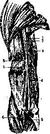
Fig. 1. Axillary and radial nerves: 1 - pectoralis minor; 2 - axillary artery; 3 - circumflex humeral artery; 4 - pectoralis major; 5 - radial nerve; 6 - thoracic long artery; 7 - upper branch to the medial head of the triceps; 8 - intermuscular septum; 9, 11 and 21 - cutaneous branches; 10 - medial head; 12 - anconeus muscle; 13 and 17 - lower branch to the medial head; 14 - lateral cutaneous branch; 15 - deep brachial artery; 16 - lower branch to the lateral head; 19 - upper branch to the lateral head; 20 - lateral head; 22 - branch to m. deltoideus; 23 - m. deltoideus. The brachial artery gives off three branches: the first is the deep brachial artery (a. profunda brachii), which passes between the medial and lateral heads of the triceps muscle; the second is the superior ulnar collateral artery (a. collateralis ulnaris superior), which in the substance of the medial head of the triceps muscle reaches the vascular network of the elbow; and the third branch is the radial collateral artery (a. collateralis radialis), which arises somewhat below the middle of the arm between the lateral and medial heads of the triceps muscle and enters the cubital network (rete cubiti). In the lower part of the groove, the deep artery pierces the lateral intermuscular septum and enters the anterior compartment of the Brachium in the region of the cubital fossa. The accompanying deep veins carry blood either to the axillary or to the brachial veins. There are no lymph nodes in the posterior compartment; lymphatic vessels, superficial and deep, drain into the nodes of the axillary fossa. In the posterior compartment of the arm pass two large nerve trunks: the radial nerve (n. radialis) and the ulnar nerve (n. ulnaris). The radial nerve, passing in front of the tendon of the latissimus dorsi muscle, goes to the posterior surface of the Brachium through the space between the surgical neck of the humerus, the long head of the triceps muscle, and the latissimus dorsi muscle. In the posterior compartment, the nerve runs obliquely from within outward from top to bottom in a groove, directly on the periosteum of the humerus, enveloped in loose adipose tissue. In the upper part of the groove, the nerve passes between the origins of the medial and lateral heads, covered above by the long and below by the lateral head of the triceps muscle; in the lower part of its course, the nerve pierces the lateral intermuscular septum and penetrates into the anterior compartment of the arm and further into the region of the elbow joint, where it divides into two main branches. On the Brachium, the nerve gives off: the posterior cutaneous nerve of the arm (n. cutaneus brachii post.), the dorsal cutaneous nerve of the forearm (n. cutaneus antibrachii dorsalis), muscular branches (rami musculares) for the triceps and anconeus muscles, and after piercing the lateral intermuscular septum, branches for the brachioradialis muscle and for the long radial extensor of the wrist; a thin branch to the brachialis muscle is often encountered. The ulnar nerve passes along the medial border of the arm along the projection line from the apex of the axillary fossa to the posterior surface of the medial condyle of the Brachium, a few millimeters posterior to the brachial artery and median nerve, behind the medial intermuscular septum; the nerve is accompanied by the superior ulnar collateral artery and its paired veins. Like the median nerve, the ulnar nerve gives no branches on the Brachium.
a. Syrotkin - Pathology of the Brachium. Congenital anomalies of the Brachium, like congenital malformations of other parts of the limbs, have as their cause abnormalities in the embryonic rudiments or mechanical factors—pressure from the amnion and uterus, the significance of which is particularly emphasized by the French school. Complete absence or significant underdevelopment of the arm in combination with a similar anomaly of the forearm, with the hand developed to a greater or lesser degree, is found in: perobrachius or phocomelus (see Phocomelia). In these cases, the developmental defect consists of the absence or hypoplasia of the embryonic main ray of the upper limb. A very rare case of complete absence of the humerus with normal development of the forearm and hand is described by Brandenberg. Spontaneous amputations and amniotic constrictions of the arm are observed significantly less frequently than in the distal parts of the upper and in the lower limbs. In some cases, there is the presence of a painful conical stump where the bone is covered only by scar tissue. Reduction in size of the humerus and congenital deformities of it are part of the picture of diseases associated with dystrophic processes—such as chondrodystrophia, osteogenesis imperfecta, osteopsathyrosis, congenital athyreosis and hereditary syphilitic osteochondritis. In congenital hypertrophies of the arm, as in other parts of the limbs, changes in muscular, bony and soft tissues are observed, which are often combined with dilation of vessels, elephantiasis and the formation of delimited lipomas. Traumatic injuries to the soft tissues of the Brachium, open and closed, are caused by direct and indirect action of various types of traumatic force, depending on which the nature of the injury is determined. Bruises from blunt instruments without violation of the integrity of the skin cover, when the injury is localized on the inner surface of the arm, lead to significant subcutaneous hemorrhages. The usual treatment consists of a pressure bandage and rest for several days. In large hematomas with separation of the skin from the underlying tissues, puncture is used to aspirate the extravasated blood. Violation of the integrity of the skin cover is caused by injury from sharp instruments, machine injuries, thermal and chemical burns. In treatment in appropriate conditions, the method of choice is: primary suture for incised wounds, primary treatment followed by an immediate suture in machine injuries, in the case of large skin defects—with the addition of skin plastic surgery (see Wounds). In extensive burns, subsequent transplantation of skin flaps taken with a broad base from the skin of the back or chest is often indicated. In the case of superficial skin defects, one can be limited to transplantation of epidermis according to Reverdin. In injuries to the inner surface of the Brachium, and especially to the axillary region, correct positioning of the limb in the position of abduction and slight external rotation is important for further function, reducing the possibility of scar stricture. Deeper open injuries to the Brachium lead to wounds of the muscles and are often combined with violation of the integrity of vessels and nerves. Closed ruptures of muscles and tendons, which are the result, for the most part, of indirect action of traumatic force, are caused by a special mechanism: Rupture of healthy muscles occurs from excessive progressive stretching, excessive active contraction or from sudden passive stretching of a muscle in a state of active tension (for example, when attempting to support a weight falling from above). Direct trauma to a tense muscle, sudden impact, compression of it and damage from bone fragments in closed fractures can also cause a subcutaneous muscle rupture. The injury can be localized in the muscular belly, at the point of transition into a tendon or in the area of attachment of the latter to the bone. A predisposing factor for muscle ruptures are previous changes in them, associated with transferred infections, alcoholism, inflammatory processes, such as the bursa of the biceps muscle in rupture of its peripheral tendon or Jacertus fibrosus (Ledderhose). Special importance is given to changes in the sulcus intertubercularis with involvement of the tendon of the biceps passing through it in deforming arthritis. (Ledderhose, Gurlt). In frequency of subcutaneous ruptures of arm muscles, first place is taken by the biceps muscle, injury to which, according to Mauyl, occurs in 22% of all ruptures. Pischler reports 222 cases of rupture of the biceps, with the long head being predominantly injured. Rupture of the short head, as well as m. coracobrachialis, is often observed in laundresses when wringing laundry (Weber). Ruptures of m. triceps and m. deltoidei occur less frequently (thus, Regard found injury to m. deltoidei in 14 out of 128 cases of ruptures). The typical picture of a rupture consists of suddenly occurring pain at the site of injury when the traumatic force acts, accompanied by a clear crackling sound, and loss of function. When the injured muscle contracts, a larger or smaller dense tumor forms, bulging below the site of rupture and disappearing when the muscle relaxes. In rupture of the biceps, the symptom described by Hueter has diagnostic significance—pain in the shoulder when flexing the limb in the supinated position, while flexion of the pronated limb is impossible. In complete ruptures of muscles and tendons, surgical intervention is indicated in most cases, in fresh cases—muscular or tendon suture. In cases with a defect in muscular substance and decreased function of the limb, muscular flap plastic surgery is used. In tendon ruptures, according to the proposal of Paug, the suture can be additionally strengthened with a fascial plastic. Subsequent systematic physio-mechanotherapy and elimination of early loading of the injured muscle are of essential importance to avoid recurrences. The prognosis for restoration of working capacity is quite favorable with timely therapeutic intervention on unchanged muscles. In the presence of pathological conditions of muscles and tendons or with late surgical treatment, when scar changes are already present, there remains significant decrease in function and tendency to recurrences.—So-called muscle hernias, arising from rupture of the fascia, are often observed in the area of the biceps muscle. Lexer and Baus differentiate hernias in damaged and undamaged muscle; the latter are rare; they are characterized by bulging of the muscle through the fascial slit only in the resting state and usually do not require treatment. If a muscle hernia is determined in a contracted muscle, then in these cases there is damage to the muscle as well. Closed traumatic injuries to the Brachium, especially those caused by significant compressive force, can be accompanied by subcutaneous rupture of vessels, which has clinical significance only when large vessels are injured. Violation of the integrity of a vessel is also observed when it is injured by bone fragments in fractures and dislocations of the shoulder, more often during their reduction (see Shoulder joint). Vessels with altered walls are naturally more vulnerable and are damaged by relatively minor injuries. Both complete rupture of a vessel, mostly transversely, and partial damage to its wall are observed. Herzog collected 61 cases of injuries to the media with subsequent formation of true traumatic aneurysm, in 18 cases there was injury to a. brachialis, in 7 cases—a. axillaris. Clinical symptoms and prognosis in closed vessel injuries depend, on the one hand, on the caliber of the vessel, on the other—on the nature of the injury and outcome of the wound—formation of a thrombus, false or true aneurysm (see). Even complete rupture of such large vessels of the Brachium, as a. brachialis and a. axillaris, does not always lead to necrosis of the limb, which depends on the rich blood supply and formation of collateral circulation. The prognosis for viability of the limb worsens with crushing of the surrounding soft tissues with damage to the vascular network and infiltration with extravasated blood.—Open injuries of vessels are encountered significantly more frequently in stab, incised, contused, crushed and mainly gunshot wounds of the Brachium. In gunshot wounds of arteries, in approximately half of cases there is also simultaneous injury to a vein (Häbeger), leading to subsequent formation of an arteriovenous aneurysm. Open injuries of large vessels—a. axillaris and brachialis—often cause fatal bleeding. In some cases, however, even with transverse injury to an artery, spontaneous cessation of bleeding can occur, due to closure of its lumen by the coagulated intima with subsequent formation of a thrombus. In lateral injuries, closure of the wound opening by neighboring tissues is sometimes observed.—In the treatment of open vessel injuries, first place is given to combating primary bleeding by applying a ligature (see Blood vessels—surgery). According to Wolff's statistics, when ligating a. subclaviae and a. brachialis, necrosis of the limb occurs in 5%, when ligating a. axillaris—in 15%.
Especially unfavorable is the ligation of the a. brachialis above the origin of the a. circumflexae humeri, since in these cases the anastomoses with the a. mammaria and with the ramus descendens a. transversae colli are excluded. The prognosis is worsened by crushing of soft tissues and primary severe wound infection. Open injuries to blood vessels, as well as closed injuries to them, in later stages, depending on the nature of the injury, lead to the formation of a pulsating hematoma, false, true, and arteriovenous aneurysm with their typical symptoms and require appropriate surgical treatment (see Aneurysm). Along with open and closed injuries to blood vessels, traumatic injuries to nerve trunks are also often observed in Brachium. The mildest form of injury are pressure paralyses, among which a group of paralyses after anesthesia is distinguished, occurring from pressure of the edge of a table on the n. radialis when the arm is hanging down, from pressure of the head of the humerus on the nerve bundle of the axillary fossa when the arm is maximally raised and abducted (Braun), or from compression of the n. brachialis between the clavicle and the first rib. The n. radialis is often compressed by fragments of the humerus in their dislocation, as well as by the resulting excessive callus. According to Bruns' data, out of 189 cases of injury to nerve trunks in fractures, 77 cases were of the n. radialis, with 69.2% of them with fracture localization in the middle third of the humerus. Scar processes in suppurations, especially in open fractures on the basis of gunshot wounds, often lead to the development of secondary, late paralyses due to nerve compression. A special form of traumatic injury is the dislocation of the n. ulnaris, which occurs either as a result of a fracture of the internal condyle of the humerus or on the basis of strong sudden flexion in the elbow joint with existing congenital flattening of the sulcus ulnaris (Dubs). In the latter case, a habitual dislocation of the ulnar nerve may develop, which requires surgical intervention: strengthening with a suture through the perineurium (Tillmanns), plastic strengthening of the posterior wall of the canal with a periosteal flap (Muller), or deepening of the sulcus ulnaris. Traumatic in nature is also obstetric paralysis in newborns, in which the abductors and muscles rotating the shoulder outward are most often paralyzed, the so-called upper-radicular type of Duchenne-Erb.
Similarly to vessels, traumatic damage to nerve trunks most often occurs with gunshot wounds to the extremity. The brachial plexus and nerves of the arm are damaged primarily by the projectile, bone fragments, and as a result of the indirect effect of the traumatic force. According to American statistics (Toby Conn), among nerve injuries, the ulnar nerve is damaged in 31.6%, the radial nerve in 25.8%, and the median nerve in 23.4%. Impairments to varying degrees of motor and sensory functions, trophic and vasomotor disorders, paresthesias, and causalgias are diagnostic signs of nerve injury, and their combination depends on the nature and localization of the injury (see Nerves). Clinical symptoms do not always make it possible to determine the degree of nerve damage, since, on the one hand, the anastomoses of the upper extremity are not fully studied, and on the other hand, anomalies of muscle innervation are often encountered, especially in relation to the ulnar, median, and musculo-cutaneous nerves. With injury to the radial nerve, motor symptoms predominate, with injury to the ulnar nerve, a main en griffe often develops, damage to the median nerve gives a pain syndrome, trophic and vasomotor disorders (P. Marie). Based on work from the clinic of Polenov (Lyustritsky), in case of injury to the nerves of the upper extremity, symptoms should be placed in the following order according to diagnostic value: data on electrical excitability, motor disorders, and changes in the sensory sphere. - Treatment. In mild nerve injuries from compression, physio-electrotherapy can be successfully applied. Compression of the nerve by scar tissue, bone callus, or displaced fragments of the humerus requires surgical intervention (see Neurolysis). In case of complete interruption of the nerve trunk to avoid secondary traumatic degeneration, early nerve suture is indicated, which however does not always give success (according to Forster's data - 38%, Staffer's - 57.1%, Perthes' - 45%) (see Nerve suture). Fractures of the humerus constitute about 7% of all fractures according to Bruns, of which 53% are diaphyseal fractures; epiphyseal fractures, leading to severe joint disorders, constitute one of the main pathologies of the shoulder and elbow joints (see Elbow joint, Shoulder joint). Oblique fractures of the humerus are most often observed. In the mechanism of fracture formation, the direct effect of force plays the primary role, less often - indirect, for example, falling on the elbow or outstretched hand. So-called 'sports fractures' of the humerus usually occur in individuals with well-developed musculature when throwing hand grenades, fencing, playing tennis and gorodki. In their occurrence, the main role is played by the discoordinated action of muscle traction with a fixed shoulder joint. According to the nature of the fracture, they are divided into 'flexion fractures' with localization of the fracture line immediately below the attachment of the deltoid muscle and torsion fractures (Kütner, Levashova). Diagnosis of diaphyseal fracture is easy, as usually all typical symptoms of fracture are present. Dislocation of fragments depends on the direction of the acting force and the disruption of equilibrium in the tension of certain muscle groups. With localization of the fracture above the attachment of the deltoid muscle, the upper fragment is displaced backward and inward, the lower one - forward, upward and outward. In the most common fractures in the middle third, below the attachment of the deltoid muscle, due to the traction of the latter, the upper fragment moves upward and outward, the peripheral fragment moves upward and backward. Displacements of fragments can be ad longitudinem, ad axin, ad latus, ad peripheriam and combined. Complicated open fractures occur with the direct action of a significant traumatic force due to rupture of the skin by sharp bone fragments, most often with gunshot wounds, which, according to Seidel, constitute 1/5 of all gunshot wounds of the upper extremities, with the humerus being predominantly damaged. Simple gunshot fractures are rarely observed; mostly comminuted and crushed fractures occur. Displacement of fragments depends more on the direction of the acting force, and since damage to the musculature at its attachment is often present, the influence of muscle traction on dislocation in gunshot fractures plays a significantly smaller role than in peacetime fractures. Simultaneous damage to nerves, especially the radial nerve, is often observed, constituting 8.4% according to Bruns, 4.1% according to Rietmis. A serious complication in open fractures is wound infection, occurring in 50% of cases according to Seidel's data in gunshot fractures. Treatment of fractures without dislocation consists of holding the extremity in the correct position by some means with the application of early movements. In the presence of significant displacement, reduction of fragments is performed under general anesthesia or local anesthesia (20 cm³ of 2% novocaine solution according to Bohler) followed by the application of extension splints or treatment is conducted in the supine position with the application of adhesive traction in the physiological position* of the extremity according to the Bardenheuer principle. Wegner emphasizes that the so-called physiological position is the abduction of the extremity from the trunk at an angle of 45°, in a plane standing at an angle of 45° to the frontal plane of the trunk. The transverse axis of the lower end of the humerus, i.e., the linea intercondyloidea, should be turned inward relative to the horizontal plane. In fractures with significant displacement along the length, it is not always possible to eliminate dislocation by adhesive traction, which is why traction is applied directly to the distal fragment of the humerus in fractures of its upper and middle third, transitioning to skeletal traction (Codivilla-Steinmann nail, wire traction according to Klapp). In fractures of the humerus, a wire is passed through the ulna 2-3 cm distal to the olecranon tip. During the past imperialist war, about 50 different types of splint bandages for the treatment of humeral fractures were proposed (collected by Seidel), which can be divided into temporary, holding splints, serving mainly for the transport of patients, and extension splints, of which, in Sitenko's opinion, the most rational are the Böhler and Borchardt splints (see also Fractures, Traction). Indications for the use of circular plaster bandage in the treatment of humeral fractures are extremely limited; it can be mainly used for immobilization of the extremity after surgical intervention and then only in exceptional cases (for specific indications).-Early surgical treatment of humeral fractures-osteosynthesis-is indicated in cases of simultaneous injury to blood vessels and nerves, interposition of soft tissues or in cases where conservative treatment does not achieve a satisfactory position of the fragments with preservation of the normal axis of the extremity. In open fractures, primary wound treatment with primary bone suture gives good results according to Koch and Rimann-up to 70% primary healing, with the key to success being as early intervention as possible (within the first 6 hours after injury). Late complications of humeral fractures are union in an incorrect position with impairment of extremity function, formation of a false joint or large defects, which is often observed in open fractures with suppuration, when large areas of bone are removed.-According to Bruns, humeral fractures have a certain tendency to form pseudarthrosis; thus, of 681 cases of ununited fractures, 33.1% fall on the humerus. Methods of surgical treatment for pseudarthrosis vary: free positioning of fragments, application of wire suture (from various, preferably non-oxidizing metals), formation of internal and external splints by means of auto- and heteroplastic material. In bone defects of the humerus, free transplantation of fresh autoplastic bone material gives quite satisfactory results (Lexer, Eden, Hofmeister), and the transplanted bone segment should be sufficiently long.-Spontaneous fractures of the humerus occur in osteomyelitis, syphilitic changes in the bone, bone cysts, ostitis fibrosa, neoplasms, diseases of the central nervous system and syringomyelia; especially often, according to Bruns, spontaneous fractures are observed in the mentally ill. Acute inflammatory processes can affect either the humerus or the surrounding soft tissues. The infectious agent penetrates into the soft tissues either from the outside in traumatic injuries, especially in injury to bone and joint, or per continuitatem, sometimes the inflammatory process develops due to ascending infection along lymphatic pathways.
Acute lymphangitis is accompanied by the typical symptoms for it, with red painful stripes predominantly located in the area of the medial and lateral groove of the biceps muscle. In severe purulent lymphangitis, accompanied by the formation of abscesses along the lymphatic vessels, it is possible to propose making transverse incisions of the soft tissues of the arm up to the fascia to interrupt the lymphatic pathways. Together with the lymphatic vessels, the lymphatic glands of the axillary fossa are also involved in the inflammatory process - lymphadenitis simplex et purulenta (see Lymphadenitis). Tuberculous inflammation of the shoulder joint with rupture of pus through the joint capsule can lead to the formation of a typical cold abscess in the area of the Brachium. Primary tuberculous myositis is extremely rare; more often it is of a secondary nature. Tuberculous nodes with caseous decay are located either subfascially or directly in the muscle, splitting its fibers. The diagnosis presents considerable difficulties. It is necessary to differentiate from gumma, neoplasm, echinococcal cyst, hematoma, and other diseases. Diffuse syphilitic myositis occurs in the early period of the disease. Among other skeletal muscles, it is more often localized in the biceps and triceps muscles of the Brachium. Sometimes a symmetric lesion of them is observed (see Muscles). Among other chronic inflammations in the area of the shoulder, there is a specific inflammatory granulation process caused by the appearance of free and splitting fat in the tissue, so-called oleogranulomas (see). They appear in the form of solitary or multiple painful nodules of various sizes, sometimes having a symmetric arrangement. Oleogranulomas were especially often observed after the epidemic of typhus at the site of injections of oil of camphor, but also often independently of them. Abricos divides them into injection, traumatic, perifocal, and spontaneous oleogranulomas. From parasitic diseases, individual cases of localization of echinococcal cysts in the area of m. biceps (Dupuytren), m. brachialis (Gerdy), and m. triceps (Neiaton) have been described. Acute purulent osteomyelitis of the Brachium arises on the basis of exogenous infection with open injuries due to the spread of the process from the shoulder or elbow joint, but mainly by the hematogenous route of infection, and is caused, as in lesions of other bones, most often by Staphylococcus pyogenes. According to the statistics of Gangolphe, the lesion of the upper metaphysis of the Brachium occupies the third place in frequency; according to Haager, it constitutes 11% of all osteomyelitic diseases of long tubular bones. The symptomatology of the disease does not present any peculiarities, nor do its outcomes, however, it should be noted that the shoulder joint is involved in the process less often than other joints, which is explained by the extra-articular position of the epiphyseal line. The rupture of pus outward most often occurs at the anterior edge of the deltoid muscle or in the area of the bicipital sulcus. Affecting mainly children, the osteomyelitic process often leads to significant disturbances in the growth of the limb. The observed epiphyseal dislocations cause deformities of the upper end of the humerus, and in spontaneous diaphyseal fractures, pseudoarthrosis often forms. The danger of general septic infection in lesions of the Brachium is less than with localization of the process in the area of the thigh and tibia, accordingly improving the prognosis of the disease. The treatment of acute osteomyelitis of the Brachium consists in opening the bone marrow cavity or in more conservative intervention - incision of soft tissues and periosteum, which is especially recommended for osteomyelitis in childhood (Krasnobaev). As an accompanying method of treatment in many cases, vaccinotherapy (polyvalent or autovaccine), proposed for the treatment of osteomyelitis by Wright, gives good results; it is especially effective for forms not accompanied by general septic phenomena. For acute cases of osteomyelitis of the Brachium, an incision in the middle third of the Brachium is recommended, starting from the attachment of the deltoid muscle along the outer edge of the biceps; the incision can be extended upward if necessary with the splitting of the fibers of m. deltoidei. When extending the incision downward to free the lower epiphysis of the humerus, there is a danger of damaging the n. radialis, for which reason an additional incision can be made on the inner surface of the Brachium. With extensive lesions of the Brachium, total subperiosteal removal of the diaphysis is performed according to Ollie (Petrov, Roman-tsev). The considerable regenerative capacity of the periosteum, especially in childhood, leads to complete restoration of the bone defect. In chronic forms of osteomyelitis with the formation of purulent cavities and sequestration of bone, necrotomy and sequestrectomy are usually performed by an incision along the outer edge of the biceps muscle. Among conservative methods of treatment, the use of a mercury-quartz lamp undoubtedly has a favorable effect on the course of chronic osteomyelitis. The functional prognosis of the disease depends on the localization and extent of the disease, involvement of joints in the process, and scar changes on the part of the corresponding muscles and nerves.
> Primary tuberculous lesions of the diaphysis of the brachium rarely occur as a diffuse disease, somewhat more often as a complication of tuberculous inflammation of the joint, and usually lead to necrosis of bone tissue. When the embolic focus is localized in the cortical substance of the diaphysis with its gradual destruction and simultaneous formation of compact layers from the side of the periosteum, a form of diaphyseal tbc is observed, very close to spina ventosa. For radiodiagnosis of diaphyseal tbc, it is characteristic that periosteal proliferations are expressed less than in acute inflammatory processes. In addition to general conservative treatment, it is possible in limited forms to perform curettage of the cavity with subsequent introduction of iodoform preparations, and in some cases also resection of the affected area of bone. Syphilis of the brachium occurs more often in congenital disease (Stromeyer, Schreiber), more rarely in late stages of acquired suffering in the form of periostitis, diaphyseal and epimetaphyseal osteitis and specific osteomyelitis. Osteochondritis syphilitica is observed in the area of the upper end of the brachium and leads to significant epiphyseal changes (see. Shoulder joint). Among osteodystrophic processes in the humerus, bone cyst (osteodystrophia fibrosa cystica localisata) is often observed, which according to Rehberg's data in frequency is only slightly inferior to true bone sarcoma. Occurring in young age, usually up to 20 years, the bone cyst is localized in the metaphysis of the proximal end of the humerus, appears elongated along the axis of the limb and never occupies more than 1/4, 1/5 of the length of the brachium. The clinical picture has no characteristic symptoms and is limited for the most part only by uniform thickening of a certain area of bone. In approximately 60% of all cases, spontaneous fracture occurs, which also gives indistinct clinical symptoms: slight hemorrhage and pain and relatively limited function of the limb. At the same time, complete fracture is observed relatively rarely, mainly in diaphyseal cysts of the brachium. Another type of localized fibrous osteodystrophies, called 'giant cell tumor' (Neiaton) ('braune Tumoren' of German authors), in contrast to true bone cysts rarely affects the humerus. Surgical treatment of bone cysts consists of opening and curettage with subsequent smearing of the cavity with cauterizing substances. Lexer, Kaposi (Lexer, Karpozy) perform resection of the affected area of bone with preservation of the periosteum, which as a rule remains unchanged; in place of the defect an auto- or heterotransplant is transplanted; The prognosis of the disease is quite favorable, surgical treatment gives excellent results. - "Changes in the humerus observed in generalized osteodystrophic processes, such as Recklinghausen's disease, Paget's disease, are part of the general picture of the disease, and in the first form the humerus is affected as often as the tibia, occupying the second place in frequency; in Paget's disease the changes mainly concern the lower limbs. - The humerus among other tubular bones is a favorite site for parasitic cysts. Often the disease goes unnoticed and is diagnosed only when spontaneous fracture occurs and an abscess forms, in the contents of which cholesterol crystals and remains of the chitinous shell are found. Treatment consists in removal of the entire affected area of bone with subsequent replacement of the bone defect. New formations of soft tissues of the brachium do not present any particular features. Most often lipomas are encountered, which sometimes reach large sizes and originate from subcutaneous, subfascial and intramuscular fatty tissue. The area of the brachium is after the back the most common site for localization of lipomas. Intramuscular tumors are observed more often in the area of the biceps, less often in the triceps, appear either strictly delimited or have a more diffuse character. Their removal is performed easily, mostly by blunt method. To benign metaplastic processes of soft tissues should also be attributed the myositis ossificans circumscripta arising on the basis of acute or chronic trauma. In addition to the process of ossification after acute trauma found in m. brachialis, muscular calcifications have been found in m. biceps (in gymnasts) and m. deltoideus. Surgical intervention is indicated only in cases of negative results of conservative treatment where there is impairment of limb function. Among malignant tumors of soft tissues of the brachium primary muscle sarcomas are encountered, originating either from the muscle itself or from its fascia (Kuttner). Thus, individual authors have described sarcomas of the deltoid muscle, a spindle cell sarcoma originating from the fascia of the brachium. The prognosis of the disease is extremely unfavorable. Mortality despite radical surgical treatment reaches 44% (Landois). Relatively often tumors originating from lymphatic glands are localized in the axillary fossa. Among benign tumors of the humerus, osteochondromas are most often observed, differing little from osteomas and localized in most cases on the inner surface of the upper or middle third of the brachium. The tumor has a broad base, a bumpy surface and sharply defined contours. If the tumor occupies the epiphyseal end of the brachium, it never grows towards the joint, but always eccentrically. Multiple chondromas, which mostly affect the metacarpal bones, can also occur in the area of the epiphysis of the brachium; in their growth chondromas do not give penetration into the joint. Among malignant new formations of the humerus, sarcomas are in first place in frequency. Gross (Gross) in 165 cases of sarcomas of long tubular bones had 25 cases of involvement of the humerus, Kocher (Kocher) in 65 cases - 10. Osteogenic sarcomas, i.e. originating from primitive bone cells, are encountered significantly more often than periosteal fibrosarcomas. Relatively often osteogenic sarcomas develop from chondromas and cartilaginous exostoses. The most frequent localization is observed in the metaepiphyseal end of the bone, but for the humerus a typical site is also the area of the roughness of the deltoid muscle. The clinical picture consists of increasing pains, rapidly growing tumor, often observed spontaneous fractures and functional disorders from the side of muscles, vessels and nerves due to pressure or infiltration by the tumor. The X-ray picture can be extremely diverse depending on the size of the tumor, the nature of its spread, the predominance of the destructive process or osteoblastic factor, the greater or lesser reaction from the periosteum. Differential diagnosis, both clinical and radiological, in the initial stages presents considerable difficulties. The most common surgical intervention in sarcomas is amputation or exarticulation of the brachium within healthy tissues. With high localization and spread of the sarcomatous process, amputatio interscapulo-thoracica is indicated (see). According to Berger's data out of 64 cases of sarcoma of the brachium, in which amp. interscapulothoracica was performed, death occurred immediately after the operation in 3.12%, in 48.38% recurrences of the disease and metastases were noted. In some cases, mainly in the presence of central sarcoma without periosteal infiltration (Koenig), it is possible to perform extensive resection of the brachium over its length, after which cases of prolonged recovery were observed. According to Kocher's data out of 46 cases of sarcomas of the limbs, where amputation or exarticulation was performed, recovery was noted in 7 cases, out of 5 cases of resection 2 recoveries were noted. The prognosis in osteogenic sarcomas is threatening quoad vitam, and despite the most radical surgical treatment the overwhelming majority of such patients die from recurrences and metastases of sarcoma to internal organs. Another type of malignant new formations, having a multiple character and localized in the diaphysis of the bone, is the so-called Ewing's sarcoma, occurring at the age of 18-25 years and belonging histologically to endotheliomas. The humerus along with the tibia and ulna in frequency of involvement occupies the next place after the tibia. The course of this suffering is more benign than that of osteogenic sarcomas, radiotherapy gives a definite effect. Among more rarely occurring malignant new formations should be mentioned multiple myelomas, when the humerus is affected along with other bones of the skeleton (see Myeloma). Among secondary metastatic new formations in the humerus, tumors are observed; the primary focus of the tumor is most often localized in the prostate, thyroid or mammary gland. The metastatic cancerous process mainly affects the bones of the trunk, from the limbs their central parts are affected more, therefore in the humerus cancer metastases are encountered significantly more often than in the bones of the forearm, where they are observed only in exceptional cases.
Among other metastatic tumors, mention should be made of hypernephroma, which can give solid metastases with a relatively benign course. The operative approaches to the shoulder have been developed depending on the purpose of the surgical intervention—exposure of the vessel, nerves, or humerus. For exposure of the a. brachialis, there are two paths: direct and indirect. In the first, the incision is made exactly along the projection line of the artery, along the inner edge of the biceps muscle. The landmark is the median nerve, which in lean people with the arm abducted can be easily palpated and seen through the skin. The n. medianus in the upper third of the arm lies laterally, in the middle third anteriorly, and in the lower third posterior to the artery. Anomalies are relatively rare, when the artery lies in front of the nerve, as well as high divisions of it, where the weaker a. radialis lies in front, and the more powerful a. ulnaris behind the nerve. Since the posterior wall of the biceps sheath forms the outer wall of the arterial sheath, the indirect approach is convenient, in which the incision is made 1 cm outward along the projection line of the brachial artery. After incising the skin and superficial fascia, the sheath of the biceps muscle is opened and the latter is retracted outward; the inner wall of the sheath is incised over the n. medianus, and the artery is located depending on the level at which the operation is performed. For exposure of the n. radialis on the H., in its upper half, the incision is made from the determined depression between the posterior edge of the deltoid muscle and the long head of the triceps and continues downward toward the anterior surface of the medial condyle of the H. After incising the soft tissues and exposing the edge of the m. deltoideus, the interval between the long and lateral heads of the triceps muscle is bluntly dissected, and on the bone the nerve is found next to the a. profunda brachii. From here, the nerve can be partially bluntly and partially sharply dissected along its further course. In the lower third of the arm, the incision is made from the lateral epicondyle toward the medial edge of the m. supinator longus; the nerve is found through the muscular interval between the m. supinator and m. brachialis internus and is accompanied by the a. collateralis radialis. In the upper two-thirds of the H., the n. ulnaris is exposed by the same incision as the a. brachialis, since it lies somewhat posterior to the n. medianus. In the lower third, it curves around the medial condyle of the H., passing through the sulcus ulnaris. When exposing it in this course, the incision is made between the olecranon and the medial condyle of the humerus; the easily palpable nerve lies under the aponeurosis. Exposure of the humerus is performed both for trepanation in purulent processes and for resection due to neoplasms, bone cysts, etc. The incision is made along the lateral edge of the biceps muscle; after separating the periosteum, the mm. biceps and brachialis are retracted medially, the mm. triceps and supinator longus laterally. If it is necessary to expose the upper third of the H., the incision can be extended upward along the medial edge of the deltoid muscle; when extending the incision downward, it is made between the mm. supinator longus and brachialis, taking into account the possibility of damaging the n. radialis here. In the lower third, it is possible to make the incision from the lateral condyle of the humerus upward along the palpable edge of the humerus. High amputations of the humerus (Fig. 12), even at the level of the surgical neck, are more advantageous for prosthetic purposes than exarticulation, as they provide, although very short, but a movable stump. The scar is placed either on the surface of the stump or in the area of the axillary fossa. The most common is the single-flap method of Farabeuf. The incision begins along the sulcus deltoideo-pectoralis with transection of the tendon of the pectoralis major muscle, followed by liberation and ligation of the vessels between two ligatures. The flap skin incision is made according to the outline of the deltoid muscle, the points of the incision are connected through the axillary fossa, taking into account the certain contractility of the skin. Along the edge of the contracted skin, the deltoid muscle is transected from outside inward, sparing the n. axillaris. Along the inner edge of the incision, all muscles are transected to the bone, which is sawn through.
Figure 13. Schematic representation of a kinesthetic stump on the shoulder according to Sauerbruch: a—after the operation; b—ready. Figure 14. Formation of a loop from the biceps muscle. In children, to avoid the formation of a conical stump, it is recommended to remove the epiphyseal cartilage. Amputation on the diaphysis of the H. can be performed either by a two-moment circular method or with the formation of two flaps, with it being better to fashion medial and lateral flaps than anterior and posterior flaps. By suturing antagonist muscles, their contractility is reduced, especially that of the biceps muscle. For the kinematization of the stump for active prosthetics, plastic operations have been proposed by Sauerbruch, Anschtitz, and Spitzy, based on the principle of forming muscle canals (Figs. 13 and 14).
Bone-plastic operations for pseudarthrosis and defects of the H. are performed according to methods generally proposed for plastic operations. For large defects of the upper end, a freely transplanted bone-periosteal transplant can be directly wedged into the fossa glenoidalis. Without resorting to subsequent shoulder plastic surgery after resection, Gersuny proposed the following methods, which improve the function of the limb in the future: 1) transfer of the attachment points of the mm. deltoidei, pectoralis majoris, and m. latissimi dorsi, and 2) fixation of the sawn surface to the axillary edge or the inferior angle of the scapula, which is indicated for more extensive resections (up to HALF of the arm).
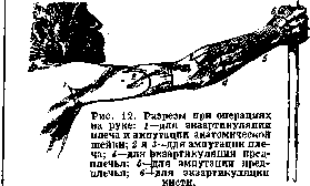
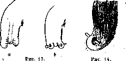
Related articles
Mentioned in
Cite this page
“Brachium.” Soviet Medical Encyclopedia. English translation of Bolshaya Meditsinskaya Entsiklopediya, 1st ed. (Moscow, 1928–1936), ed. N. A. Semashko. https://sovietmedicalencyclopedia.pages.dev/article/brachium/