Traction
Historical document, translated for reference. It reflects medical knowledge of the 1920s–30s and is not medical advice.
Summary
An overview of extension and traction methods in medicine from ancient times to the early 20th century, covering their use in fractures, dislocations, and joint diseases, detailing various apparatuses, adhesive techniques, and functional treatment principles.
Encyclopedia article (1928–1936)
TRACTION (extensio, distractio), a mechanical therapeutic procedure used for elongation, displacement, alignment, immobilization, and unloading of certain body parts or individual tissues. Traction can be produced by living muscular force, the weight of one's own body or attached weights, the elastic force of a rubber tube, a steel spring, or a rope twist (see). Traction can be either a short-term surgical intervention or a prolonged orthopedic method of treatment. Most commonly, traction is used for dislocations, fractures, and contractures. Under the name of the 'distraction' method, it was proposed in the second half of the 19th century for the treatment of subacute and chronic joint inflammations. In the history of medicine, traction as a method of treatment is first mentioned in the writings attributed to Hippocrates. For the reduction of dislocations, at that time, besides various apparatuses operating by lever force, machines with pulley blocks and other devices for traction were also used. The first device specifically for the treatment of traction in fractures, in particular fractures of the lower leg, is described in the writings of Galen, almost seven centuries after Hippocrates. This device, known as the 'glossokomon', had the appearance of a long, open wooden box in which the diseased leg was placed and subjected, after the reduction of the fracture, to traction in one direction and counter-traction in the other by means of straps gripping the joints above and below the site of the fracture. As the limb lengthened, the loops were pulled closer to the walls of the box. The shortcomings of the apparatus are obvious. They consisted, apart from the danger of pressure sores and harm to the function of healthy joints from the pressure of the straps, in the intermittent nature of traction. A further improvement, which first realized the idea of 'permanent', unremitting traction, consisted in the use of a suspended weight as the force for traction. This method of treatment for fractures is mentioned for the first time in the 11th century by the famous representative of Arab
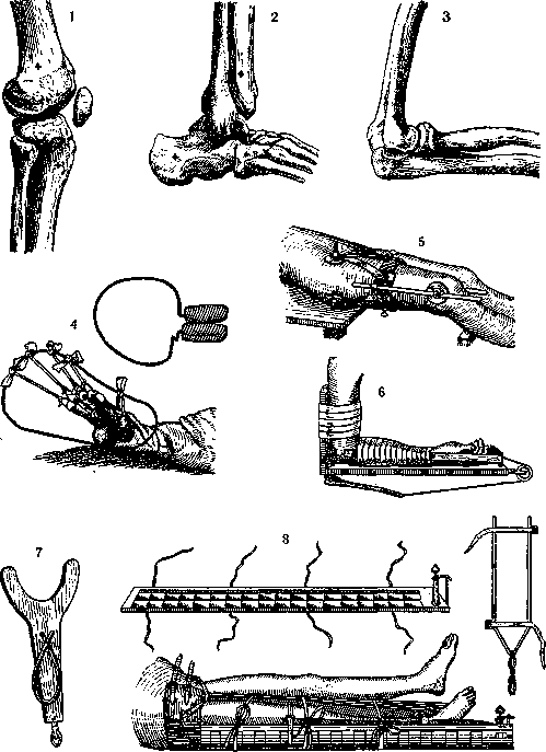
Figure 1-3. Puncture sites for traction with a Steinmann nail. Figure 4. Traction by the fingers
according to Sinclair. Figure 5. Splint-nail traction according to Kirschner. Figure 6. Borchgrevinck splint for forearm fractures. Figure 7. Borchgrevinck splint for arm traction. Figure 8. J. L. Petit glossokomon for traction in femoral fractures. medicine, Avicenna, and in the 14th century by the papal physician Guy de Chauliac. The latter's book, later named 'Chirurgia magna', served as a textbook in higher medical schools for several centuries. Nevertheless, the treatment of fractures by traction was little practiced and was soon forgotten. In the splints of Petit (J. L. Petit; see figure 8) in the 18th century, the idea of Galen's glossokomoi was revived. Traction was achieved by winding a rope loop, attached by a cuff to the ankle joint, onto a rotating shaft. Later, a technical detail gained decisive significance: the adhesive plaster attachment of the traction to the broken limb. It must be emphasized that only since the use of American rubber plaster (James, 1839) has continuous traction become a harmless and truly expedient method of treatment for fractures. In the second half of the 19th century, American surgeons (Swift, Gordon Buck, and Crossby) contributed to its widespread use more than others. In Germany, Volkmann proposed a special sled apparatus (see figure 18) to reduce friction between the dressing and the bed. The lower leg, together with the strips of adhesive plaster attached to it, was bandaged to a tinned gutter-shaped splint, and the latter rested by a transverse bar on a board with longitudinal runners. Thanks to this device, sliding was facilitated, a smaller weight was required, and at the same time, lateral instability of the position of the fractured limb was eliminated. Subsequently, Bardenheuer played a major role in the history of fracture treatment by traction; starting from the 1880s on a very large clinical material, he detailed the technique of traction for all kinds of fractures and in 1907 at the Congress of German Surgeons presented results of fracture treatment that had previously been unattainable. The fundamentally new aspect in Bardenheuer's technique lay not so much in the combination of longitudinal traction with transverse traction, but in the use of the largest possible surface for the application of force and in the extension of plaster traction always also to the proximal fragment. Based on the conviction that the displacement of fragments develops and is maintained exclusively or predominantly by muscle contraction, it was natural to demand action on the soft parts from all sides and throughout the entire extent over which the influence of the muscles on the broken bone manifests itself. Therefore, the longitudinal loop of adhesive plaster was always applied starting from the central end of the diseased body part. Spiral turns of plaster fixed this longitudinal loop, but in the region of joints and bony prominences, the plaster was not adhered to the skin, in order to avoid pressure sores and to facilitate active movements in the joints. Longitudinal traction was usually combined with traction in various transverse directions (see Plate II, fig. 24). Bardenheuer attached great importance to muscle exercises with simultaneously continuing traction, but lost sight of another requirement put forward quite definitely in the 1890s by Zuppinger. The latter proved that large weights (10-20 kg) used by Bardenheuer to stretch the limb to its normal length are superfluous if the musculature is relaxed by a 'physiological', semi-flexed position. In his opinion, not only shortening of the leg, but also displacements in general in any fracture are easily eliminated, almost automatically, if instead of full extension, a middle position of flexion of the neighboring joints is used. In such a case, the normally harmful influence of the leg's weight can be used as a force for traction and reduction. The realization of this idea was two splint apparatuses proposed by Zuppinger in 1905 for the thigh and lower leg (see figures 9 and 10). In Russia, Wegner at the Kharkov Medico-Mechanical Institute systematically applied the so-called 'functional' treatment of fractures since 1910, the essence of which consisted in the combination of continuous traction with immobilization of the limb in the middle 'physiological' position and in active joint exercises starting from the first days. Adhesive plaster loops were always applied separately to the thigh and lower leg, and in the case of a fracture of the upper extremity, separately to the arm and forearm, and the traction of each part was performed independently, in a special direction, with a separate weight (see figure 19). A technical detail common to all these traction methods is a spacer board inserted into the loop and protecting the region of the condyles and malleoli from excessive pressure. A rope is attached to the center of the board, which is then passed over 1-2 pulleys, and to the free end of which a bag of sand or another weight is suspended. Instead of adhesive tape or other kinds of rubber plaster, cambric strips can be successfully used by gluing them to the skin previously smeared with a resinous solution. The idea and first recipe for such a solution belong to Heusner. The best composition was proposed by Pifk in Kharkov according to the following prescription: Terebinthinae venetae 15.0, Masticis 12.0, Colophonii 25.0, Resinae albae 8.0, Spiritus vini (95°) 180.0, M., filtra, S. Kleol. This solution, under the name 'cutaneous glue', is now prepared by the Kharkov Sanitary-Bacteriological Institute. In compound fractures, sterile cambric bandages are used, and holes are left or cut out in the dressing glued for traction corresponding to the wounds. In wartime, with the frequency of severe primary infection and due to the comminuted nature of gunshot fractures, it is especially difficult to combine traction with wound dressing and the transportation of the wounded. In femoral fractures, the standard long lateral Liston-Esmarch splint for military sanitary supply is usually used (see figure 22). During the imperialist war, British field surgeons achieved excellent results by systematic primary immobilization of such fractures using the Thomas splint. The ring of this splint
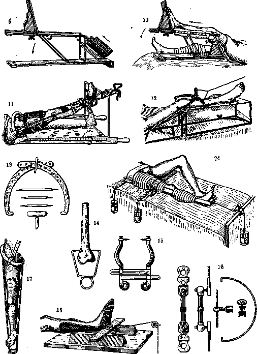
Figure 9. Zuppinger apparatus for femoral traction. Figure 10. Zuppinger apparatus for lower leg traction. Figure 11. Adaptation of the Thomas splint to a stretcher. Figure 12. Nail traction on a Loffler splint with a Steinmann frame. Figure 13. Steinmann frame, nails, and wrench. Figure 14. Schmerz clamps. Figure 15. Dorrance American clamps. Figure 16. Hackenbruch apparatus. Figure 17. Thomas splint for ambulatory unloading. Figure 18. Volkmann sled apparatus. Figure 24. Adhesive plaster traction according to Bardenheuer.
abuts against the ischial tuberosity, and the stirrup serves for attaching to it a loop enclosing the ankle joint over the boot and holding the fractured limb in a state of extension. Setting the lower end of the splint in a raised position (see Figure 11) facilitates the extension of the fractured femur by its own weight and reduces pressure on the perineum. Later, when fracture consolidation is still insufficient, the splint provides unloading while walking (see Figure 17). In the treatment of gunshot comminutions complicated by infection, extension should not be strong, but then it is essential to provide the limb with complete rest in conditions most favorable for blood circulation. Suspension in combination with extension best meets this requirement. The related technical devices can be easily improvised, at least in the form of the so-called Balkan frame (see Figure 20). The systematic organization of such treatment for complicated fractures in hospitals was the merit of many surgeons during the imperialist war. The more extensive the skin wounds or inflammatory changes accompanying the fracture, the more difficult is the usual technique of extension by strips glued to the skin. Under such conditions and in all cases of severe displacement of fragments along the length, extension directly to the bone according to Steinmann comes into its own. Codivilla was the first in 1903 to publish a method of forced femoral extension by traction using a nail driven transversely through the calcaneal tuberosity. Steinmann's merit lies in the fact that he, independently of other authors, invented and developed nail extension as an independent method of treatment for fractures. A cylindrical nail, about 3 mm thick, four-sided near both sharpened ends, is driven with drilling movements through the metaphyseal parts or apophyses of the bones in the places indicated in Figures 1-3. For drilling, a handle is used, fitted to the end of the nail and resembling a railway wrench. Instead of one long perforating nail, one can use two short ones penetrating only into the superficial bone layers. Then the danger of infection of the bone canal when pulling out the through nail is excluded, but less stability of the nails is obtained or, with a very oblique direction, the possibility of the nails advancing into the joint. To eliminate these shortcomings of Steinmann's nail extension, a frame of two semi-arches was constructed (see Figures 12 and 13), the extension by the crossed ends of which turns at the other ends into a force that drives the nails deeper towards each other across the bone. When there is no need for great force and duration of extension, one can use the simplified technique of bone extension by means of brackets or forceps (see Figures 14 and 15). Direct extension to the bone must be recognized as safe if it is performed within healthy tissues and lasts no more than 3-4 weeks. The so-called splint-nail extension according to Kirschner requires double perforation of the same bone at the proximal and distal ends (see Figure 5) and is a method of reduction and fixation of fractures by distraction and lateral displacement of fragments. The Hackenbruch apparatus (see Figure 16) is equipped with distraction brackets improving this method (application in a bridging plaster cast - see Figure 21). As for the extension of various parts of the body separately, the special technique of spine extension should be noted. Extension in this case is satisfactorily performed only by the head by means of the Glisson loop (see Figure 23) enclosing the back of the head and chin. The force of extension can be the weight of the patient himself, suspended by the head in a standing position or lying on an inclined plane. The greater the slope, the stronger the extension. In the horizontal position, it is necessary to suspend a weight to the rocker arm holding the loops of the Glisson collar; in addition, it is necessary to take care of counter-extension by another weight suspended from the pelvic belt (worse - to the legs). For the impossibility of using the submandibular or occipital region, bone-wire extension by the zygomatic arches was performed. Attachment of traction to the spinous processes has special indications. Finger extension presents some features; for attaching traction, either smearing the skin with one of the resinous solutions indicated above and knitted caps (see Figure 4), or silk sutures gripping the nail or metal serphines serve. As an active force for extension while walking, especially in fractures of the upper extremities, the elasticity of a rubber tube serves (see Figures 6 and 7). Extension should also be used systematically and persistently to prevent persistent cicatricial contractures.
Related articles
Mentioned in
Cite this page
“Traction.” Soviet Medical Encyclopedia. English translation of Bolshaya Meditsinskaya Entsiklopediya, 1st ed. (Moscow, 1928–1936), ed. N. A. Semashko. https://sovietmedicalencyclopedia.pages.dev/article/traction/