Dupuytren
Historical document, translated for reference. It reflects medical knowledge of the 1920s–30s and is not medical advice.
Summary
This article describes Dupuytren's disease, a condition characterized by flexion contracture of the fingers due to thickening and shortening of the palmar fascia. It covers the historical background, pathogenesis, clinical features, differential diagnosis, and treatment approaches as understood in the 1930s.
Encyclopedia article (1928–1936)
DUPUYTREN, Guillaume (Guillaume Dupuytren, 1778-1835), famous French surgeon and scientist of the early 19th century. Already at the age of 12, D. began to study anatomy in Paris, and at 16 became a prosector in anatomy and gave lectures. In 1802 he received the title of surgeon at the famous Hotel-Dieu in Paris, becoming head of the surgical department in 1812. At the same time, he took the chair of operative surgery at the university, presenting for the competition a work on lithotripsy. As head of the surgical department at Hotel-Dieu, D. showed extraordinarily energetic and fruitful activity, amazing his contemporaries and students with his rich erudition. D. was such a brilliant lecturer and scientific supervisor. Extraordinary attention to patients, diagnostic and operator abilities soon made him famous far beyond his homeland. D. is the author of many operations; resection of the lower jaw (first performed by him), subcutaneous incision of the sternocleidomastoid muscle, ligation of the external iliac and subclavian arteries. The fractures of the lower third of the tibia and radius described by him still bear his name, as well as the contracture of the palmar aponeurosis. Among the first, D. took up the question of reducing old dislocations. D. can quite deservedly be called one of the founders of modern scientific surgery. A significant part of his large fortune D. bequeathed to the University of Paris for the establishment of a chair in pathological anatomy, but the Paris municipality took this expense upon itself, using the amount bequeathed by D. to establish a museum of D. The "Leçons orales de clinique chirurgicale faites à l'Hôtel-Dieu de Paris" (v. I-IV, R, 1832-34), published by the medical society and translated into many languages, were widely known. DUPUYTREN'S CONTRACTURE (Dupuytren), is characterized by 1) palmar adduction of one or more fingers, 2) the appearance of nodes and cords in the palmar fascia, 3) typical changes in the skin of the palm. 1. The adduction mainly affects the IV and V fingers (Fig. 1), less frequently the middle finger and extremely rarely the index and thumb.

The contracture develops imperceptibly; first the patient feels as if there is roughness in the palm, preventing full extension, then sequentially palmar adduction occurs: first more often in the metacarpophalangeal joint, then in the first interphalangeal and very rarely (in late stages) in the second interphalangeal. 2. Usually the disease begins with the appearance near the transverse skin groove corresponding to the fourth metacarpophalangeal joint of small, sometimes painful nodes, from which later develop dense cords going to the fourth finger, well palpable as round cords when attempting to straighten the finger and disappearing along the middle phalanx and the center of the palm. 3. The skin of the palm in the direction of the affected finger forms a series of indentations in the form of arcs, concave toward the finger (Fig. 2).-The disease

Figure 1.
Figure 2. was first described by Plater (Plater; 1614), but the true cause of the disease remained unclear both for him and for other authors until Dupuytren. Thus, Cooper attributed the disease to inflammation of the tendon sheaths, Bonnet to muscle spasm, etc. Dupuytren, in 1831, performing an autopsy on a patient he had long observed with the described disease, through precise dissection established that the basis of the disease lies in pathological changes of the palmar aponeurosis. Dupuytren's observation caused a whole series of objections and gave impetus to a whole series of studies by anatomists, pathoanatomists and clinicians, who in the end joined his teaching that the anatomical substrate of the disease is the lesion of the palmar aponeurosis. There is extensive literature on the pathogenesis of D. c. Attempts were made to see in D. c. dystrophic phenomena and intoxication; at the same time, arthritism and gout were given pride of place, drawing parallels with scleroderma and tophi; D. c. was also explained by lead poisoning, diabetes, syphilis, even tuberculosis of the Poncet type (Perdrizet). Schubert and Sherstov see in D. c. a congenital-constitutional disease, while König and Kocher see a hereditary predisposition. Bunch reports a case of transmission of D. c. in one family along the male line for 300 years. Bilateral D. c. was tried to be associated with lesions of the central nervous system, as they were observed in tabes, in mental 6S4 patients and epileptics (Neutra, Biegansky, Caspari). Eulenburg, Dejerine, Reichel, Novotelnov report cases of D. c. after damage to the ulnar nerve, finding in them elements of trophoneurosis, as also mentioned by Vyalyaminov. Dupuytren, Cooper, Golyanitsky, Nedokhlebov and others consider D. c. a professional trauma that creates chronic inflammation of the palmar aponeurosis (Despres), subcutaneous tissue (Madelung) and skin in carpenters, rowers, peasants, etc. after the development of calluses. The most convincing evidence of the professional nature of this disease in workers, in Golyanitsky's opinion, is the correspondence of the localization of D. c. to the sites of greatest traumatization of the palmar aponeurosis in a given profession. Opponents of traumatic theories of D. c. (Adams, Janssen, Rosenburg, etc.) point out a) the small percentage of workers affected by D. c., b) the relatively significant spread of D. c. among people engaged in mental work, c) the involvement of both hands, while usually one hand is subjected to the greatest traumatization, d) the often less pronounced process in the working hand. The nature of the patho-anatomical process itself Kocher defines as follows: "It must be classified among the so-called chronic plastic inflammations, such as we observe in the area of other active and passive organs of movement with mechanical ^harmfulness". Most of the existing histological studies of the affected areas of the palmar aponeurosis in D. c. define the disease as focal hyperplasia of connective tissue, which originates from the walls of small vessels and causes wrinkling. As a result of this wrinkling, shortening of the fascia and contracture of the fingers occurs, preventing extension. From the palmar fascia, finger processes extend to four fingers, which, approaching the base of the finger, divide into three cords - two lateral ones going to the lateral surface of the proximal phalanx, and the middle one, from which bundles spread over the entire palmar surface of the finger to the very last phalanx (Vogt). A process also extends to the thumb, but less pronounced. In addition, throughout the palm and on the fingers, numerous processes extend from the palmar aponeurosis, going directly to the skin. The wrinkling of the finger processes explains the contractures of the fingers themselves

Figure 3. Lines of incisions for exposing the palmar aponeurosis: ab- longitudinal (Hardie); lih- angular according to Busch; L- flap with base on the ulnar side; abed and abef- with formation of flaps according to Lexer. (From Bier-Braun-Kümmell.)
and the wrinkling of the processes perpendicular to the skin gives the characteristic local indentations of the skin. In differential diagnosis, one must keep in mind: 1) griffe cubitale- atrophy of the thenar and interosseous muscles + + sensory disorders, 2) scar contracture after burns and injuries, 3) tendon contractures after inflammatory processes, in which not only extension but also flexion is impossible, 4) Volkmann's ischemic and reflex contracture. Treatment. Massage, hot air, mud baths, injections of fibrinolysin (Quenu), ionotherapy (Hartmann) somewhat slow but do not stop the further, usually gradually progressive development of D. c., which is why one has to resort to operations. Surgical
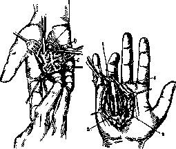
Figure 4.
Figure 5. Figure 4. Excision of the scarred and transformed palmar aponeurosis: a - the tendon sheath of the IV finger exposed from the scarred aponeurosis; b - the scarred aponeurosis; c - growth into the skin. (From Bier-Braun-Kummell.) Figure 5. Operation for Dupuytren's contracture: 1 - scar at the site of the aponeurosis; 2 - flexor tendon of the IV finger; 3 - blood vessels; I - displacement of the digital nerve by the scarred aponeurosis. The interventions should be divided into less and more extensive ones. The former include a) subcutaneous incisions of aponeurotic cords (Cooper), b) multiple punctures and incisions (Adam), c) transverse incisions of cords passing through the skin (Dupuytren), d) excision of the affected aponeurosis in the form of a triangle with its base toward the affected finger. All these methods have only historical interest; truly radical operations are those of Kocher, Muller, Lexer, and Berger (Kocher, Muller, Lexer, Berger). Figure 3 shows appropriate skin incisions. Basically, these operations consist in transverse excision of the dense palmar aponeurosis in its entire thickness and its separation from the underlying tendons or their sheaths as far as the middle of the proximal phalanx of the affected finger, where it is again cut transversely (figs. 4 and 5). Basically, the methods of Muller and Lexer differ from Kocher's method only in the careful separation of the affected cords as far as the end of the middle phalanx and the replacement of the skin thinned by dissection by means of grafting flaps from healthy skin of another area. Lit.: Golyanitsky I., Surgical Professional Diseases of the Motor Organs, M., 1927; Nedokhlebov V., Contracture of the fingers of the hand in tram conductors of Kharkov as a professional disease, Works and Materials of the Ukr. Inst. of Labor Medicine, vol. 1, Kharkov, 1925; Dupuytren &, Lecons orales de clinique chirurgicale faites a l'Hotel-Dieu de Paris, v. I, P., 1832; Kocher Th., Behandlung der Retraction der Palmaraponeurose, Centralblatt f. Chirurgie, 1887, № 26-27; Lexer O., Zur Pathologie der Aponeurose des Fusses u. der Hand, Arch. i. klin. Chirurgie, B. LV, 1897; Muller W., Operationen an der oberen Extremitat (Chirurgische Operationslehre, herausgegeben v. A. Bier, H. Braun, H. Kummell, B. V, Lpz., 1923).
Related articles
Mentioned in
Cite this page
“Dupuytren.” Soviet Medical Encyclopedia. English translation of Bolshaya Meditsinskaya Entsiklopediya, 1st ed. (Moscow, 1928–1936), ed. N. A. Semashko. https://sovietmedicalencyclopedia.pages.dev/article/dupuytren/