Burns
Historical document, translated for reference. It reflects medical knowledge of the 1920s–30s and is not medical advice.
Summary
This article provides a comprehensive overview of burns, including their classification by degree, pathological anatomy, and systemic effects. It details various classifications of burns (Boyer, Dupuytren, Kreibich) and describes the tissue changes at different burn depths.
Encyclopedia article (1928–1936)
BURNS (combustio), injuries to tissues that occur under the action of high temperature (thermal burns) or chemical substances (chemical burns). The first type of burn can be caused by any agent capable of emitting or giving off heat—sun rays, electric arc, flame, heated or molten metals, hot vapors (in boiler explosions), gases (in explosions), boiling or highly heated liquids (scalding) and oils, and finally electric current, lightning, X-rays, and radium. The second type of burn occurs under the action of a number of chemical substances (caustics)—acids, alkalis, salts of heavy metals and many others, used in various industries, laboratories, in war (mustard gas), etc. The degree of tissue destruction and the nature of burns vary depending on the factor causing the burn (steam, boiling water, molten metal, burning kerosene), its temperature, duration of action, distance. Much depends on the circumstances in which the burn occurred and on the victim himself: the degree of protection by clothing, the presence of skin folds at the burn site, the condition of the skin (callousness, thickening, endurance of this area to high temperatures under the influence of prolonged professional training, constant protection by clothing, etc.), age, etc. All these peculiarities, combining in various combinations, can cause extraordinary diversity in both local and general phenomena accompanying burns. Classification of burns. Attempts have long been made to develop a certain classification that could cover all types of burns. However, the extraordinary diversity of local patho-anatomical changes, the different expression of them in different areas, the difference in the body's reactions to one or another degree of spread and depth of burn, in advance doom all attempts to develop an exhaustive classification to failure. However, practical considerations have forced to retain some of the simplest of the many proposed and to use them in daily practice. To these should be included the classification of Boyer adopted in the USSR, in Germany and in a number of other countries, according to which all burns are divided into three groups or degrees. First degree, combustio erythematosa, is characterized by diffuse, intense, fading to the periphery redness, slight swelling and painfulness. In the second degree, c. bullosa, to the signs characteristic of the first degree of burn is added the formation of blisters of various sizes, appearing immediately or after several hours, more rarely—on the second day. Third degree burn, c. escharotica, is the result of particularly intense action on the tissues of the burn-causing factor. In it there is necrosis of tissues to a greater or lesser depth. To these three degrees of burn is added one more, when tissues or entire limbs under prolonged action of high temperature are charred. The classification of Dupuytren, adopted in France, enjoys somewhat less popularity, according to which six degrees of burns are distinguished: 1) redness, 2) formation of blisters, 3) superficial necrosis of the skin, 4) death of the entire thickness of the skin, 5) gangrene of the thickness of soft tissues to the bone and 6) complete charring of any part of the body or limbs, including bones. In the recently proposed (1929) classification by Kreibich, an attempt is made to somewhat clarify the depth of skin damage and to link it with subsequent clinical symptoms, as well as with the paths of healing and restoration. Kreibich proposes to distinguish 5 degrees. The first two correspond to those given above. 3rd degree with or without blisters, when necrosis spreads to the tops of the skin papillae. In the 4th degree there is necrosis of the entire papillary layer and finally the 5th degree—charring. The first two of the above classifications have received particularly wide distribution, although they suffer from the above-mentioned shortcomings. In them only part of the visible surface changes is taken into account, mainly the depth of the process, and the extent of the burn, which plays a decisive role in prognosis, does not find its reflection. Therefore, to the numerical designation is added the term "widespread", "limited", which makes it possible to some extent to judge the degree of danger to life from the received burn (see below). Pathological anatomy. Changes in the skin in first degree burns are insignificant and correspond to the picture of aseptic inflammation. At the site of indistinct redness and swelling there is dilation of blood vessels and significant exudation with greater or lesser outpouring of polymorphonuclears. The exudation explains the existing swelling at the site of the burn. In second degree burns, the phenomena of inflammation are expressed much more significantly, especially in the deeper layers of the skin. Through strongly dilated vessels, a large amount of blood plasma is secreted, separation of the horny layer from the underlying layers occurs, as a result of which blisters are formed. In the exudate of the latter, a small amount of red blood cells, white blood cells and fibrin are found. The bottom of the blister, or the exposed Malpighian layer, represents a uniformly red, loosened surface with pinpoint hemorrhages. Under the microscope, significant degenerative changes are found here. The cells are displaced, lose the correctness of their contours, the nuclei are poorly or not at all stained. The vessels are strongly dilated, small vessels are thrombosed. The subcutaneous tissue is severely edematous. A characteristic feature of the patho-anatomical picture of third degree burns is local necrosis of tissues of varying depth, mostly in the form of dry gangrene. The affected area represents a more or less hard, dry, whitish-yellowish, sometimes brown or black insensitive crust. Necrosis occurs immediately after the burn as a result of coagulation of tissue proteins, clotting of blood plasma under the action of high temperature and thrombosis of vessels. The process destroys tissues to a considerable depth, completely destroying all layers of the skin without remainder.
All of the above refers to the local changes in areas of skin that came into contact with the factor causing the burn. In adjacent areas of skin, not subjected to the direct action of the harmful factor, significant dilation of vessels is also observed as a result of the reflex influence of the burn on the vascular nerves. In limited burns, occupying small areas of skin, these local changes are limited to this. In extensive burns, the patho-anatomical picture is complicated. Under the influence of a number of causes (see below) changes occur in the central nervous system and parenchymal organs, found in the bodies of persons who died in the first days after the burn. In general, however, these patho-anatomical changes are not characteristic and not sharp. As a rule, the vessels of the brain and its membranes are strongly dilated, the brain itself and especially the soft meninges are edematous, small hemorrhages and foci of inflammatory infiltration are noted in the brain substance. In autopsies of those who died in fires, epidural hematomas (between the bones of the skull and the dura mater) are sometimes found. Usually the hematomas are located where the heat had the greatest influence. The origin of the hemorrhages is not exactly clarified. Most likely it is the result of rupture of sinuses and small veins when the dura mater shrinks under the action of heat. Changes in the solar plexus in burns have been noted for a long time (Korenko; 1897). In recent years, changes in the nervous system have been studied by Lozinsky (1927). In ganglion nodosum n. vagi and in the upper thoracic sympathetic nodes, two types of pathologically altered cells are observed: shrunken, with pyknotic nuclei, intensely staining protoplasm and swollen, light, with nuclei displaced to the periphery. In the glia, a picture of neuronophagia (see Nerve cells) is observed, death of glial cells. Significant changes are also determined in the nerve fibers passing through the nodes and a number of degenerative-atrophic processes with death of glial cells and sharp changes in nerve fibers in the peripheral and central autonomic nervous system. In the heart, most often there is an overflow of the right ventricle and atrium with liquid blood and small hemorrhages in the pericardium. All lymph glands are enlarged, swollen. The spleen is enlarged, brown-red in color, dense. In the liver, dilation of vessels and degenerative phenomena, not reaching, however, high degrees. Somewhat more significant changes in the kidneys in the form of degenerative processes from the side of the epithelium, sometimes in the form of inflammatory infiltrates. The earlier death occurs, the more intense these changes. In the tubules in early death, cylinders of red blood cells are found. However, the described changes occur to varying degrees and not always. Some authors, e.g. Dohrn, did not see clearly expressed changes, as in nephritis. In recent years, special attention is paid to changes in the adrenal glands in burns. Older authors did not note special changes in these organs. However, G
More detailed examinations in recent years have established a number of constant deep changes in the adrenal glands, which leads some to speak of these changes as the primary cause of death from B. Indeed, autopsies find these glands greatly enlarged (by weight 3-5 times) (Christopher). Parallel with the severity of B. there is a loss of chromaffin substance (negative reaction with chrome), depletion of lipoids, and development of hemorrhagic infarcts, as in anaphylactic shock. The vessels are usually greatly dilated. These changes, however, are not constant, but are very frequent, especially when death occurs within the second week. They are most pronounced in children. On the mucous membrane of the gastrointestinal tract, diffuse infiltration is noted, resembling the picture of acute inflammation. Particularly important are the changes in the duodenum, where in some cases of severe B. typical ulcers develop. When the mucous membranes of the mouth, pharynx, larynx, especially the vocal cords (for example, when inhaling flames of flammable liquids, gases) are burned, they have a whitish scalded appearance, and later superficial ulcers and phenomena of scarring develop. Sometimes the burned mucous membranes are shed in entire layers. In the clinical picture of B., local and general phenomena should be distinguished. The latter are almost absent in B. of small areas of skin. Instead, they come to the forefront in the picture of the disease in extensive B., covering more than 1/5 of the body surface. The severity of general phenomena and the degree of their danger depend relatively little on the degree of B. They are sufficiently strongly expressed and invariably dangerous even in B. of the first degree, if the latter occupy large areas. Locally, in B. of the first degree, patients experience a feeling of heat, severe pain, especially from touching with hands or clothing. These pains depend on the compression of the terminal nerve apparatus by inflammatory exudate impregnating the skin. Already after 2-3 days they disappear completely, the redness gradually fades, the swelling disappears. On the 4-5th day, peeling occurs, and the superficial layers of the epidermis fall off in very thin, large flakes. In place of the B., a pigmented spot sometimes remains, which often disappears only after several months. In B. of the second degree, when blisters of various sizes appear immediately on the burned surface or after several hours or within the first two days, the pains and feeling of tension are much stronger. Usually the pains last 3-4 days and gradually disappear. The pains are especially tormenting if the blisters burst on their own or are opened and the exposed surface comes into contact with clothing or poorly fixed bandages. The contents of the blister are partly absorbed, partly evaporate. The separated and deadened epithelium dulls and falls off after the process of epithelialization is completed on the surface of the exposed papillary layer, which proceeds partly from the periphery to the center, partly from the preserved hair follicles and ducts of the sebaceous and sweat glands. If the contents of the blister are not infected from the outside or by microbes preserved in the skin glands, epithelialization occurs quite quickly. In place of the B., a pink spot with normal skin remains. After 2-3 weeks, the skin takes on the usual color for that part of the body or loses its usual pigmentation for a long time. In some cases, a pigmented stripe remains at the borders of the former B. In cases where infection is added, the healing process is prolonged for significantly longer periods. Granulations appear on the surface of the B., slowly covered with epithelium. The resulting skin cover in such cases has the character of a scar, juicy and hyperemic at first and tendon-white and thin later. Severe pain in the first days after B. of the third degree more often depends on the accompanying B. of the second and first degrees. The resulting scab itself is painless. Extremely severe pain occurs from contact after the necrotized area is shed, when in place of the B. there remains an extremely sensitive wound surface covered with granulations, slowly epithelializing from the edges. The infection that often joins during the period of sequestration severely complicates healing and manifests locally in abundant discharge of pus, lush growth of easily bleeding granulations and significant slowing of the epithelialization process. In extensive B., the prolonged existence of suppuration leads to severe phenomena in the form of amyloid of the kidneys, liver, general exhaustion, and in some cases to pyemia and septicemia. Depending on the location of B., on the 2-3rd week with the added infection, perforations into joints, into the abdominal and pleural cavities may be observed. B. on the hairy part of the head tend to be complicated by erysipelas and can lead to meningitis. B. of the chest sometimes cause pleurisy. The proximity of large vessels threatens necrosis of their wall with subsequent, sometimes fatal, bleeding. Extensive tissue defects that form after B. of the third degree heal by scarring. The very dense, radiating scars that form at this time have a great tendency to contract. Especially severe scars form in cases where tissue destruction at the moment of B. or subsequently reached the fascia or muscles. Certain parts of the body (neck, axillary regions, groin area) are particularly prone to the development of scars, due to either the peculiarities of tissue structure here or for mechanical reasons. If B. is localized near joints, especially on flexor surfaces, or extends to surfaces facing each other, the condition usually ends with the development of contractures or dense membranes sharply limiting movement in the corresponding joint. On the face they lead to severe disfigurements in the form of eversion of the eyelids, lips, wrinkling of the wings of the nose, etc. The form, position and extent of these adhesions are extremely diverse. The epithelium covering the scar is extremely unstable. Friction of clothing and stretching lead to the frequent development of persistent ulcers, the struggle with which is not an easy task. Prolonged irritation of the scar and repeated ulceration in some cases lead to the development of cancer on it, especially if the healing process was delayed at the time. Cancer sometimes appears after a very long time (up to 60 years, Iwamoto) after the former B. However, the superficial position, good demarcation and rarity of metastases reduce the degree of its danger. Often on a scar after B. in predisposed persons, keloid develops (see). Extensive B., occupying more than 1/5 of the body surface, cause severe general phenomena. From the very first moment after B., two forms can be distinguished in the behavior of patients - torpid and erethic. The first form is more common in severe B. of the third degree; in it, patients are very lethargic, lie quietly, immediately fall into a semi-conscious state. The erethic form is observed in extensive B. of the second degree and is characterized by extreme restlessness of patients. Billroth gave a classic description of the clinical picture of severe B.: 'immediately after B., if it is not very deep, patients are usually extremely agitated; they cry from pain... They only ask to drink. Consciousness is preserved. In children, vomiting often follows B., of food residues, bile, rarely blood. Several hours after the injury, yawning, deep sighs occur. The patient is apathetic. If he was not vomiting before, now he has belching and wheezing, and in between vomiting. Then delirium develops, patients toss about in different directions, not paying attention to the burned places; clonic convulsions appear, often opisthotonus, consciousness is lost, and patients fall into a soporose state several hours after B. or on the first or second day'. The patient lies pale, the skin is cold to the touch, pupils are dilated, pulse is frequent, soft, passing into thread-like. Urine is excreted in negligible amounts or there is none at all. In some cases, not even a drop of urine can be obtained with a catheter. Temperature is 1-2° below normal. In extensive, but not fatal B., all the described symptoms are less pronounced. Vomiting is often absent, there is no clouding of consciousness, patients endure their suffering more calmly. Urine is excreted in an amount close to normal or somewhat less. The temperature curve is quite characteristic at this time: reaching high figures by the end of the 1st day, it remains so for 6-12 days and falls by lysis. In urine - protein, hemoglobinuria, in many cases in the first days indicanuria. Blood shows peculiar changes. Initially, a sharp increase in the number of red blood cells - up to 7,000,000, especially in severe cases. Soon a decrease in their number to 3,000,000 occurs. Sharp leukocytosis from the 2nd day (up to 50,000). Fragments of various sizes separate from red blood cells, microcytes, poikilocytes appear, but in general the destruction of red blood cells never reaches significant sizes. Lymphocytosis up to 36% and myelocytes. Eosinophils appear together with improvement in the general condition. The changes in the morphology of blood are accompanied by a significant increase in its concentration with a decrease in the amount of chlorides.'
With extensive burns, a sharp acidosis appears both locally and generally in the first days. The clinical picture of the disease changes if infection or other complications such as pneumonia, thrombosis, or gangrene of the lung develop after burns of the oral mucosa and pharynx. Among complications, ulcers of the duodenum should be noted. In most cases, ulcers appear after abdominal burns as early as the second day, more often in persons suffering from skin diseases on the basis of idiosyncrasy to certain food substances. These ulcers are localized in the upper part of the duodenum and often give rise to severe complications in the form of perforation and hemorrhage. They are observed more often in children, but in general in 6% of cases with fatal outcome. As a very rare complication of burns, tetanus should be noted, observed with extensive second-degree burns, more often when the latter are localized on the head. The prognosis for burns can only be made taking into account a number of factors. Burns are poorly tolerated by children, who sometimes die after burns involving 3/4 to 1/5 of the body surface. In old people and women, burns run more severely and dangerously than in men of middle age. Individuality of the patient plays a great role. Tuberculous patients, nephritics, sclerotics, and malaria patients poorly tolerate burns. The degree of burns and their localization are important. First-degree burns are dangerous if they occupy 2/3 of the body, with second-degree burns - 1/2 and 1/3. Burns of the abdomen and chest, partly of the head, give a poor prognosis due to the possibility of developing peritonitis, pleurisy, meningitis in the future. The more extensive the burns, the sooner death occurs. Generally, the effect of burns is proportional to their extent and depth, taking into account the growth of the victim. Finally, the nature of the agent causing the burn has a great influence on the course and outcome. Kerosene burns are extremely painful. Flaring gasoline, ether usually causes first-degree burns, which run comparatively favorably if the case is not complicated by burns from burning clothing. Burns from explosions are especially dangerous, as their severity is aggravated by concussion and inhalation of poisonous gases developing at that time. However, it should be noted that even with the most extensive, seemingly fatal due to their extent, burns, recovery can occur. The mortality rate varies greatly for obvious reasons, in particular depending on therapy and its timeliness. Generally, with extensive burns in children, it varies between 10 and 40%. In 80% of cases, death occurs from acute toxemia, in 15% - from sepsis, and in 2.5-10% - from shock. Theories of death from burns. Two types of death from burns should be distinguished - early, occurring on the first or second day, and late. Secondary death from burns is also distinguished, when it occurs from complications that have joined (pneumonia, infection). Many theories have been proposed to explain the causes of the first type of death from burns; some of them have retained their significance to this day. In 1879, Sonnenburg proposed the theory of reflex shock as the cause of death from burns. Severe pain at the moment of burn causes excessive irritation of nerve endings and reflex lowering of vascular tone with subsequent paralysis of vital nerve centers. Some experiments with a new technique have somewhat shaken the value of the reflex theory. Thus, the experiments of Heyde and Vogt with parabiosis showed that when one of the conjoined animals is burned, the second one dies with the same phenomena as if it itself had received extensive burns. The intoxication theory explains death from burns as poisoning of the body by products of decay of cellular elements. At the site of the burn, massive cell death and protein decay occur. The larger the surface of the burn, the more significant the decay. Large quantities of toxic protein breakdown products enter the bloodstream at once, phenomena of more or less pronounced, often fatal intoxication occur. Unlike third-degree burns, phenomena of intoxication are especially pronounced with burns of milder degrees. Wilms explains this by the fact that with deep burns with necrosis, thrombosis of blood vessels occurs, the circulatory system at the site of the burn is to some extent excluded from the general circulation, and the absorption of decay products from the site of the burn is "difficult" in this case. Aiello and Parascandolo believe that ptomaines develop at the site of the burn, causing death of the organism after absorption. Kyanitsyn believes that the source of ptomaines is either blood that has undergone mortification under the influence of high temperature, or that they represent a product of the vital activity of damaged tissues and blood. Giampaolo believes that the occurrence of toxic protein breakdown products is due to the inactivation of blood serum under the influence of high temperature. The unstable complement is destroyed, functional insufficiency of blood serum develops, and anaphylatoxins develop. From the point of view of this theory, cases of survival of experimental animals, to whom burned areas were excised, become understandable, as well as the death of a healthy animal after transplantation of a flap of burned skin to it. It also becomes understandable why animals conjoined by the parabiosis method both die after one of them is burned. It should be noted that to this day neither the nature of the poison nor the place where it is produced has been established - whether at the site of the burn, at the border with healthy tissue, or in the blood. The retention theory explains death from burns as the retention in the body of metabolic products normally excreted through the skin, and disruption of cutaneous respiration. The acute exclusion of a large surface of skin should negatively affect gas exchange in particular. Both the intoxication and retention theories have met with many objections. Experiments on animals with injections of urine and blood serum of burned animals do not always confirm their toxicity. The respiratory function of the skin is not as great as was previously thought, and is relatively easily compensated by the work of other organs. A number of theories are based on changes in blood in burns. According to Baraduc, significant thickening of the blood occurs due to the loss of large amounts of moisture from the body at the site of the burn and difficulty in blood circulation at the same time. However, according to the research of Troyanov, thickening of the blood in burns does not reach significant degrees at all and is most sharply expressed in the first 6 hours after the burn, in order to subsequently disappear. For a long time, mass damage and decay of red blood cells, hemoglobinemia, blockage of urinary tubules by precipitated hemoglobin, development of thromboids, which occur under the influence of high temperature, have attracted attention. Some theories of death from burns are based on this. However, subsequent experiments have established that changes in red blood cells are not so significant as to be the cause of death. Heyde and Vogt consider early death from burns as a result of anaphylactic shock. Kolisko and Riehl believe that in the first moment after the burn, due to the rapid flooding of the body with toxins, the chromaffin elements of the adrenal glands die. The resulting insufficiency of them is the cause of late death from burns. Thus, toxins are not the main cause of late death from burns, but the loss of adrenal function, leading to an irreversible drop in blood pressure by the end of the second week. The decrease in heat production and disturbances in carbohydrate metabolism observed with extensive burns should also be connected with changes in the adrenal glands. None of the theories proposed so far can explain all cases of death from burns. The variability and often insignificance of patho-anatomical changes in the central nervous system, parenchymatous organs, and endocrine glands also do not provide sufficient data to support any of them. Treatment of burns. Reduction of pain, prevention of infection, acceleration of epithelization, and smooth scar formation are the tasks of treating small, localized burns. The matter is considerably complicated with extensive burns with life-threatening general phenomena. The fight against the latter comes to the forefront, which of course does not exclude the need for therapy of the burn itself, aimed at preventing infection. Therapy varies depending on the degree of burn. With first-degree burns, the main task is pain relief. Dusting with bismuth, soda, or combined powders (Bismuti subn. 6.0, Zinci oxyd. 1.5, Amyli tr., Talci veneti aa 15.0) is used. It is better to leave such burns open, without applying dressings. Various ointments (Bismuti subn. 5.0, Vaselini amer. 25.0. or Bismuti 3.0, Zinci oxyd. 1.5, Vaselini Lanolini aa 15.0) are also used, applying these ointments in a thick layer to the surface of the burn. Lime ointment (see) is widely used among the population. Methods for treating second-degree burns are especially diverse. Here the main task is protection from infection. The following method is widely used. The entire surface of the burn is carefully wiped with alcohol or 3% hydrogen peroxide, torn fragments of burst blisters are cut off, tense large ones are punctured at the base, so that the separated epidermis would lie on the denuded surface. Small blisters are not opened.
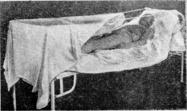
A sterile dressing is applied over the burn, covered with a thin layer of cotton wool, and secured with several turns of bandage. Such a dressing is left in place for 7-10-14 days if there is no excessive soaking. In the latter case, only the cotton wool and upper layers of the dressing are usually changed to avoid damaging the new epithelium. Some apply to the burn gauze with powder rubbed into it consisting of bismuth with starch, bismuth, zinc, starch, dermatol, etc. These powders, mixing with the exudate, form a scab under which epithelialization occurs. Ointment dressings are also used, made from sterilized vaseline or lanolin or Resorcini 0.5, I 01. Eucalypti 1.0, 01. Olivar. 2.5, Paraffini I 46.0. Wet dressings are also used, made from aluminum acetate (soothing), zinc chloride (1:500), and finally from a solution of picric acid (Ac. picrinici 2.0, Alcohol 1.5, Aq. destillata 100.0) or a saturated solution of methylene blue, 5-15% Kali hypermanganici, rivanol, etc. The Davidson method (proposed as early as 1879 by Nikolsky) is widely used at present in America - smearing the burn with a tannin solution. This method consists in wiping the entire burned area with alcohol or ether, removing the blisters (under anesthesia), and applying a 2.5-5-15% aqueous solution of tannin to the burned surface with a brush or atomizer. This smearing is repeated every hour until a dark crust forms. Subsequently, no dressings, especially wet ones, should be applied. The patient should not be moved so as not to damage the integrity of the formed film. Wet wicks are placed over the eyes, ears, and nostrils during spraying. The application of this method has reduced mortality from burns in children, according to Wilson, from 38% to 11%; this method reduces healing time to 24 days even with extensive burns; skin grafts are rarely needed; scars and contractures develop very rarely. However, this method gives good results if applied immediately after the burn and if no other methods have been used before it. The proposal to treat wounds by the open method has also found supporters in the therapy of burns. With this method, the general reaction is significantly weaker, the affected organ is given rest, the rejection of necrotized tissue occurs faster; mummification is accelerated and facilitated. Healing proceeds much faster, giving 'ideal plastic regeneration'. Disfiguring scars do not result. There is a saving of dressing material. Technique: wiping the periphery of the burn with alcohol, emptying the blisters, removing pieces of epidermis. Smearing the burn surface with a thick layer of vaseline. Sterile linen in bed. Over it, a canopy is set up with a frame of iron hoops or wire or mesh, covered with gauze for the hands, face [see separate table (art. 18:5 - 184), fig. IV.2-3]. Good ventilation. Some combine the open method with smearing with a 2-3% aqueous solution of methylene blue (A. F. Gorbachevsky). Wittek recommends daily hot air baths with the Fenn apparatus for 5-10 minutes. Goldblatt exposes the burn to the sun: starting from the 3rd day, he begins to apply insolation for 20 minutes daily without dressings. Oeconomos notes very good results from the use of heliotherapy in burns, tested by him on 500 patients. Ultraviolet rays, quartz lamps are also used, especially in cases of suppuration. Treatment of third-degree burns is conducted mainly by dry methods or by applying dressings with antiseptic ointments. After the separation of necrotic tissues, a granulating, very painful surface remains. The treatment of complications such as phlegmons, etc., is carried out by conventional methods. With burns of the mouth and pharynx, stenosis of the larynx may occur, requiring tracheotomy. The prevention of contractures presents a rather difficult task. Early skin grafting to surfaces where epithelialization is delayed, extension of the limb in the appropriate position applied from the very beginning, gymnastics, mechanotherapy can to a large extent prevent their development. The fact that scars develop more frequently where local nutrition is disturbed makes it necessary to pay special attention to the application of dressings so as not to impair the blood supply by tight bandaging. General treatment of burns. With extensive burns, when severe general phenomena such as shock, cardiac failure, uremia, etc., come to the forefront, the fight against them is especially important. Therapy should be carried out systematically and requires great attention. The burned patient is placed in a warm room or ward, surrounded with hot-water bottles, bottles with hot water. In the stage of primary shock, morphine is indicated as an analgesic and cardiac agents such as camphor, digalen, ether, caffeine are administered subcutaneously from the very beginning, especially in children, for whom the first day is critical. Copious drinking (tea, coffee), subcutaneous and rectal injections of physiologic solution ('washing' of the body). The latter should be continued in the second, toxic stage. Intravenous administration of 150-200.0 of grape sugar solution every 5-6 hours is recommended. For acidosis, solutions of soda or sodium phosphate are given orally or administered in drop enemas. If there is no collapse, warm constant baths according to Hebra are very desirable. As early as 1876, Avdakov put forward the proposition that with extensive, life-threatening burns, a powerful means of combat is blood transfusion. In recent years, this method has been applied quite widely with good results. Wilson uses the 'depleting transfusion' method: in small children, 100-300.0 g of blood is aspirated by puncture of the sinus longitudinalis and immediately replaced with the same amount from a corresponding donor. For threatening uremia, intravenous administration of uro-
182. In conclusion, it is necessary to say the following. Whatever method may be used in the treatment of extensive burns, proper organization of care is an extremely serious matter. Observation of cardiac activity and pulse, urination, abundant and timely drinking, rational nutrition, observation of dressings and ensuring that the patient, when using the open method of treatment or when using tannin, is constantly lying on a sterile surface, as well as a number of other measures noted above—all this is of the highest importance in the treatment of burns. Sunburn. It is not yet possible to distinguish the action of thermal and chemical rays on the human organism. In any case, different parts of the spectrum act differently. Thus, short-wave rays cause hyperemia, while long-wave rays cause erythema. Already during irradiation, redness of the skin and considerable warming are observed. The redness disappears along with the cessation of irradiation. If the action of the rays was sufficiently intense and prolonged, then after 6-10-20 hours (latent period), purplish-red redness, swelling, and significant pain (inflammation) reappear. The skin is hot to the touch. Every touch causes sharp stabbing pains and a burning sensation—erythema solare. Within 1-3 days, peeling occurs, and the painful phenomena disappear. In some cases, the redness persists for several weeks. In people with delicate skin, with longer and more intense action of sunlight, blisters appear on the skin, but they do not reach the size of those in ordinary second-degree burns. If a large area of skin or the entire body is exposed to irradiation (improper use of sunlight, without preliminary training), then along with local phenomena, general ones also appear, similar to those that occur with extensive burns, scalds, etc.: restlessness, severe thirst, elevated temperature, decreased amount of urine, protein in the urine, sometimes diarrhea. These phenomena can last 3-5 days. Even from a single exposure to sunlight, after the inflammatory phenomena disappear, pigmentation—tanning—appears. In its appearance, the temperature of the rays is less important than their intensity (tanning in polar travelers, mountaineers).—Pathological anatomy: dilation of the smallest capillaries, slowing of blood circulation, stasis, diapedesis mainly of white blood cells, cloudiness of tissues, edema phenomena. After 48 hours, the reverse development of symptoms begins, blood circulation is restored.—Treatment of sunburn is the same as for ordinary thermal burns. Prevention is based on the property of the skin to get used to the action of sunlight. Repeated exposure to them with proper dosing causes pigmentation, which, significantly changing the optical properties of the skin, at the same time protects it from burns and increases the body's resistance to many infections.
Chemical burns. Acids, alkalis, some salts of heavy metals, upon coming into contact with the skin or mucous membranes, cause burns. The degree of damage and its character depend on the type, concentration, and duration of action of the damaging factor, as well as on the type of tissues with which it comes into contact. The action of acids is based on their ability to remove water from tissues and to combine with tissue proteins. Different acids act on tissues with different strength and speed. The most destructive and rapid action is that of a mixture of hydrochloric and nitric acids—"aqua regia". Nitric acid acts faster but more superficially than sulfuric, hydrochloric is weaker, then acetic, lactic, carbolic. Some acids (hydrochloric) act strongly on the mucous membranes, affecting the skin to a slight degree. The higher the concentration, the stronger and faster the action. Weak solutions cause changes in the superficial layers of the skin, similar to those observed in first-degree thermal burns. Higher concentrations or prolonged action of weak solutions lead to blister formation. Finally, concentrated solutions cause necrosis. The action of acids, especially concentrated ones, is rapid but does not extend to great depth. In this case, crusts of various characters and colors are obtained: sulfuric acid gives a white-colored crust, gradually darkening, nitric acid gives a light yellow crust, etc. Burns from acids are accompanied by severe burning pains, sometimes with widespread paresthesias. The pains usually do not occur immediately after the injury, but after some time. The latent period is the longer, the weaker the concentration. The forming crust gradually hardens and is slowly undermined by growing granulations. The separation of the crust is sometimes accompanied by severe bleeding if any vessel has undergone necrosis. The picture of burns is significantly intensified if a thermal factor acts simultaneously with the chemical one. The effect of action of some acids is not limited to local changes. Being absorbed, they act on the entire organism as a protoplasmic poison. At the site of the burn, a wound surface remains, healing with a scar. The latter may be either delicate and smooth or rough and drawing together adjacent tissues, depending on the depth of the lesion. Alkalis act on tissues more slowly but more deeply. The more concentrated the solution, the stronger its action. With moderate-degree burns, the skin swells, becomes mucous and slippery (saponification of fats). Subsequent reactions in the form of crust formation and healing with alkali burns are also delayed.—Salts of heavy metals, in their action on tissues, are closer to acids. Common to chemical burns is their strict limitation corresponding to the place of action of the damaging factor. On mucous membranes, acids and alkalis act much more destructively than on the skin.—Treatment. In all forms of chemical burns, it is necessary as early as possible to resort to abundant irrigation of the affected area with water. Subsequent energetic application of neutralizing substances significantly reduces the destructive effect on tissues in chemical burns. With acid burns, alkalis are used for neutralization and vice versa. With burns from alkaline metals, washing with weak solutions of vinegar is indicated, with acid burns—solutions of soda, ammonia. When blisters or crusts form, treatment is carried out by the same methods as for burns of thermal origin.—Burns from combat substances—see Combat toxic substances, Mustard gas.

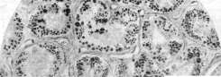
Burns from electric current and lightning. The severity and danger of damage to the body when included in an electrically conductive circuit depend on the strength of the current, its type (direct or alternating), voltage, duration of action, frequency of oscillations, changes in voltage. "The more amperage, the higher the voltage, the longer the time of action and the smaller the surface of contact, the stronger the action" (White). Furthermore, the uniformity of the conductor's contact with the body surface is of great importance. On the other hand, the place of application of the current, the degree of moisture of the skin at the point of contact, the method of connection (unipolar or bipolar), the moisture of the soil on which the victim was standing at the moment of connection, etc., are important. The intensity of local damage from electric current ranges from ordinary first-degree burns to severe necrosis and charring of entire limbs and is the result of both purely thermal and electrolytic action of the current. Passing through the human body, the current encounters enormous resistance from the skin at the points of its entry and exit. At these points, the current strength is transformed into the so-called Joule heat, reaching 3,000°-4,000° depending on the properties of the current and the strength of the resistance. Along with the action of heat, the electrolytic properties of the current begin to manifest themselves, under the influence of which acid and alkaline ions in the tissues move to different poles. Locally at the entry and exit points of the current, the so-called "current marks" (Strommarken) appear in the form of small, pearl-skin areas of parchment-like density, usually of round shape, sharply outlined, without reactive changes in the surroundings and painless. A characteristic symptom here is the twisting of the ends of hairs, which remains even in severe burns. In some cases, the skin at the point of contact is pierced by numerous small holes. Longer action or higher voltage currents are accompanied by necrosis extending far into the depth, sometimes in the form of narrow channels resembling bullet wounds with burned edges. In some cases, exposed bone is visible at the bottom of the channels. The closer the points of entry and exit of the current, the more severe the damage to the skin cover. The appearance of the damage is different at both poles: at the point of contact with the positive pole—a yellowish focus surrounded by a slightly edematous band of greenish color; at the negative pole—the skin is colored dirty gray with blisters of second-degree burns. Sometimes on the surface of the burn, one can see the smallest metallic particles impregnated into the thickness of the skin and brought here at the moment of short circuit and melting of the conductor. In severe cases of damage from high voltage current (2,000-10,000), death does not occur, "*" -", "Figure 1. Degenerative fatty degeneration of the epithelial tubules (a) of the kidney. Drops of fat are stained black by osmium; b—part of the glomerulus!" "
Figure 2-5. Open method of treating burns. Protective dressings

To the article Burns, Osmium. not only of individual phalanges or fingers, but of entire limbs. Finally, in cases where the wire or cable through which the current passes had b. or m. long been in contact with one place on the body, amputation of the limb may occur (general phenomena see below). The histological picture of the described lesions is peculiar. At the site of "current marks" the superficial layers of the epidermis are wrinkled. In the horny and papillary layers, small blisters (honeycombs) are scattered. The cells of Malpighi's layer and their nuclei are spindle-shaped and stretched out and arranged perpendicular to the surface of the skin like a brush; the boundaries between the cells are blurred; the nuclei barely stain. The vessels also undergo significant changes, especially in cases with deep tissue destruction. Their walls become brittle and fragile, mural thrombi form. The bones also undergo significant changes. A characteristic feature is the formation of bone pearls due to the melting of calcium phosphate under the action of high temperature. These pearls are usually embedded in adjacent tissues.-The course of E. from electricity is very characteristic. "Current marks" together with adjacent tissue areas are sometimes rejected after several weeks. A surface covered with good granulations remains, which quickly epithelializes without a tendency to form disfiguring scars. Suppuration during the healing process is the exception. Even very severe cases of necrosis of entire limbs follow the pattern of aseptic necrosis. Increases in temperature and disturbances in the general condition are very rare. One should remember the possibility of bleeding, which often occurs during complete well-being in the case of electric burns. These bleeding, due to the fragility of the vessels, not only at the site of the E. itself but also in nearby areas, are difficult to stop and often require ligation of the main feeding vessel along its length.- Among the complications, the possibility of thrombosis of important vessels in the brain, lungs, persistent limited angiospasm, slowly disappearing edemas (local paralysis of vessels) should be noted. Complications from the peripheral nervous system in the form of unstable lesions, neuritis, paresis, paresthesia and a number other functional disorders are common. Treatment. To the present time, conservative methods have been used in the treatment of electric burns. The generally favorable course even of very extensive injuries, the rarity of suppuration and complications in the form of sepsis, the impossibility of determining the true boundaries of tissue damage, the fragility of vessels sometimes over a large distance from the site of the E., spontaneous amputations that occur without complications, make active interventions not only unnecessary but also unsafe. In questions of amputations and exarticulations, "double caution is required due to the specific features of electrical injuries, which in most cases show a completely exceptional tendency to favorable healing". (Explanations to the "Guidelines for providing first aid to victims of electric current", approved by the XI All-Union Electrotechnical Congress.) With increasing signs of increased intracranial pressure-spinal puncture. E. from voltaic arc or sparks are treated like ordinary E. of the third degree. Similar in nature to E. from electricity are the injuries caused by lightning. In the latter case, we are dealing with currents of extremely high frequency and voltage. For these injuries, changes in the skin coverings are characteristic, in particular "lightning patterns"-red-colored stripes (local paralysis of vessels), more often in the form of a zigzag line with a large or

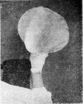
Damage from lightning.
with fewer branches (see "fig."). The entry and exit points resemble bullet wounds with severely burned edges. These streaks usually last 2-3 days. Point-like hemorrhages in the skin persist longer. Associated injuries include hemorrhages in the conjunctiva of the eyes, iritis, formation of cataracts, sometimes paralysis of the eye muscles, and deafness. A milder form of lightning damage is the singeing of hair, more often on the head, in the form of a limited area with small hemorrhages along the edges of the resulting hair defect.-General phenomena in injuries from electric current and lightning vary greatly depending on a number of conditions in which the accident occurred and the nature of the current. In severe cases, unconsciousness, cold pale skin, weak pulse, shallow breathing, and incontinence of feces and urine are observed. In cases where death did not occur immediately after the injury, patients slowly recover from the described phenomena of a specific shock. Subsequently, a severe traumatic neurosis often remains. (The effect of electric current on the body as a whole, the cause of death, as well as prevention and first aid for persons affected by current-see Electric current.) Industrial burns. In hot shops in metallurgical plants, in the chemical industry, in glass factories, in electrical enterprises, laboratories, burns occupy one of the most important places among the group of injuries. Just as industrial injury in general, the frequency and severity of burns in production vary in different shops and depend on both the degree of technical equipment of the enterprise and the extent of safety measures and labor protection in a given plant, shop. According to materials by S. G. Granovsky, concerning the Stalin plant, for 1925-26, 30,772 injuries were registered, of which 18,718 resulted in loss of work capacity; thus, burns in 1926 constituted 40.8% of all morbidity in general and 28.3% of diseases with loss of work capacity. At the same time, burns account for 16.2% of all injuries among workers in hot shops, and in rolling mills 20.2% of all injuries. By localization, the upper extremities occupy first place among burns-8.9% out of 16.2%, with 7% resulting in loss of work capacity, averaging 18.2 days. Molders in blast furnaces suffer particularly-150.4 (per 100 workers on average per year), slag removers-129 and furnace operators-148.1. Next come packers in the open-hearth shop-57.6, hook operators in the rolling mill-55.8%. When considering professional experience, a sharp decrease in injury in general and burns in particular is striking with increasing professional experience: up to 1 year of professional experience-51.4%, from 3 to 5 years-6.8%, from 10 to 15 years-2.6%, over 25 years-1.5% of all injuries. According to Kagan's data for the Bryansk plant and the "Sirius" plant for 1915-1918, out of 1,151 injuries, 19.7% were burns (45% of victims - foundry workers). The consequence of burns was temporary loss of work capacity in 59.2%, with an average duration of 19.7 days. Small burns "are an ordinary everyday phenomenon in the life of a foundry worker". The data from the Moscow First Aid Station for 1926 (A. S. Puchkov) are of no little interest. Out of 674 calls for industrial injuries, 120 were for burns, or 19.8%. The figures given force one to speak of burns as a type of industrial injury that requires vigorous and immediate measures to combat them. For burns from X-rays-see Radiotherapy and Radiotechnique. Burns of the eyes. Isolated burns of the eyes are observed in various industries (metallurgy, chemical industry, among stonemasons, etc.). The eyes more often suffer from extensive burns of the face. However, even in these cases, the eyes suffer significantly less compared to surrounding tissues or remain intact due to reflexive closure of the eyelids at the moment of injury. The severity of patho-anatomical changes in eye burns depends on the intensity, quantity, duration of action, and nature of the burn-causing factor. Three degrees of eye burns are distinguished: I degree is characterized by pronounced hyperemia of the conjunctiva with greater or lesser edema of it and the subcutaneous tissue of the eyelids. In burns caused by flame, the eyelashes and eyebrows are singed. Usually, the redness disappears by the 3rd day.-In II degree burns, the conjunctiva swells significantly. After a short time, small blisters filled with clear fluid and quickly bursting appear on it. In this case, blisters do not form on the cornea, but it is covered with white, delicate films. In this form of burn, the phenomena of edema, hyperemia, and pain last much longer. After epithelialization of the affected areas of the conjunctiva (5-10 days), the edema and redness disappear, and the normal state of the conjunctiva is restored. Disfiguring phenomena after II degree burns are usually not observed.-III degree burns are much more severe in both course and consequences. Causing necrosis of both eyelids, this type of burn leads to severe disfiguring changes in the palpebral fissure up to its complete overgrowth. These cases are particularly severe when infection is added. In milder cases, adhesions of the eyelids to the eyeball, partial adhesion of the eyelids to each other, etc., remain. Corneal burns accompanying these forms are particularly severe in their consequences.-In milder cases, after the slough is rejected, an ulcer of greater or lesser size remains on the cornea, after healing of which a scar remains. In severe cases, the eye is completely lost. Treatment of II and III degree burns requires strict asepsis. Usually, in the first hours, cold compresses from weak boric acid solutions are applied. Subsequently, ointment or simply aseptic dressings are used. To eliminate disfiguring scars and restore the main fissure, it is necessary to resort to a number of new complex plastic operations with transplantation of mucous membrane and skin.-Special attention should be paid to burns of the eyes, in particular the cornea, by splashes of molten metal or boiling oil. Very small splashes, getting into the eye, manage to cool in tears without causing significant destruction. The entry of larger splashes onto the conjunctiva, especially into the lower conjunctival sac, leads to the appearance of limited II and III degree burns. In cases where a drop of metal falls on the cornea, an ulcer of greater or lesser size forms on it, after healing of which a scar remains. Treatment of corneal burns is carried out as in simple ulcers.-Prevention of eye burns-see Protective equipment in production. Chemical burns of the eyes. What has been said above regarding the action of acids and alkalis on tissues should also be applied to the organ of vision, taking into account the specificity of the tissues from which it is built. In burns with acids, there is a process limited only by the place of action of the reagent, more or less pronounced depending on the concentration, duration of action, and amount of substance that entered. Here, too, I degree burns are distinguished, when some time after the action of weak solutions of acids, a more or less uniform in intensity redness of the conjunctiva and skin of the eyelids appears, disappearing after 2-3 days. II degree burns are not sharply separated from III degree burns. The glassy edema that forms here takes on various shades depending on the type of acid that caused the burn. At the same time, a limited, dense, grayish-colored slough forms. The latter lasts quite a long time, causing irritation of surrounding tissues. After its rejection, epithelialization of the mucosal defect occurs with subsequent development of scar tissue deforming the eyelid.-Burns with alkalis differ in many ways from those just presented. Symptoms of such burns often begin to manifest only 1-2 days after the injury. Furthermore, tissue destruction is not limited to the place of direct action of the reagent, but spreads sideways and deep. The slough formed under the action of alkalis is never dense, as after acid burns. The granulations that develop after its rejection have a tendency to overgrow and often require repeated curettage or cauterization with a thermocautery. Burns with lime and ammonia are particularly severe. Even after the entry of small amounts, pronounced glassy edema and redness of the entire conjunctiva are observed. Often, small firmly and deeply seated pieces of lime can be seen in its thickness. At the same time, the cornea becomes dry, cloudy, and milky-white. Burns with ammonia are particularly severe. In this case, the affected organ resembles a "boiled fish eye." Not causing special concern in the first days, the disease takes a very rapid course on the 6-8th day, and in most cases, the eye is lost. The sooner measures are taken to remove or neutralize the reagents that entered, the less subsequent destruction and the greater the chance to save the eye. Abundant washing of the affected eye with cold water or milk (in alkali burns) is recommended.-Prevention of eye burns-see Acid production, a. Lidsky. In forensic medicine, 4 degrees of burns are distinguished: 1) erythema or reactive redness, 2) formation of blisters, 3) formation of a slough, and 4) charring.
Erythema of the skin due to burns usually disappears on the corpse when hypostasis appears; however, if death did not occur too quickly, sometimes swelling remains, but more often there is desquamation of the upper skin layer in the area of the former erythema. After second-degree burns, either blisters filled with serum are found, or the latter burst, the upper skin layer shrivels at the edges of the burned areas, hangs in shreds, and the underlying tissue is exposed. Sometimes on the hands and feet the skin layer comes off together with the nails like a glove. On the corpse, such areas lacking skin (epidermis) initially appear moist, pale (rarely, mostly in the underlying areas, reddened), and then under the influence of air they dry out, become dense, parchment-like, are difficult to cut, and take on various shades of yellow, brown, brown-red color—in short, the same changes develop as are observed in abrasions. Third-degree burns are recognized by a whitish, grayish-brown scab penetrating more or less deeply into the thickness of the skin, which has the appearance of having been boiled or, under the action of flame, slightly roasted. In general, external changes on the bodies of burned persons depend on the degree of burns, their origin, and the time that has passed from the moment of burning until death. Death from burns is mostly accidental. Suicides by self-immolation are also known; these are predominantly found among the mentally ill or persons who have no other means of ending their lives (in places of confinement, etc.). Cases of murder are relatively rare: there are cases of scalding children, setting sleeping persons, especially intoxicated ones, on fire with kerosene. Much more often, bodies of persons killed by some other method are set on fire to conceal traces of the crime. There are also cases of sprinkling with caustic liquids (e.g. sulfuric acid) out of revenge, jealousy. In former times in Siberia on the Terek there were sects of 'self-immolators' as one of the manifestations of religious fanaticism. Some external changes in burns have significance for determining their origin. Flame causes all degrees of burns, in particular soot is noted on the skin, singeing, charring of hair, nails. Burns spread on the body in the form of tongues, stripes, along the direction of which it is often possible to judge the position of the body at the moment of action of the harmful agent. Thus, if clothing catches fire and a person runs through a column of flame, the stripes of burns have a longitudinally ascending direction; conversely, when a sleeping person or a corpse is set on fire—transverse. On the other hand, the action of caustic liquids (acids, alkalis) does not cause charring, and in scalding (with water, steam) usually only two degrees of burns are observed; soot and singeing of hair are absent; burns spread in the form of descending stripes depending on the flow of the liquid causing the burns. However, there are exceptions, e.g. if scalding occurred from falling into a hot liquid. Destruction of hair can also be caused by the action of hot bodies, and sometimes by caustic liquids. Burns penetrate into the cavity of the nose, mouth, pharynx, etc., when the subject was surrounded by burning gases. In some cases, the substance causing the burns (lime, varnish, etc.) is found to be adhering to the skin. For the correct solution of a number of questions arising in forensic practice, the expert must have a good knowledge of the changes in tissues from the action of high temperature, especially of flame, on a living person and on a dead body. It is often important to determine whether a person entered the flame alive or whether the corpse was subjected to the action of fire. At autopsy, the following indicates that the burns were antemortem: 1) erythema, which unfortunately after death fades or completely disappears. 2) The presence of blisters, which upon microscopic examination contain fluid with the characteristics of an exudate. Only in rare cases from the action of fire on a corpse was the formation of blisters filled with a small amount of serous fluid, which moreover did not have the properties of an exudate. In particular this is possible, according to Reuter, on edematous corpses, when under the influence of heat fluid is displaced from the tissues into neighboring parts and raises the upper skin layer (epidermis) in the form of blisters. 3) The presence on scorched areas outside the area of location of livor mortis of a network of vessels, noticeable to the eye (or better through a magnifying glass), filled with clotted blood, and sometimes hemorrhages in the substance of the scab crust. In unclear cases, microscopic examination of the changed areas should be performed. Charring does not provide a basis for determining whether the burns were antemortem, and if it occupies a large surface area of the body, it generally forms only postmortem. If a person was alive and breathing in the flame, products of incomplete combustion—particles of coal and gases—can be found not only in the respiratory tract (soot) but also in the blood (CO). Soot can be detected even in the pulmonary alveoli, and sometimes in the stomach due to swallowing. The presence of carbon monoxide in the blood is conclusive if the blood was taken for examination from deep parts, e.g. from the heart. The red color of the blood in itself has no significant importance, since blood upon heating can generally take on a light-red color. With further action of high temperature on the corpse, fluids are lost, the skin becomes hard, shrivels, and cracks in less stable places (joint bends, perineum); sometimes such cracks occur when attempts are made to straighten the limbs; externally they can be mistaken for incised wounds. The dead body under the influence of heat assumes a peculiar position—the 'boxer's pose' or that of a fencer (Fechter-stellung), with the arms and legs more or less bent. It is believed that this phenomenon is of postmortem origin and depends on the shrinking and shortening of muscles; the displacement of the limbs in the direction of bending is caused by the more developed flexor muscles. From the action of high temperature on the bones of the skull, cracks, holes may form in them due to expansion of the bone and swelling of the organic substance contained in it, as well as partly from the pressure of water vapor in the cranial cavity. Then the bones calcinate, i.e. lose their organic basis, become brittle, from which postmortem fractures may occur. From the action of high temperature, postmortem hemorrhages occur, in particular when the soft tissues of the skull are charred—epidural hematomas; under the influence of heat blood is displaced, as it were, escapes into neighboring parts, the blood filling of which increases, which creates favorable conditions for rupture of vessels with subsequent hemorrhage. Such a finding can be erroneously attributed to a preceding head injury. In cases of severe charring of the body, it may be found completely without a head and limbs, with cavities exposed; at the same time, due to evaporation of fluids, there is a significant decrease in the volume of the body and internal organs, which have the appearance of children's organs. This circumstance can lead to errors in determining age. If injuries are found, one should not forget that they may have occurred accidentally, e.g. from falling a burning beam of a house or because the deceased, wanting to escape, jumped and fell from a significant height. These injuries and the accompanying hemorrhages can be recognized even on severely charred corpses. In cases where someone was first killed and then burned, signs of another manner of death can be found. In particular in strangulation with a cord, when the tightly drawn rope is left on the neck, the strangulation groove is well preserved. With sufficient experience and attention, it is usually possible to avoid confusing burns with other processes. It would be an unpardonable error to take for changes due to burns those detachments of the upper skin layer (epidermis) and blisters that are observed in significant putrefaction of the corpse. Burns can more likely be confused with the redness and blisters arising from certain skin diseases (pemphigus, dermatitis exfoliativa). Resemblance to burns is also presented by postmortem extensive detachments of skin in persons who died during the eruption of acute exanthems. Sometimes a bedsore was mistaken for a burn. Observations have shown that during fires people first suffocate in smoke without yet receiving burns. Non-fatal burns in terms of their properties and consequences are classified according to the existing rules on the severity of injuries.
V. Vladimirsky.
Related articles
Cite this page
“Burns.” Soviet Medical Encyclopedia. English translation of Bolshaya Meditsinskaya Entsiklopediya, 1st ed. (Moscow, 1928–1936), ed. N. A. Semashko. https://sovietmedicalencyclopedia.pages.dev/article/burns/