Wounds
Historical document, translated for reference. It reflects medical knowledge of the 1920s–30s and is not medical advice.
Summary
This article defines wounds as any damage to body tissues involving the breaking of skin or mucous membrane integrity, describing their characteristics including gaping edges, bleeding, and pain, and how these vary based on tissue type and direction of injury.
Encyclopedia article (1928–1936)
WOUNDS, INJURIES. A wound (vulnus) is any damage to the tissues of an organism associated with violation of the integrity of the skin or mucous membrane. However, even with closed injuries, if the integrity of the covering of any organ is violated, one speaks of its injury, for example, with a closed injury to the abdomen, in case of violation of the integrity, for example, of the liver or stomach, one can speak of wounds of the liver, stomach, etc. The immediate consequences of injury are: 1) gaping (divergence) of the wound edges, 2) bleeding and 3) pain. Only very superficial injuries do not have gaping; then they are called scratches or excoriations. The degree of gaping of wound edges depends on the structure of the damaged tissue and on the direction of the wound. In the skin, gaping depends on the degree of development of elastic fibers in a given area and on the relationship of the direction of the wound to their course. Wounds located transversely to the course of the fibers give the greatest gaping, the least - parallel. The degree of gaping of skin wounds in different areas of the human body is determined by the so-called lines of Langer (see vol. XIII, art. 210, fig. 1). A muscle wound also gapes to the greatest extent with transverse incision of the muscle fibers and hardly gapes with longitudinal damage. Organs where there are few or no elastic fibers gape little (for example, liver, spleen, pancreas, etc.). Bone and cartilage do not gaping by themselves; the divergence of their edges depends either on the defect of tissue during injury or on the traction of fragments by muscles attached to them. The gaping of a wound can be very severe if during injury a section of tissue is excised and a defect is formed. Finally, the nature of the injury can greatly affect the gaping of the wound depending on the damage to the wound edges. Thus, when they are crushed, the contractility of the tissue is lost and gaping may hardly occur.
Bleeding from wounds depends on damage to blood vessels. It is absent only when bloodless tissues are wounded (for example, certain areas of the eye). Its intensity depends on the caliber of the damaged vessels, their quantity, and the nature of the damage (see below). Depending on the nature of the vessels, arterial, venous, capillary, or parenchymatous bleeding is distinguished. In practice, due to simultaneous injury to different vessels, bleeding is usually mixed. (For details, see Bleeding.) Pain depends on damage to sensory nerves. It may be absent only temporarily in cases of so-called local shock (see) or when there is no pain sensitivity in the given area (for example, the surface of the gastrointestinal canal and others). The degree of intensity of pain sensations depends on the anatomical area of injury. For example, pain, which is very severe on the fingers of the hands, in the area of the anus, and in the genital organs, is much weaker on the back and neck. There may be no pain sensations at all in injuries in patients with lesions of the central nervous system (for example, syringomyelia) or large peripheral trunks. Usually, the intensity of pain sensations is maximal immediately after injury, subsequently it begins to decrease, and as a rule, by the end of the first day, pain is replaced by tenderness, i.e., the appearance of pain sensations from any new irritating factor affecting the damaged area, for example, pressure, palpation, etc. The resumption of pain in W. often serves as a sign of complication. Classification of W. Various signs can serve as the basis for the classification of injuries; thus, W. can be divided into surgical and accidental, aseptic and infected, penetrating into any cavity and not penetrating, etc. But the most accepted classification is according to the instrument with which the W. was inflicted, and in connection with this, according to the nature of the tissue damage. Depending on this, cut and incised (vulnus caesura), punctured (v. punctum), torn (v. laceratum), contused (v. contusum), crushed (v. conquassatum), and gunshot (v. sclopetarium) W. are distinguished. Flap-like W. are those where part of the coverings is separated in the manner of a flap; if this part is completely removed during injury, then a tissue defect results. In this classification, the nature of the damage to the wound edges has the greatest practical importance, from which regeneration subsequently proceeds, i.e., the healing of W. It is clear that the less damage to the wound edges, the faster the regeneration processes proceed; the more these damages, the slower and more complex these processes, since processes of rejection of non-viable areas are also added to them. On this basis, it is advisable to simplify and clarify this classification by dividing into W. with a small zone of damage and W. with a large zone of damage. The first group includes W. where the damage to the edges is not noticeable (surgical, cut wounds) or very little noticeable (punctured, incised, certain bullet gunshot wounds), the second group includes wounds where the wound edges appear damaged to the naked eye (hemorrhages, crushes, crushings, etc.). To the latter belong all contused, torn, crushed W. and the majority of gunshot wounds, especially artillery wounds. Both in relation to the healing processes and in terms of therapeutic effects and clinical course, the two groups differ significantly from each other and have no fundamental differences within the group. W. with a small zone of damage, in terms of gaping, generally follow the conditions mentioned above. Punctured and bullet gunshot wounds usually gape little due to the rapid filling of the wound cavity with clotted blood. Bleeding in cut and incised W. is greatest because vessels are usually completely divided by a sharp instrument and gape. Pain in all W. of this group depends on the area of injury, but gradually subsides after injury. Due to the good viability of their edges, W. with a small zone of damage give more rapidly proceeding regeneration processes, heal by first intention if their edges are brought close together, and are less prone to complications from infection. W. with a large zone of damage have edges poorly inclined to regeneration, often give visible areas of necrosis, which are subsequently rejected; their edges are nourished worse due to damage to vessels and hemorrhages, they are prone to complication by infection, and usually heal by second intention. Gaping in them depends not only on the location and direction of the injury but also on the degree of damage to the wound edges, which, losing elasticity under the influence of crushing, may diverge from each other little but do not give hope for first intention even when sutured; bleeding also depends on the degree of damage to the vessels of the wound edges: it is relatively small, as the vessels are crushed and easily thrombose. Even relatively large arterial trunks in crushed W. bleed little due to twisting and flattening of the vessel walls. If in W. with a small zone of damage the source of pain sensations is the sites of nerve damage in the W. itself, then in W. with a large zone of damage the area where nerves are damaged greatly increases due to damage to them in the thickness of the wound edges. Therefore, the latter W. hurt more intensely and for a longer time than W. with a small zone of damage. It is clear that as a result of all these properties, a wound with a large zone of damage gives a worse prognosis and requires more attention from the surgeon than a wound with a small zone of damage. Healing of wounds. The process of wound healing belongs to the inflammatory process, as all signs of inflammation are present in it. However, the phenomena of regeneration in the healing of W. are so predominant that they somehow overshadow all other signs of inflammation, therefore pathologists often add to characterize such a process the definition of inflammation as aseptic (Maksimov), restorative, reconstructive (Aschoff, Lubarsch). Clinically, this process is also clearly distinguished from the picture of inflammation familiar to the surgeon. A distinction is made between healing of W. by first intention (sanatio per primam intentionem; this expression is often shortened and they say 'per primam') and healing of W. by second intention (sanatio per secundam intentionem, abbreviated 'per secundam' or 'per granulationem'). Healing of W. begins from the moment bleeding stops and can be divided into three periods: 1) preparatory period, 2) period of preliminary regeneration, 3) period of final regeneration (Girgolav). Healing by first intention. Only W. with directly adjacent edges, even if this contact was caused artificially (for example, by suturing), and consequently not having a macroscopically noticeable cavity, heal by first intention. The course of the healing process by first intention is easiest to follow on a sutured aseptic cut W., i.e., a surgical one. In the first moments after bleeding stops, the wound slit, examined under a microscope, reveals between the adjacent wound edges clots of blood and lymph, fibrin clots, and groups of tissue cells that separated at the moment of injury. In such a state, from the point of view of the morphological picture, the W. remains for the first hours after injury. But even observation of the active chemical reaction in the W. shows that during this time, i.e., during the first period of healing, a series of changes occur that prepare for further tissue regeneration. After 2-21/2 hours, it is already possible to establish a significant shift in the pH of the wound toward acidity-6.8 instead of the normal 7.4 (v. Gaza believes that simultaneously in the W. the formation and accumulation of new chemically active substances of an enzymatic nature occurs). Experiments of Haberlandt on plants, subsequently confirmed on animals by Stark and S. S. Girgolav, show that apparently the factors changing the chemical composition of the W. and causing regenerative phenomena are the dead, dying, and destroyed cells as a result of injury. The end of this period is determined under the microscope by more or less pronounced phenomena of emigration of white blood cells and changes in local mobile elements toward their rounding and increase in volume; simultaneously, figures of cell division appear. Regenerative phenomena are most clearly manifested in cells of mesenchymal origin and less in covering epithelium. Among the cells of外来 (from vessels), lymphocytes, leukocytes, and partly polymorphonuclear neutrophils are noted; among the cells of mesenchymal origin, fibroblasts, endotheliocytes, and histiocytes are observed.

Figure 1. New formation of vessels (after 48 hours). Multiple vascular sprouts originating from the wall of the capillary of the subserous membrane of the intestinal loop, directed toward the fibrinous exudate at the site of injury. In the exudate there are many fibroblasts. (According to Vereshchinsky.)
of cells - elements of the reticulo-endothelium (noted in the literature as Maximov's polyblasts, Marchand's adventitial cells, etc.) and fibroblasts. Thanks to emigration, some of these cells are located at the wound edges, while others move into the clots of fibrin and blood that adhere the wound edges (primary adhesion of W.). The clots of blood and lymph in the space between the wound edges play a very important role for the cellular elements, providing them with a supporting environment and creating the possibility for cells that are emigrating and dividing to come into contact, in particular fibroblasts from both wound edges. Thus, by the end of the first day, the wound space is already filled with viable elements. By the beginning of the second day, the growth of capillaries begins, which grow endocellularly from the undamaged capillaries of the wound edges (Fig. 1); when the capillaries connect, blood circulation is restored. By the 5-6th day, fresh scar tissue is already formed from fibroblasts and new vessels, which connects both wound edges. By this time, the epithelialization of the skin surface also occurs. Part of the epithelial covering at the sutures often bends inward into the wound space (Fig. 2), in which case this area usually undergoes degeneration, but individual groups of cells can persist for quite a long time and even give off processes (Zavarzin). In the areas adjacent to the W., epithelial cells increase in volume, swell, and begin to less intensely take up the staining of the protoplasm. By division, the epithelial layer advances toward the wound space and, as mentioned, by the 5-7th day, epithelialization is completed. By this time, the increased acidity of the tissues also begins to decrease, but it has not yet reached normal. Thus, the second period of healing ends with the restoration of living fresh scar tissue in the place of the former wound space. However, the entire healing process by this time is by no means finished, but only enters its third stage, the stage of final regeneration. According to the research of a number of authors, only by the 14th day does the wound area of the skin, stomach, fascia reach its original strength (to rupture). By this time, according to the data of S. S. Girgolav, the active reaction of the wound area also gives normal figures (pH 7.4). The third period, the period of final regeneration, is characterized by the transformation of the newly formed tissue into a form that remains constant in the future, and by those regenerative phenomena that develop at later stages and also have a permanent value. Young scar tissue consisting of fibroblasts gradually transforms into fibrous scar tissue (see Scar). The third period also includes the regeneration of elastic fibers, which occurs rather imperfectly, and the regeneration of nerve tissue, which is represented
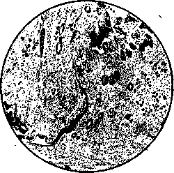
Figure 2. Skin wound after 9 hours. On the right top, the folded epithelial covering, the wound cavity is glued with exudate, in which the emigrated elements are located.
sti; part of the epithelial covering at the sutures often bends inward into the wound space (Fig. 2), in which case this area usually undergoes degeneration, but individual groups of cells can persist for quite a long time and even give off processes (Zavarzin). In the areas adjacent to the W., epithelial cells increase in volume, swell, and begin to less intensely take up the staining of the protoplasm. By division, the epithelial layer advances toward the wound space and, as mentioned, by the 5-7th day, epithelialization is completed. By this time, the increased acidity of the tissues also begins to decrease, but it has not yet reached normal. Thus, the second period of healing ends with the restoration of living fresh scar tissue in the place of the former wound space. However, the entire healing process by this time is by no means finished, but only enters its third stage, the stage of final regeneration. According to the research of a number of authors, only by the 14th day does the wound area of the skin, stomach, fascia reach its original strength (to rupture). By this time, according to the data of S. S. Girgolav, the active reaction of the wound area also gives normal figures (pH 7.4). The third period, the period of final regeneration, is characterized by the transformation of the newly formed tissue into a form that remains constant in the future, and by those regenerative phenomena that develop at later stages and also have a permanent value. Young scar tissue consisting of fibroblasts gradually transforms into fibrous scar tissue (see Scar). The third period also includes the regeneration of elastic fibers, which occurs rather imperfectly, and the regeneration of nerve tissue, which is represented
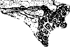
Figure 3. Branching of newly formed nerves in the scar after healing of the wound by secondary intention.
is an important circumstance connecting this newly formed area with the nervous system of the entire organism. The processes of regeneration of nerve fibers and endings, as shown by the research of V. M. Nazarov and Kolosov, proceed slowly and end only within several months, and the appearance and structure of these endings do not always correspond to those that were previously present here (Fig. 3). During this same period, the transformation of young connective tissue, connecting the bone fragments into callus, and the evolution of this callus tissue into its final form (see Fractures) occurs, which has primary functional significance. Thus, the third and final period of wound healing ends with the formation of final tissue that remains permanently in this area. Healing by secondary intention. Any wound that has a macroscopically noticeable cavity located between the wound edges heals by secondary intention; a characteristic feature of this type of wound healing is the macroscopically observable development of so-called granulation tissue. This same type of healing occurs for wounds if there is any mechanical obstacle between the wound edges that prevents their direct union, such as a foreign body, blood clot, hematoma, etc., and finally, a wound also heals in the same way when complications are present (e.g., in case of wound infection). The first, preparatory period, in an open wound, is also expressed in preparation for the regenerative processes that will subsequently arise. The entire wound surface is covered with a thin fibrinous exudate, in which groups of blood cells (hemorrhages), blood clots, and lymph accumulate locally. Under the influence of the same factors described above, a shift in the wound reaction to the acidic side occurs and new enzymatic substances are formed. Then, the migration of white blood cells into the mentioned marginal exudate and into the wound edges begins, the cells of the reticulo-endothelium acquire a rounded, succulent appearance, cell division occurs mainly of mesenchymal origin, and new blood vessels are formed. Macroscopically, such a wound appears as if veiled, the boundaries between individual tissues seem to be smoothed, ceasing to be clear and distinct. From the elements mentioned, granulation tissue (see) is formed, which by the 4th-5th day usually completely covers the wound cavity. If one recalls the significance of granulation tissue, its poor ability to absorb, and its resistance to infection, then the great clinical importance of this fact becomes obvious. On the other hand, the need for careful handling of the granulation covering during surgical manipulations also becomes understandable, since damaging it, as if inflicting a new injury, facilitates the possibility of wound infection. Finally, the presence and development of the granulation covering, taking into account general factors (see below), serves as the basis for determining the time of injury. As the granulation tissue develops, it gradually fills the wound cavity, and the healing times here now depend on the size of the wound cavity, and often weeks and months are required to fill large cavities. Simultaneously with the edges of the wound, with the same changes in the epithelium that were described earlier, the epithelial covering advances. The formation of granulations and their filling of the wound cavity represents a phenomenon of preliminary regeneration; but if the wound is large, then while its center is still being filled with granulations, the edges are already undergoing the restructuring of granulation tissue into scar tissue, i.e., the phenomena of final regeneration; thus, the third period of wound healing, as if following the second, occurs simultaneously but in more peripheral areas of the wound. This circumstance has great clinical significance, since the transition of granulation tissue into scar tissue leads to shrinkage, dehydration, and reduction in size. As a result, the size of the entire wound cavity decreases, and thereby accelerates the filling of the remaining central cavity by granulations. Epithelialization is constantly somewhat delayed due to the lesser regenerative abilities of epithelial tissue, however, its completion still does not mark the end of the healing processes. In a number of cases, with very extensive granulation surfaces and depending on general factors, epithelialization may not occur at all, then plastic measures are required (see Plastic operations). A fresh, more or less extensive scar, depending on the size of the wound, subsequently undergoes the same changes as described above - it shrinks, becomes poor in blood vessels, nerve fibers grow from the nerves remaining in the wound edges, and new endings are formed. As a result of healing by secondary intention, a more or less extensive scar is formed, replacing the tissues that were previously present here; naturally, functionally such a scar is not complete, and even the strength of scar tissue, despite its density, turns out to be less than that of normal tissue, and the scar, if it is subjected to constant pressure, is capable of stretching in the future and thereby further weakening the corresponding area (see Abdominal wall). In addition, scars cause a number of other functional disorders (see Scar). From the review presented, it is clear that there is no fundamental difference between healing by primary intention and secondary intention; even the characteristic feature of secondary intention - granulation tissue - can form on small areas of a sutured wound where it is not possible to achieve complete approximation of the wound edges, but practically both types of healing differ to a significant degree. In healing by first intention, the entire wound heals simultaneously over its entire length and by the end of seven days it is already epithelialized, whereas in healing by second intention, the filling of the cavity with granulations requires a significantly longer time, sometimes measured in months. As a result of healing by first intention, a narrow, minimal-size scar remains, while after secondary intention, the scar is extensive and often so significantly disrupts the function of this area that it necessitates further surgical interventions or reduced work capacity of the individual. Furthermore, healing by second intention sometimes may not end at all due to the extent of the wound and may require plastic measures. Finally, extensive scars, especially in areas subjected to pressure or friction, often ulcerate and require either time or surgical aids for treatment. All this taken together compels, in surgical operations where possible, to strive for healing by primary intention. Healing under a scab. Relatively small wounds, as a result of the drying of the wound exudate of blood and lymph, are covered by a brownish dry crust, a scab, which remains firmly attached to the tissue throughout the entire healing period, and then falls off by itself and reveals a healed, epithelialized surface. This type of wound healing, especially in the pre-aseptic era, was distinguished as a special type of healing - under a scab. However, an analysis of the phenomena occurring in such a wound shows that in this case too, with close approximation of the wound edges, the process is one of healing by primary intention, and in the presence of a wound cavity, even if not noticeable under the scab, it is a matter of formation of granulations with their subsequent covering by proliferating epithelium, i.e., healing by second intention. In other words, there is no basis to single out healing under a scab as a special form. Practically, the scab acts as a dressing, and the dryness and mechanical structure of the scab protect the wound from infection and thereby ensure the course of healing without complications. However, such protection is not always sufficient, infection can still develop under the scab; under these conditions, the scab is rejected by the wound discharge and a wound cavity covered with granulations is found beneath it. Further healing in such cases proceeds as in the healing of any infected wound (see below). Conditions for wound healing. The healing of wounds in the same individual on different areas and in different tissues and organs occurs at different speeds and with different degrees of completeness. It is known that on the head and face, healing (mainly of skin wounds) is more successful than on other areas, which is connected with the abundant blood supply of these areas and probably with the nervous system, better trained on areas not covered by clothing. But the difference in wound healing on organs covered by peritoneum and those not covered by it (esophagus, urinary bladder) is much more pronounced, due to the great adhesive (plastic) ability of the peritoneum. Healing of wounds with scar tissue sometimes precedes final regeneration for very long periods, as for example in bone wounds (see Fractures), nerve wounds (see Nerve suture), but in general, the functional completeness of wound healing is determined by the completeness of the regenerative processes.
Some connective tissue formations, as a result of healing W., lose little functionally, as has already been mentioned (e.g. bone, fascia, peritoneum), but a number of other tissues also of mesenchymal origin show very little tendency to regenerate as a result of the factors that arise in W. Thus, the lymph gland heals with a scar and gives almost no regeneration of lymph tissue, just as the spleen does. Organs whose main functional part is epithelium also give very imperfect regeneration, e.g. liver, kidneys, salivary glands, mammary gland (where the milk ducts regenerate partially; Protasiev). In healing of W. of the lung, only the epithelium of small bronchi regenerates to a small extent (Aranson). However, all this regeneration is so slight that it has no practical significance. Even less practical significance has muscular regeneration, both of striated and smooth muscle. Kolber established only regeneration of muscle fibers going into a scar for a few mm in the uterus. Practically, therefore, it should be recognized that W. of muscle heal with a scar, and at this site, obviously, all ability to contract is lost forever. As a result of this, an inscriptio is formed in the muscle, as it were, dividing the muscle into two bellies, or a 'dead' non-contracting space of a hollow muscular organ (e.g. intestine). It is clear that in this way the function suffers to one degree or another. Regeneration of the central nervous system in relation to cellular elements is zero; healing occurs only through elements of mesenchyme (Ramon y Cajal, Spielmeyer and others). Even regeneration of fibers in the central nervous system, although it takes place, is subsequently partially accompanied by their death and proceeds to an extent devoid of practical significance (Samarin). The matter is different in nerves: here the nerve fibers (axis cylinders), being processes of nerve cells, show a very great tendency to regenerate, which has very important practical significance, since it is accompanied by restoration of sensitivity or disappearance of paralysis. However, to obtain such an effect, it is necessary that the W. of the nerve heal by first intention and that there be minimal development of scar tissue at the site of the injury (see Nerve suture).--The combination of all the conditions discussed can therefore, in the best case, after healing of the W., give an incomplete but functionally little-influencing on the general life processes of the organism area of tissue; in the worst case, the matter can go to a complete loss of function of the injured area. Treatment of W. Treatment of aseptic W. The most favorable conditions for healing are usually created in the operating room during surgical work when W. are inflicted. These conditions not only give the maximum possibility to protect the W. from infection, but also to inflict the W., taking into account the best conditions for tissue regeneration and for healing of the W. Therefore, the best results in all respects are given by operative wounds. But to obtain these results, it is necessary to create the most favorable conditions also throughout the entire period of healing of the W. (see Postoperative period).--Treatment of W. begins from the moment of applying a dressing to it. If the W. is tightly sutured, then the need for a dressing is in essence not great. A number of W. on the face not only can but should be left without a dressing, since applying a dressing near the eyes, nose, mouth easily causes it to be moistened with tears, nasal discharge, saliva, etc., and not only does not protect the W. from infection, but, on the contrary, promotes it, while leaving the W. without a dressing leads to rapid drying of the wound discharge along the line of the wound, which is already sufficient protection. In the future, it is only necessary not to traumatize such a W.--and its healing will be ensured. A number of observations in treating patients without dressings and on other areas also confirms the full practical possibility of this method, however, it is necessary in the first period until drying and formation of a crust to cover the W. with a sterile piece of gauze to avoid contact with non-sterile objects (linen, blanket, etc.). Therefore, it is quite appropriate to protect by applying a gauze roller along the line of the sutures and tying its ends with threads from the skin sutures. Such a roller protects the W. from contamination. Instead of it, a piece of gauze covering the entire line of the sutures can be glued at the edges with collodion, mastisol, rubber cement, cleol, or strips of adhesive plaster, etc. For drying, to prevent excessive sweating and the associated danger of secondary infection, the middle of the gauze pad in the area of the W. itself should never be attached with a fixing substance. Extensive dressings with gauze, cotton, and bandages are currently applied to tightly sutured W. only for special indications. The main task--to protect the W. from secondary infection--is fully achievable with the light dressings described. The second task--to provide rest to the wounded area (and of course to the whole body)--is achieved in extensive and penetrating W. by the patient being in bed and the appropriate position of the limb if the W. is on a limb (placing on a pillow or in a splint, hanging, etc.). In the first hours (up to 24 h.) after injury, operation, rest should be more strict due to the danger of secondary bleeding; later, immobility is no longer necessary, nor is bed rest, unless there are special indications for it depending on the location of the W. and its depth, the patient's strength, the nature of the disease for which the operation was performed, etc. (see Postoperative period). In case of danger of secondary bleeding, it is useful in the first hours after the operation to apply pressure to the area of the W., for which sandbags, cold, or pressure bandage dressings can serve (see Desmurgy).--Fighting against painful sensations in the W. is usually required during the first day and is expressed in the administration of analgesics (morphine, pantopon), usually in the form of subcutaneous injections. Otherwise, the W. requires less therapeutic measures than observation, in order to promptly detect complications if they arise. A light dressing significantly facilitates such observation. Mention should also be made of observation of the temperature of the wounded. It is very common on the day of injury and in the following days for small rises in temperature not exceeding 1° to occur without any clinically determinable complications of the course of the W. occurring later. This fever is known as aseptic or Volkmann's fever; the absorption of tissue breakdown, clots of blood is undoubtedly able to cause rises in temperature, by which this fever is usually attributed, but it is also undoubtedly true that despite all aseptic precautions, pathogenic microorganisms in the W. as a rule do get in, however, not finding there a suitable medium for development and not having high virulence, they do not cause suppuration, but the general reaction to such an invasion in the form of a rise in temperature is still observed. A number of authors (Wright and others) believe that the latter circumstance is more often the source of the rise in temperature than the conditions causing true 'aseptic' fever (see Fever). The general management of the wounded in the absence of complications from the W. is given in the Postoperative period. It was stated above that epithelialization of the W. and the development of fresh scar tissue in it ends by the 5-7th day. By this time the sutures usually also become unnecessary and are to be removed (see Dressings). A slightly different attitude is required for an aseptic, but not sutured, and therefore healing by secondary intention W. The reasons for leaving the W. unsutured are the presence of infection in it or well-founded fears of its development, inability to close the defect formed as a result of trauma or operation, stopping of bleeding by tamponade, communication with a cavity requiring drainage, etc. (on treatment of an infected W. see below). But even an uninfected extensive wound cavity gives a certain amount of discharge, and above all needs very careful protection from infection; this purpose is served by a dressing (see). After formation of a granulation covering, the W. requires systematic dressings (see). If the wound edges allow, it is rational to reduce the cavity of the W. by drawing the edges together with strips of adhesive plaster or even by applying a secondary suture. After the W. is filled and a flat granulation surface is formed, it is advisable to take measures to accelerate epithelialization. As little as we have in our hands the possibilities to accelerate the entire process of healing of the W., i.e. to influence the very factors of regeneration, so practically achievable is to accelerate the growth of epithelium. The latter is achieved either by acting on the growing epithelium or by transplantation of epithelium. From non-plastic methods, tiling the granulations with plaster is used, under which epithelium develops well, ointment (5-10%) Scharlaehrot (amidoazotoluol), dressings with liquid paraffin, etc. However, all these methods are inferior in results to epithelial transplantation.
Epithelial transplantation is a very effective method and is currently used very frequently, both on granulation tissue and on fresh flat wound surfaces, for example, after nail removal (Danelidze), etc. (see Plastic operations). - The question of accelerating the development of granulations and filling the wound cavity is currently in the development stage. It is possible that in addition to chemical substances, radiant energy in the form of ultraviolet rays and X-rays may find application here. Since the main task of managing an aseptic open wound is to protect it from infection, the question naturally arises about the use of antiseptic agents in dressings for such wounds. A number of surgeons (Lexer and others) answer this question affirmatively, however, all antiseptic agents that damage granulations are still, for the reasons mentioned above, inapplicable here. In Girgolav's opinion, there is no reason to apply even gently acting antiseptic substances systematically, since with their constant application it is still difficult to avoid harmful effects on the growth of granulations. Great importance should be attached to careful care of the skin around the wound, which as a source of possible secondary infection has great significance. Treatment of accidental wounds. An accidentally inflicted wound in its subsequent course either becomes infected or, despite contamination, still heals without complications. In the first case, it requires treatment against infection (see below), in the second case it is managed as a surgical wound, with the main difference in measures being the moment of primary wound treatment. First aid for injury is divided into 1) first aid at the place or in the immediate vicinity of the injury site, and this help can be provided by the injured person himself, by nearest persons of non-medical and middle medical personnel, and 2) primary surgical aid in appropriate conditions. The task of initial aid is to stop life-threatening bleeding and to protect the wound from infection. For measures regarding the first, see Bleeding. The second can be achieved by the simplest measures, even accessible to persons with very limited knowledge: it is quite sufficient to cover the site of injury with a sterile dressing and in case of skeletal injury of the limbs to immobilize and immediately transport the injured person to a medical institution. To provide these conditions, it is necessary for enterprises and institutions to have appropriately equipped points, on railway and water transport to have in first-aid kits sterile dressings of the type of individual packets, and in cities to organize 'emergency aid'. In rural areas, it is also necessary to have a network of health points (see) and especially transport suitable to the given road conditions. Since the most favorable conditions for wound healing are created in surgical wounds, the main task of primary treatment is to strive, as it were, to turn the wound into a surgical one. This striving should not be limited only to the area of the wound, but should extend to the entire injured person, who at the same time, as it were, turns into an operated patient. Such a task is fundamental, and from this point of view, all manipulations in treating both the wound and the injured person should be considered. Therefore, every injured person should be regarded as a patient requiring an operation on that part and in that area where he has an injury. Thus, the first concern in providing initial surgical aid is sufficient exposure of the wound area according to the same rules as required for performing an operation. Further, the injured person should be given an appropriate position, namely, if the wound is small, not deep, and manipulations on it do not exceed a minor outpatient operation, one can be satisfied with a sitting position, but in most cases it is necessary to be in a position on the operating table. Along with preparatory measures, anamnestic data are collected, which in connection with the position, size, shape, and character of the wound and the general phenomena in the injured person provide grounds for the diagnosis of injury. Finally, they proceed to the treatment of the wound itself. The wound, closed during the preparation of its surroundings with sterile gauze, is opened and examination is begun. If there are indications of a penetrating wound, damage to internal organs, etc., then the measures, in addition to manipulations on the wound, result in appropriate surgical intervention, for example, laparotomy with various operations on abdominal organs, opening of the chest cavity, skull, etc. The measures on the wound itself aim, as stated, to turn it into a surgical wound, which is achieved by removing contamination and creating the best possible conditions under the circumstances for regeneration, i.e., healing of the wound. Obtaining primary tension, as well as after an operation, is the best result that can generally be achieved. Experiments by Friedrich, confirmed by Brunner and his colleagues, as well as by Policard, have shown that microorganisms that enter a wound remain in the wound cavity and its edges for approximately 6 hours, without being absorbed deeply into the tissues. Later, their entry into the lymphatic pathways begins, then into the glands, and after 24 hours, microbes are usually found to have penetrated far beyond the limits of the wound. This circumstance shows that measures for cleaning the wound can be effective only if they are taken in the first 6 hours after injury. Such a period has important significance for organizing aid to the wounded in peacetime and wartime; it is therefore necessary to arrange the distribution points of surgical aid and transport to them so that every wounded person can reach the operating table no later than 6 hours. The 6-hour period, obtained experimentally on wounds heavily contaminated with virulent microbes, can practically be extended in any case to 12 hours for not very contaminated wounds; some surgeons consider it possible to extend this period even to 24 hours; later on, one has to reckon with the presence of infection, unless the wound by chance turns out to be completely uninfected, despite sometimes visible contamination. The second important condition for wound healing is the viability of its edges, depending on the zone of injury (see above). Above we saw that wounds with a large zone of injury, even without infection, do not heal by primary tension due to the subsequent rejection of necrotized areas; in the presence of infection, such tissue areas, saturated with blood and poorly nourished, easily become a medium where infection finds favorable conditions for development. Therefore, it is possible to set as a goal the achievement of primary tension only as a rule in wounds with a small zone of injury, that reach the operating table within 6-12 hours after injury. Other wounds can only be approximated to surgical wounds left open. At the disposal of the surgeon for influencing wounds there are mechanical and chemical means. In wounds with a small zone of injury and without a visible cavity, such as stab and gunshot wounds with a small stuck entrance and exit wound, there are no indications for mechanical interventions, unless these indications are created by internal injuries. Contamination in them rarely penetrates inside the wound canal, bleeding outward is usually small and easily stops on its own. All kinds of mechanical interventions from outside to inside are not only pointless but can easily transfer contamination from the outer part inside the canal and thus infect tissues that are not yet infected. Therefore, in such wounds, it is impermissible to introduce instruments inside the canal - probing. The direction of the wound canal, its depth, etc., are determined on the basis of general methods of examination, and not on the basis of examination of the course of the canal. If there is suspicion of damage, for example, to internal organs of the abdomen, it is much more advisable to leave the wound untouched and to make an incision in one of the usual places for trial laparotomy. Finally, if it is necessary to make an incision through the area of the wound, it is advisable to excise the entire wound canal or at least its significant outer part (operations in injury to the heart, lung with profuse bleeding, etc.) for the reasons given below. Thus, for the indicated wounds, the approach is conservative, but in treating the surroundings of the wound, as well as the operating field, the application of an antiseptic substance, most often tincture of iodine, to the wound opening itself is quite permissible, with the aim of acting on the superficially located microbes. Then a light dressing of the postoperative type is applied, unless there are any other indications, such as a gunshot fracture, etc. In wounds with a small zone of injury and with gaping, it is clear that primary tension cannot be achieved without mechanical interventions; elimination of gaping is necessary, i.e., suturing the wound, since in its regenerative properties such a wound is quite capable of giving primary tension. The decisive factors here are therefore the degree of contamination of the wound and the time elapsed since the moment of injury.
The earlier a wound comes under the observation of a surgeon, the less reason there is to suppose that infection has penetrated deeply into the wound edges; the presence of bleeding also favorably influences the cleansing of the wound. Examination of the wound cavity must always be performed visually, therefore the first moment of examination is the spreading apart of the wound edges and inspection of its cavity; if foreign bodies are discovered in the form of parts of clothing, fragments of the weapon with which the wound was inflicted, parts of earth, sand, etc., all of this must be removed; simultaneously with this, the depth of the injury is established, and damage to deeply lying parts is determined, such as: vessels, tendons, nerves, etc. It is understood that simultaneously with the examination of the wound, additional examinations are performed for loss of function of the damaged parts, such as: paralysis is established, abnormal mobility is determined, and the exact tendons and nerves that are damaged in this case are identified. Through examination of the wound and data from general examination, the extent of the intervention required in this case is established. In case of severe contamination of the wound and danger of contamination being carried into the depth, the primary concern is the elimination of this contamination and only then work in the depth of the wound. Examination can be performed without anesthesia, it makes it possible to conclude about the extent of the necessary intervention and the need to apply one or another type of anesthesia. Indications for putting the patient to sleep in general do not differ from the usual ones. The presence of the wound somewhat complicates local anesthesia, however both infiltration and especially regional anesthesia are performed without difficulty: injections are first made in the area around the wound, avoiding advancing the needle into the wound cavity; injections from the wound cavity are undesirable, as they can contribute to contamination of the thickness of the wound edges. To some extent, morphine injections also have an anesthetic effect, which finds application in the first moments of aid to the wounded. Since accidental injury is often associated with a series of severe experiences (injury at the workplace, street accident, etc.), morphine, by weakening reflexes, raises blood pressure and acts beneficially on the general condition of the wounded. In case of abundant blood loss, simultaneously with the treatment of the wound, administration of physiological solution or blood transfusion is begun (see Blood Transfusion). Based on the experiments of Friedrich, the ideal method for converting a wound with a small zone of damage into an operative one can be considered excision of its edges and the application of subsequent layered sutures. The practice of numerous cases of such treatment both here and in the West has shown the complete appropriateness of this method, however its practical implementation due to the location and conditions of the wound is not always possible, and in addition such treatment requires very carefully performed technique. Taking into account that often the greatest contamination is localized on the outside, the excision method can be applied even where the depth of the wound does not allow for complete excision. To perform excision of the wound, it is necessary to have many identical instruments, namely several scalpels and surgical forceps, so that at the slightest contamination of each of them it can immediately be sent for sterilization and replaced with a fresh one. Excision can begin at any part of the wound, taking several millimeters of tissue. It is completely incorrect to begin the incision from inside the wound cavity: in this way the scalpel, passing through infected tissues, will then come into contact with uninfected tissues and contaminate them. To prevent this, it is necessary to grasp one of the wound edges with a surgical forceps and, inserting the scalpel into it at a distance of several millimeters from the edge, to begin excision from here around the wound. The scalpel should constantly be turned with one side toward the wound cavity and the other toward the remaining tissues. If for some reason this seems inconvenient (for example when cutting away from oneself), the scalpel should be changed. Similarly, the forceps that grasp the part to be removed, and therefore the contaminated part of the wound edge, should by no means come into contact with the remaining, i.e., aseptic tissues. Further, the part to be removed should be held suspended to avoid its contact with the remaining tissue of the wound. If the part to be excised were torn or found to be cut through, then the scalpel should be changed and excision continued, always observing the above rules. Only when strict observation of the part being excised is maintained does excision achieve its goal, i.e., converts a traumatic wound into an operative one. When applying the primary suture, those parts of the wound edge that seem to have little viability and may later become a medium favorable for the development of microorganisms should also be excised; this should especially apply to muscular tissue. In wounds with a small zone of damage, such parts are encountered as an exception. Deep layers of the wound are also excised just as carefully and methodically. Damaged tendons found in the depth can be sutured; before the suture, they are carefully refreshed at the site of injury with a scalpel. Forceps used to hold the parts to be removed are immediately replaced with clean ones. Cutting tendons with scissors is undesirable, as is working with them during excision of the wound, since scissors crush tissues and slow down future regeneration of the wound edges. If the central parts of the tendon due to contraction are not found in the wound, then from the evaluation of each particular case it should be determined whether cuts are permissible to search for the central segment or whether it is better to abandon the suture. In the evaluation, the time elapsed since the injury, the functional significance of the tendon, the degree of contamination, the presence of other injuries, etc., are taken into account. A similar approach is taken when indicating nerve suture (primary suture). Indications for vascular suture are rare, mainly in injuries of very large vessels; much more often the matter is about ligation of vessels. Bleeding, stopped by applying clamps, may resume during excision; it is stopped according to general rules. Overloading the wound with a large number of silk ligatures is undesirable; ligatures from absorbable material (catgut) are preferable, especially for small vessels. When everything that was disrupted by the injury has been restored by one means or another, layered suture of the wound is begun. Material for skin sutures is most often silk. In injuries to the face, where especially thin and barely noticeable scars are desirable, and also where the wound may become moist (lips, nose), Linberg highly recommends horsehair; the clinic of Bier (Bier) used very fine sutures from aluminum-bronze wire. The shorter the time from the moment of injury to treatment of the wound, the less contamination there was, the more perfectly restoration in the depth of the wound and stopping of bleeding were achieved, the more often external sutures can be applied. In doubtful cases, sutures should be applied rarely to prevent hematomas, and therefore in a number of cases one has to be satisfied with only situational sutures, especially with an irregular line of the wound. Light postoperative-type dressings are permissible in tightly sutured wounds where there is no fear of the light dressing getting wet; otherwise, bandage dressings are preferable; in wounds with tendon or nerve suture—splint dressings [in injuries of bones and joints after the application of immobilizing (sometimes final) dressings or traction, X-ray control of the position of the fragments is necessary]. With favorable course, which to a large extent depends on the time of injury and the degree of contamination, the method of primary excision and subsequent suture gives the best results. According to Gabaev's data, the percentage of injuries healing without complications reaches 92.6. However, some localizations of the wound do not allow or at any rate greatly complicate the excision method. Thus, on the palm and sole due to the structure of the skin and subcutaneous tissue, the drawing together of the wound after even small excisions of the wound edges seems very difficult, sometimes impossible. Also on some parts of the face, excision of the skin can lead to functional disorders (eversion of the eyelid) or external disfigurement. Excision of the wound is a purely mechanical method; since it is not always applicable or not always entirely feasible, there naturally arose a desire to combine it with chemical effects, i.e., mainly with antiseptic agents. The use of strong antiseptic agents that sharply affect tissues, such as carbolic acid, sublimate, Tinctura Iodi, strong solution of Arg. nitrici, etc., is contraindicated. If one adds to this that there is no guarantee that microorganisms in the tissues die along with the tissues, then it is clear that the use of antiseptic agents hinders regeneration, thereby slowing the healing of the wound, and not only does it not sterilize the wound, but even to a certain extent promotes the growth of microbes, giving them a favorable nutrient medium for development. Therefore, the use of antiseptic agents in the indicated wounds should be limited to agents that do not reduce the viability of tissues; of these agents, rivanol in a concentration of 1:500-1000 has been most frequently used in recent times.
These solutions, as is known, are used for surface application and for intratissue application, by injection into the tissues. Such solutions can find application alongside the excision of W. However, moistening of W. with wet gauze balls should follow excision, and not vice versa. Along with rivanol, hydrogen peroxide is used, especially for freeing W. from clots in its depth, as well as when there is suspicion of contamination with anaerobic infection. Exposed tendons and nerves, to prevent drying, are moistened with physiol. solution. Rivanol solutions are especially readily used in joint injuries (Klapp). Good results can be obtained from weak solutions of Arg. nitrici (Pyatnitsky). Heavily contaminated W. are cleaned before excision mainly mechanically and with balls containing physiol. solution. The amount of antiseptic solution applied simultaneously should not be so large that it overflows from the W.: moistening the skin surrounding the W. is undesirable, as it increases the danger of secondary infection from the skin. In cases where the excision method is not applicable, or in cases of too late treatment, however without signs of infection already having set in, the W. is subjected to the same examination as indicated above; damaged nerves and tendons, if their ends are accessible, are still better subjected to suturing, then the W. is cleaned, moistened with one of the above antiseptic solutions, and then one should proceed to partial, and sometimes complete, suturing, but with rare sutures. In this, the localization and nature of the W. are taken into account: thus, wounds on the head and face, as they heal more easily, can be sutured sooner than W. of other areas; W. on the hand and fingers need to be approximated with sutures, keeping in mind the function of these areas, etc. The sutures can be rare or part of the W., especially contaminated or most damaged, can be left unsutured and loosely packed with gauze. Tampons or turundas should not be placed near tendons and nerves; it is better to cover them with muscles; similarly, in penetrating abdominal wounds, not sutured airtight; also tampons should not be placed near the suture site on the stomach and intestines. In the latter case, fistulas easily arise, in the former - necrosis of tendons due to their drying. Dressings in all such cases are applied with bandaging and the W. subsequently requires observation, as changing the surface layers and examining the periphery of the W. after 1-2 days is mandatory; likewise, observation of the general condition of the patient is also mandatory. Even with wounds with a small zone of damage, reaching the operating table within the first 6-12 hours, especially when contaminated with earth, street dust, etc., prophylactic administration of tetanus antitoxin is necessary. The need for gangrene antitoxin in peacetime can only be in particularly contaminated individuals with wounds contaminated by long-worn clothing or manure earth. Primary treatment of W. with a large zone of damage, which include bullet W. with burns, with a torn exit wound, as well as injuries from shell fragments, presents greater difficulties and does not promise such favorable results. Due to the nature of the wound itself, the same degree of contamination appears much more dangerous, and its elimination both mechanically and chemically is much more difficult to achieve. The excision method for W. can be applied even to W. with a large zone of damage, but only in a much more limited number of cases and often with a significant tissue defect, since leaving barely viable tissues does not allow for the use of sutures, and leaving the W. open deprives the excision method of its most valuable results - namely, achieving healing per primam. But in any case, if excision is possible based on localization and nature of the wound, it is performed, following the same rules as described above, with the modification that more tissue is excised, including even barely viable areas. In the overwhelming majority of cases, however, it is necessary to use the method of combined cleaning by mechanical and chemical means simultaneously. After the usual treatment of the periphery of the W., it is expanded with blunt hooks and subjected to careful examination, systematically from one edge to the other. All foreign bodies and freely lying tissues are removed with forceps. As a rule, fingers, even protected by gloves, should not penetrate into the W., as it is easy to contaminate the gloves and then infect tissues that are not yet infected. For the same reasons, it is recommended to change instruments frequently. Obviously non-viable tissue areas, especially visibly contaminated, are also removed. Visible dirt is removed with gauze balls moistened with physiol. solution, rivanol solution, or hydrogen peroxide. The latter can be poured directly into the W. in small portions. Particularly contaminated areas that cannot be excised can be treated with strong antiseptic agents, e.g. T-ra Jodi. There is no need to fear tissue necrosis here, as it will occur anyway due to the nature of the W. Of course, it is completely impractical to smear the entire surface of the wound with T-ra Jodi. Blood clots, especially those located at the bottom of the W. and in the so-called wound pockets, should be removed. Special attention should be paid to wound pockets. It is quite permissible and even desirable to make incisions to examine them with the eye. Since the wound cavity is to be treated openly, there should be no areas in it where wound exudate could stagnate. When the entire wound cavity has been systematically examined and treated in this way, one proceeds to clamping the bleeding vessels and final hemostasis. It should be borne in mind that W. with a large zone of damage bleed little, despite sometimes the significant caliber of damaged vessels, due to crushing of their walls. However, later, when necrotic areas slough off, bleeding may resume, especially when infection sets in. Therefore, vessels should be carefully sought out and ligated, some distance from the cut edge. Here, silk is better for ligating larger vessels, while smaller ones should be ligated with absorbable material. Then one should proceed to repair the damage that has been sustained in each particular case. If the injuries penetrate into cavities, the latter should be isolated with sutures; this often succeeds, even if suppuration occurs later, in confining it to the superficial parts. An exception is the cavities of small joints in the hands: their capsule is so thin and cannot be tightened that isolation here is not possible. Torn nerves and tendons, if their ends are viable, can be sutured after being freshened. Plastic operations on tendons are unlikely to have much chance of success. After applying a suture to a tendon, it should be immersed in adjacent muscles. Crushed muscles are not suitable for suturing, but if muscle tissue is well preserved, one or two situational sutures can be applied. However, the main attention should be paid to the accessibility of all parts of the W. during dressings and to the convenience of draining its cavity. If the cavity is inconveniently located for drainage, the question arises of applying a counter-incision. By introducing a forceps into the wound cavity, that part is pulled out from inside to outside from which the opening would give the best drainage for the exudate, and an incision is made in the skin and fascia; usually it is then possible to bluntly withdraw the instrument through the skin opening. Through this opening, a rubber or glass drain or gauze tampon is introduced into the wound cavity, using the same forceps. French authors recommend capillary drainage, i.e., drainage with a wick of threads introduced into its cavity. If the skin wound has a flap character or the skin edges are separated from the underlying tissues, such flaps can be secured with one or two sutures. The cavity of the W. is loosely packed with gauze. Lexer in such cases recommends iodoform gauze; rivanol and other antiseptic tampons are inserted into the wound. In the presence of a severely crushed wound, especially with bone comminution, it is useful, according to van Stockum and Suter, to fill the W. with Peruvian balsam. A significant layer of hygroscopic cotton is placed over the gauze, and the dressing is finished with bandaging. Immobilization by means of splint dressings to the injured area in W. with a large zone of damage is mandatory. In open fractures, in addition, reduction of fragments and immobilization or traction (see) is necessary. Immobilizing dressings must be made final, as wounds require very careful observation. Likewise, the general condition of the patient, his temperature, pulse, pain in the W., functional disorders, etc., require observation. On the 2nd-3rd day during dressing, the periphery of the W. should be examined and the surface layers of the dressing removed. The gauze lying directly on the wound surface should be changed as the granulation layer of the wound cavity develops. In W. with a large zone of damage, administration of tetanus and gangrene antitoxin is mandatory. - Gunshot W.
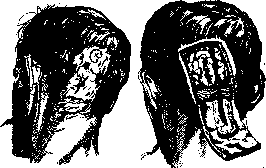
Gunshot wounds do not fundamentally differ from wounds of other origin. In peacetime, gunshot wounds are observed as injuries from revolver bullets, and less frequently from rifle bullets and shotgun pellets. Among peacetime wounds, one should note wounds from suicides, which are often inflicted with direct contact between the muzzle of the weapon (usually a revolver) and the skin of the affected area. Usually, suicides aim at the heart area, the head (right temple), or the mouth. The entrance wound in such cases is surrounded by a "burn belt" from the action of ignited gases, and in addition, by the embedding of small particles of unburned powder in the surrounding skin. The location of the wound and the character of the entrance wound provide important data from the standpoint of forensic medical examination and therefore must always be accurately marked by the surgeon providing first aid. Wounds from hunting shotguns (with pellets) depend on the distance at which the wound was inflicted: the closer, the more densely the pellets fly, and with tangential direction to the body surface, they can tear out large areas of tissue, thereby creating a defect. At a greater distance, the wound consists of more or less numerous openings with slightly depressed edges, sometimes covered with a scab. Shotgun wounds are usually blind, as the kinetic energy of each pellet is very small. Such wounds usually require conservative treatment, and only later, after healing, removal of palpable or functionally obstructing pellets. Bullet wounds (from rifles and machine guns) can be blind, through-and-through, or tangential. Through-and-through wounds have entrance and exit openings: the entrance is usually a small, round, slightly bleeding hole, plugged with a blood clot and tissue fragments, with edges slightly bruised for a short distance; the exit is always larger than the entrance, with its edges everted outward (Fig. 4). If there is no disruptive effect of the bullet, which depends on the distance and structure of the affected tissues (see "Field Surgery"), both openings are connected by a narrow wound channel. If the hydrodynamic effect of the bullet comes into play, the damage to tissues can be very great, and the bullet channel expands many times; the exit opening can reach the size of a palm, the character of tissue damage is lacerated-contused, bones encountered on the way are shattered into small fragments. In such cases, the wound at its entrance end differs sharply in character of damage from what is observed at the exit opening. Wounds from explosive and bursting bullets have exactly the same character, but already regardless of distance and
a
b
Figure 4. Wounding from a small grenade fragment: a-external view; b-view of bone and brain, x-entry wound. (After Cuenen.) of tissues: a narrow canal at first quickly turns into a cavity filled with blood and tissue fragments, bone fragments, as well as parts of the bullet. The exit wound in this case is very large, some tissues can be torn out by gases and the force of the deformed bullet. One should distinguish between the tearing action of a dum-dum bullet, i.e., with a cut-off jacket, and an explosive one, i.e., filled with an explosive substance. Diagnosis can sometimes be very difficult, only the presence of components of an explosive bullet on an X-ray or when extracted from the wound serves as indisputable proof.-A blind wound is called when a bullet gets stuck inside the human body; such a wound consists of an entry wound and a canal, at the bottom of which lies the bullet. Tangential wounds represent an open bullet channel, the bullet does not penetrate deeply into the tissues but produces a groove-like defect. In some cases the bullet passes very superficially under the skin, so that the bullet track is detected by dark, thinned skin. (For wounds from other military projectiles, organization of aid to the wounded and methods of surgery in war-see Field Surgery.) Poisoned wounds. A poisoned wound is such a damage when, simultaneously with the violation of the integrity of the coverings, a poisonous substance penetrates into the wound, which either has a general effect or causes local destruction of tissue, i.e., in other words, a combination of the effects of mechanical and toxic factors. In practice, poisoned wounds are the result of bites from poisonous animals, accidental wounding with instruments containing poisonous chemical compounds on them, e.g., infection with cadaveric poison when working on human or animal corpses, industrial injuries, for example when simultaneous damage and entry into the wound of acids and alkalis or when intentionally wounding an enemy with a weapon contaminated with a poisonous substance, as was observed earlier, and in individual cases can also be observed at present (wounds from poisoned arrows), and finally, as is practiced in modern wars, simultaneous wounding by projectiles and poisoning with combat toxic agents (OV).-For lesions caused by poisonous animals, see Snakes, Snake venom, Karakurt, Scorpions, Poisonous animals.-Lesions from so-called cadaveric poison in wounds should be considered differently. Products of tissue decomposition (cadaverine, belonging to the group of ptomaines) can cause infection; but on the other hand, when wounding while working on fresh corpses, infections with pus-forming microbes are possible, which can take the form of general infection. In such cases, abundant squeezing of blood from the wound, smearing its surroundings with tincture of iodine, as well as dressings with a hypertonic solution are beneficial. In the presence of general phenomena-usual symptomatic treatment.-In recent time, poisoned wounds resulting from pricks with an ink, so-called chemical pencil, containing certain poisonous aniline dyes have attracted attention. If during the prick, as is usually the case, the end of the pencil breaks off and remains in the tissues, a focus of necrosis forms around it, which increases in volume as the pencil melts. Such pieces of pencil must be excised together with the surrounding tissues colored blue.-As a poison for bullets, various vegetable poisons, such as curare, strychnine, strophanthus, etc., have been used in various countries and are still used in some places. Often, however, in the preparation of such poisons, mixtures of various substances are used, including snake venom and others. Both the picture of poisoning and the danger of the lesion depend on the composition used. Since usually highly poisonous substances are used in large quantities, the poisonings are very dangerous, often fatal. Help must be quick and energetic. It is most expedient to completely excise the poisoned wound and energetically suck out the poison; quick application of a tourniquet, if a limb is injured, slows down absorption and allows for more complete treatment of the wound. In the presence of general phenomena-symptomatic measures. Wounds poisoned by OV. The use in modern war of so-called combat toxic agents (see) creates conditions where mechanical damage can be combined with the effect of these substances. If an OV gets on the wound surface, conditions for a poisoned wound are created. The combination of wounding with general poisoning when the wounded person is in an atmosphere of OV that does not have a vesicant effect, although it aggravates the wound, cannot be classified as poisoned wounds. Of the OV, mustard gas (see) has the greatest practical significance, then lewisite (see) and some other arsines. Regarding the latter, mass poisonings of wounds with them have not been observed, since lewisite shells were made by Americans at the end of the world war and were not tested on the front. Thus, their effect can only be judged on the basis of individual accidental poisonings or experimental data. Already the studies of Drugov showed that a preliminary violation of the integrity of the epidermis causes a much stronger reaction when mustard gas acts on such a skin area than on intact skin. Subsequently, the process of healing a wound infected with mustard gas proceeds slowly, with the rejection of necrotic tissues. Further experiments (Prota-sievich) confirmed these data. It turned out that the presence of a poisoned fragment in the tissues causes general poisoning with mustard gas, which in large doses leads to a fatal outcome. Mustard gas that entered the wound with a projectile fragment cannot be detected by any reactions in the tissues themselves even after 1 hour. Apparently, under the influence of tissue fluids it decomposes and gives compounds of a different composition-a circumstance that must be taken into account in late treatment of wounds poisoned with mustard gas. Mechanical damage to tissues is successively greatly increased by the effect of mustard gas. The histological picture corresponds to what is observed on intact skin (see Mustard gas). In the depth of the wound, spreading areas of necrosis surrounded by infiltrated and edematous tissues attract attention. Wounds closed with stitches and containing poisoned fragments remain for several days as if in the process of healing, if the amount of mustard gas is not fatal, but then decay occurs, the wound turns into a deep ulcer, extensive necrotic areas appear and the wound itself becomes extremely susceptible to secondary infection. In the absence of secondary infection, there is extremely slow rejection of necrotic tissues followed by equally slow development of regenerative phenomena. Even after rejection of necrotic areas and with the development of usual granulations, the latter remain for a long time unusually sensitive to any irritations, as a result of which new foci of necrosis appear again. In the presence of secondary infection, the resistance of tissues and the whole organism is significantly reduced, and therefore any infection is especially dangerous.-The clinical picture of the wound in infection depends on the type and virulence of this infection, but under all circumstances, a tendency to tissue necrosis, delayed regeneration and high sensitivity to new harmful factors, for example traumatization, is characteristic. Despite the fact that mustard gas, according to S.V. Anichkov, is a protoplasmic poison, it does not have an antiseptic effect, at least on pus-forming microbes, especially in those concentrations that are possible *in a combat situation. From the data presented, it follows that a wound inflicted by poisoned mustard gas fragments is extremely dangerous in terms of general poisoning; such wound, due to the subsequent onset of chemical destruction, causes greater destruction than is determined at the first moment. Such a wound by no means should be closed, it is very prone to secondary infection* and even in favorable cases requires a long time to heal. When giving first aid for wounds poisoned by mustard gas, it should be borne in mind that this OV belongs to the category of persistent agents, therefore the wounded person's clothing and all objects that come into contact with the skin around the wound and with the wound surface become contaminated and capable of transferring the OV to another object. Such objects include balls of gauze, surgical instruments, gloves, etc. Surgical instruments require special attention: ordinary simple boiling does not free them from mustard gas, and when a wound is made with such a sterilized scalpel, a mustard gas ulcer results (Prota-sievich and Lukhanov). Therefore, the instrument must be subjected to de-mustardization before sterilization, preferably by wiping with gasoline. Both de-mustardization and treatment of the wound must be done with gloves. Primary treatment and treatment of wounds poisoned by mustard gas. Naturally, the first task in treating such a wound is to remove the mustard gas from it or neutralize it (destroy). None of the substances used on intact skin (see
Mustard gas (Poisoning) is not suitable for treating the wound surface; of all petroleum products, only vaseline, mainly liquid vaseline, can be considered. Substances that oxidize mustard gas and convert it into a non-toxic state, such as potassium permanganate and bleaching powder, are more suitable for treating W., but according to Magnitsky's data, both of these substances proved to be little effective on intact skin (see Mustard gas). From all the data about decontaminating intact skin and the peculiarities of mustard gas poisoning, it follows that the question of rational aid for poisoned wounds in a military situation primarily involves organizational difficulties, namely the necessity of immediate assistance to the victims. The matter is further complicated by the fact that the aid itself first requires removing the affected person from the poisoned zone (see Field surgery). A distinction must be made between mustard gas poisoning from injury through contaminated clothing in a contaminated zone, when mustard gas enters the W. after the injury, and injury from a mustard gas fragment, when mustard gas enters the W. at the moment it is inflicted. In the first case, all initial care for the victim should be directed at decontaminating the mustard gas, and after its completion or, if possible, in parallel with it, the W. is treated. It is understood that this is permissible for injuries that allow any delay in measures or the possibility of using temporary measures, for example, applying a tourniquet. During general decontamination, the area of the W. must be protected by all means from the solvent or washing water getting into it. If the nature of the injury is such that the wounded person can be given a general washing, then after wiping the affected surface with solvent and similarly thorough treatment of the area around the W., the latter can be sealed with rubber putty or another waterproof glue, then a general washing with warm water and soap can be performed, and immediately after that, treatment of the injury should follow. Any greasing of the skin around the W. with tincture of iodine, as an irritant, is not applicable in this case. The skin after gasoline can be treated with alcohol. In cases where there is a W. cavity, it can be washed with Dakin's solution. If a tourniquet was applied during decontamination, the area of skin traumatized by the tourniquet requires special thorough treatment, where, as is known, under the influence of trauma, poisoning phenomena manifest more intensely. Subsequently, the W. is treated according to general rules, but the circumstances of the injury and the conditions in which the W. was before medical aid must be taken into account, and on this basis, it must be decided to what extent the wound cavity can be considered as not contaminated with mustard gas. After treatment, any suspicious wound should be treated by open method. Mixed, i.e., obviously poisoned W., should be treated first, since absorption of mustard gas through damaged tissues occurs even faster than through the skin. Complete removal of mustard gas from a W. with a large area of damage, such as a W. from a mustard gas fragment, is hardly achievable even a few minutes after injury, nevertheless all measures for removal must be taken, the wound surface should be treated with wet tampons soaked in Dakin's solution or chloramine solution; the cavity can also be abundantly washed with these solutions. The most suitable is chloramine T in a 0.25-0.1% solution: its solutions are less irritating than Dakin's. Finally, liquid paraffin can be used as a solvent. Particularly dangerous is leaving in the W. pieces of clothing and metal fragments contaminated with mustard gas. All such contaminated bodies must be necessarily removed from the W. during its primary treatment. While searching for fragments in ordinary W. is not recommended, in mustard gas W. they must be found and extracted. This follows from the completely clear necessity of locating special portable X-ray apparatus as close as possible, without which in a number of cases searching for and removing fragments becomes impossible. The wound area should be examined on a screen to ensure that no fragments are left in the W.- If the most important task of primary treatment of purely mechanical damage is to prevent the development of infection, then a mustard gas W. needs this especially, since the resistance of tissues affected by mustard gas is even more reduced. Therefore, the final moment of treatment, after mechanical treatment is completed or in parallel with it, should be the application of antiseptic agents. Of these, derivatives of acridine are particularly suitable, for example, rivanol solution 1:1,000-500, and it is more expedient to leave rivanol-soaked gauze in the W. than to subject it to brief exposure to this agent (S. V. Anichkov). Besides rivanol, Eucupinum hydrochloricum 0.2-0.3% and Vuzin 0.1-0.15% can be used for the same purpose.- The question of anesthesia when necessary for persons poisoned with mustard gas cannot be considered resolved. In any case, inhalation anesthesia for persons who have been in a mustard gas atmosphere is contraindicated. Avertin or hedonal anesthesia might be most suitable, but data on this question are not yet available. Treatment of mustard gas W. can be carried out according to the principle of treating mustard gas lesions (according to Warthin and Weller), namely alternating irrigations of the W. with Dakin's solution and hypertonic (5-10%) NaCl solution, followed by washing with physiol. solution. The authors recommend carrying out the first two procedures for 2 hours each, the last one for an hour. At night, the W. is covered with a wet dressing with hypertonic NaCl solution. If there is simultaneous skin damage around the W. of limbs, irrigations are replaced with baths. If the general situation does not allow implementing this rather complex method, then according to Vedder, who obtained good results with superficial injuries from chloramine ointments, dressings with chloramine (1%) can be used in treating wounds. Finally, in treating wounds, the paraffin-wax Ambrin recommended by Sendford can also be applied, spraying it in heated form (65°) with a sprayer onto the wound surface. Of course, care for the W. in case of mustard gas poisoning includes removing all necrotic areas and observing proper drainage of wound exudate.- Parallel to treatment of the W., general measures are carried out (see Mustard gas, Poisoning). Beginning to develop granulations require the most careful handling. As necrotic areas are rejected and infection weakens, hypertonic solution can be used mainly with dressings. Delayed epithelization is stimulated by the use of Scarlet red (4-8% ointment), Azodermin (2%), and Pellidol. If the granulations are healthy and the W. cavity is filled, a transplant according to Thiersch and Davis can be performed. Regarding aid for wounds poisoned with lewisite (abbrev.), one can only speak on the basis of the properties of this chemical agent. When it enters the tissues internally through injury, the most rational approach is apparently complete excision, since leaving even somewhat significant amounts of this chemical agent in the W. threatens the death of the wounded person from general poisoning. The destruction of tissues with 5% caustic soda proposed by Vedder hardly has an advantage over excision. Excision of intact skin 12-24 hours after poisoning, however, can prevent a fatal outcome. It should be thought that in case of injury these time limits should be shortened. For small superficial injuries and simultaneous exposure to lewisite, a paste with iron hydrate (glycerin 1 part, iron hydrate 6 parts) should give a favorable result. After excision, the W. should be left open; dressings with hypertonic NaCl solution are preferable to create drainage into the dressing. All newly forming foci of necrosis are removed, thorough asepsis and the use of antiseptic agents, as with mustard gas, are necessary to prevent secondary infection, to which lewisite W. are very prone due to the weakness of regenerative processes. Only after the danger of general poisoning has passed and the W. is completely cleansed and healthy granulations have developed, attempts to reduce the wound cavity and plastic measures are indicated. It is understood that in case of limb damage, indications for amputation are more extensive than in simple mechanical injuries.- Of other arsines, ethyldichloroarsine ('Dic') and diphenylchloroarsine (D.A. according to American nomenclature) deserve mention. The combat use of either has the aim of causing general poisoning not through W., but mainly through the respiratory tract, but on the skin both chemical agents give a vesicant effect, and when absorbed through the wound surface, also a general effect. With the first of them, this is possible only when very large amounts are absorbed, so it does not pose great danger. Dressings with hypertonic solution are indicated. The same treatment is possible with D.A. with simultaneous injury. The latter, when absorbed, causes damage to the nervous system, expressed as hyperesthesias, anesthesias, and paresthesias. It apparently also does not present particular danger to life when absorbed through W.
In view of the fact that (according to Walton and Eldridge) chlorine has a beneficial effect on the respiratory tract (in battle both chemical agents are used as 'smokes'), in cases of simultaneous injuries, Dakin's solution or chloramine solution can be tried. Complications of wounds. It has already been stated above that the purely mechanical factor affecting tissues, when combined with thermal or chemical factors, causes more or less significant deviations in the course of wound healing from the ordinary, described above. Since no process, including the process of wound healing, can be considered a purely local process, it is clear that more or less clinically determinable general deviations in the body are also reflected in the process of wound healing. These influences often require special measures to eliminate certain factors that are particularly harmful to wound healing, or in extreme cases, their consideration when it is necessary to surgically inflict a wound or when it is accidentally formed. A very important factor affecting wound healing is age: childhood and especially infancy are characterized by rapid and energetic course of regenerative processes, with restoration of connective tissue elements and epithelialization occurring equally well. The third period of healing also passes more energetically and faster, for example, union of a fracture with callus formation, regeneration of nerve fibers, etc. However, it cannot be said that childhood is characterized by an easy tolerance of injuries in general; in particular, wound bleeding presents a greater danger in children than in adults. On the contrary, in old age, all three periods of wound healing pass sluggishly and slowly. Cases may be observed where wound healing is so sluggish that union does not occur and the wound opens after the sutures are removed. Hence the clinical conclusion: the time for removing sutures in children can be accelerated, while in the elderly it can be prolonged, and when in doubt, it is useful not to remove all sutures at once, but initially through one. Avitaminosis and scurvy disease have a clearly delaying effect on the course of regenerative processes. Sokolov, who studied the causes of dehiscence of postoperative laparotomy wounds, came to the conclusion that avitaminosis plays a very important role in this. The influence of diet on wound healing became a subject of study after it was established that inflammatory processes occur in the presence of local acidosis (Schade, Neukirch, Halpert), and further even in its weak manifestations, as is the case in healing by first intention (Girgov). At the same time, the idea arose to influence the pH of tissues by changing the diet; with the aim of enhancing acidosis, Sauerbruch, Hermannsdorfer proposed an acid diet; Gatsa, on the contrary, considering acidosis a deviation from the norm, intended to weaken it by prescribing an alkaline diet. These studies first established that it is possible to influence the pH of the tissue in the wound area by changing the diet with the predominant prescription of acids or alkalis, and further that the best results apparently are given by the acid diet. Girgov believes that diet therapy for wounds should be related to the stage in which the wound is located, and also depends on whether this process occurs with or without infection. However, this question is still in the study stage. - Delay in wound healing was noted by Grekov during years of famine under the influence of starvation; at the same time, a general decrease in the body's resistance of the wounded person to wound complications, particularly to infection, was also observed, since the latter was observed more frequently. Regardless of the dangers of bleeding in hemophiliacs, a delay in the healing process itself is also observed. Hesse and Gregory, observing a sharp decrease in regenerative abilities after severe acute infections (typhus, relapsing fever, paratyphus N), even warn against surgical interventions in such subjects, except of course in emergencies. Hesse observed a 12-week formation of callus at the site of a fracture, and Trekov even observed the resorption of callus. Similarly, wound healing is sharply delayed in chronic debilitating diseases,
tuberculosis, malignant tumors. The healing process of Wounds in syphilis is quite distinctive: on one hand, the formation of granulations seems to occur excessively, on the other hand, these granulations bleed easily, i.e., they turn out to be unusually vulnerable, their evolution proceeds sluggishly and slowly^ Wounds often acquire the appearance of a syphilitic ulcer and heal only after specific treatment. Delayed healing of Wounds is also observed in chronic poisonings, which depend both on the intake of harmful substances from outside (for example alcoholism) and on those formed in the body (cachexia in cancer and sarcoma, Bright's disease and others). Diabetes sharply reduces the resistance of tissues to infection, and both operations and accidental injuries in diabetics require the mandatory use of insulin. The menstrual period in women, according to recent studies by Popova, although not clinically noticeable in the course of healing, still favors the development of complications and is a contraindication for performing operations. Recently, the question of the influence of the wounded person's profession and working conditions on the course of Wounds and their complications has been studied.--In all cases where it is possible to influence the general cause slowing the healing of Wounds, this must be done necessarily, such as treatment of diabetes, syphilis, elimination of avitaminosis, improvement of general nutrition, prescription of an acid diet and others. Of the methods of local action on Wounds, only the use of radiant energy in the form of a quartz lamp, weak doses of X-rays, sunlight can be mentioned. However, these influences are still little studied and require development, especially in relation to forms of application and dosage. On granulating wounds, the course of healing is controlled by the appearance of granulations; with a favorable outcome of the measures taken, the first sign is the restoration of the normal appearance of granulations. It was mentioned above that the blood supply to the area of injury and the structure of the damaged tissue clearly affect healing.-Great importance for healing, especially for functional results after the end of this process, is the mutual arrangement of the wound edges in Wounds involving several different tissues, for example, skin, fascia, muscles, bone and others. Only the careful fitting of similar tissues to each other gives more or less complete healing. Thus, the interposition of muscle between the edges of a bone Wound (fragments)^ easily leads to the formation of false joints, the turning of the skin surface into the wound cavity clearly hinders healing and others.-Harmful role in the healing process is also played by foreign bodies; in their presence the healing process is modified, the formation of connective tissue capsule around the foreign body occurs, more extensive development of scar tissue, which weakens the functional results, not to mention the possibility of complications with infection, poisoning (see Combat Poisoning Substances) and others. The same delaying role in the healing of Wounds is played by accumulations of blood in the wound cavity (hematomas). The edges of the Wound are pushed apart and the remaining cavity, as the blood is absorbed, is filled with granulation tissue, which later gives a more extensive and massive scar than is the case in the normal course of healing. In some cases, hematomas, without being absorbed, after capsule formation, can pass into an encapsulated form—a blood cyst. Therefore, the formation of a hematoma in a Wound requires careful observation; the spilled blood, if absorption is delayed, must be removed by punctures and suction with a syringe or Potteau apparatus, followed by the application of a pressure bandage to bring the wound edges together as much as possible.-The presence of trophic disorders in lesions of the central and peripheral nervous system can reduce the regenerative properties of tissues to such an extent that the healing process not only slows down but even stops (for example in syringomyelia). With lesions of peripheral nerves, a Wound can turn into a trophic ulcer. An exception seems to be the treatment of Wounds in leprosy: in it, according to observations by Biehler, not only is there no slowing of the healing of Wounds, but the healing process seems to accelerate. Infected Wound. However, infection is of the greatest importance as a complication in the course of Wounds; this complication is the most frequent and most strongly affects the healing of Wounds. The final results of operative Wounds only corresponded to the intentions of surgeons when, thanks to antiseptics and asepsis, it became possible to prevent infection. The fatal outcome in injuries is very often associated with infection, as a complicating factor of the injury. A distinction is made between pyogenic, putrefactive and anaerobic! and specific acute and chronic infections. Pyogenic infection is caused by the so-called pus microbes, to: which belong staphylococci, streptococci, pneumococci, Pseudomonas aeruginosa, the group of intestinal bacilli, the causative agents of influenza, meningitis and others. All these microorganisms, entering a Wound, cause purulent inflammation, which, superimposed on the process of healing of the Wound, sharply disrupts it; suppuration first of all makes primary healing of the Wound impossible, since the tissue of the wound edges is melted due to proteolytic enzymatic substances and is rejected with pus, forming a wound cavity, which is then filled with granulations only when the inflammation undergoes reverse development. Thus, each infected Wound with purulent inflammation heals by secondary intention and only after the infection is localized and the inflammatory process undergoes reverse development. In the best case, this greatly delays the healing of the Wound, in the worst case, the infection spreads from the Wound by the usual routes (lymphatic and blood vessels), passes into a general form of infection and leads to fatal poisoning by toxins. The same consequences occur with infection of Wounds by putrefactive and anaerobic microorganisms, of which Proteus vulgaris, B. perfringens, B. oedematis maligni (V. septique Pasteur's) and others have the greatest practical significance, often together with streptococci and other pus microbes. Although each of the various pus microbes or anaerobic infection manifests its own clinical features, still on the basis of the picture of complications of the Wound, they speak of a group form of infection, purulent or putrefactive, with the latter often leading to the formation of gas products, detectable not only by a special smell but also by the presence of gas in the affected tissues.-The picture of complications of the Wound is quite different in the presence of specific infection; here each of the microorganisms gives either a special, unique to it picture of wound complication (as for example diphtheria of the Wound, wound scarlet fever), or the injury is complicated by the general specific to this type of microbe picture (such as tetanus, rabies, rat-bite disease), while the Wound itself can sometimes even heal. Chronic infections, such as tuberculosis, syphilis and others, usually turn the wound into an ulcer specific to this type of infection.
A distinction is made between primary infection of wounds, when it penetrates the tissues simultaneously with the injury, and secondary infection, when a wound, initially free from infection, becomes contaminated some time after the injury. The latter type of wound infection has great practical significance in cases of delayed wound treatment, with inadequate or improper first aid, etc. As for the prevention of wound infection, it has been stated above, since any initial treatment of a wound includes measures to prevent infection. To what has been said, it only remains to add that the general conditions and surrounding circumstances at the time of injury are of great importance in terms of the danger of infection. Its most severe forms occur in the presence of a sharp violation of general hygienic conditions. Where possible, attention should be paid to this (for example, working conditions and the general environment in industrial enterprises, the general conditions in which soldiers are on the battlefield, etc.). Even during the World War, it was clearly noted that baths, showers, and laundries should be regarded not only as institutions of general hygienic importance but also as preventive institutions in relation to the course of wounds. The signs of wound infection represent the clinical signs of inflammationg that are added to the clean clinical picture of the injury. It is noteworthy that the painful sensations in the wound, with the passage of time, not only do not diminish but intensify; upon examination, the edges of the wound are found to be swollen, the skin is tense and shiny, and upon palpation it is hotter than in more distant areas; upon palpation, the pain increases even more. Along with local phenomena, generalv signs are also determined, which is expressed first of all in an increase in temperaturer and acceleration of the pulse. In a closed wound, these data are already sufficient to conclude the presence of wound infection. The edges of the wound and the area of the wound, upon careful examination, may be found somewhat hardened (cellular inflammatory infiltrate), edematous, when along with the cellular infiltrate there is impregnation of the tissues with a more liquid inflammatory exudate; finally, sometimes by fluctuation it is possible to establish the accumulation of liquid exudate, i.e., pus. Only the complete absence of liquid exudate forces one to adopt a wait-and-see attitude and, by the local application of heat, especially in dry form (hot-water bottles), to hope to achieve the reverse development of the infiltrate, provided the affected area is completely at rest. As soon as the presence of a purulent accumulation is detected, the wound must be immediately opened. In doubtful cases, the removal of one or two sutures often quickly resolves the matter. The purulent exudate must be released to the outside, and the wound left open. In cases with not sharply expressed general phenomena, with low-virulence infection, which is indicated by the absence of signs of the spread of infection beyond the wound (lymphangitis, lymphadenitis), the mere opening of the wound may already be sufficient, especially when the infection develops, for example, in hematomas; the wound should be loosely covered with gauze, possibly iodoform or soaked in a solution of rivanol; tamponade or drainage of the wound may not be required; then, as the purulent exudate continues to be discharged, the wound gradually begins to fill with granulations and epithelialize from the edges. - In open wounds, the signs of inflammation of the wound edges become noticeable on the basis of the same signs: redness, swelling, and painful sensations. The discharge from such a wound sharply increases and takes on a purulent character; the properties of the pus depend on the pyogenic microbe that caused the infection in this case. In the treatment of such wounds, antiseptic dressings and dressings with hypertonic solution are used; the injured area should be ensured rest (on the limbs, splint dressings are beneficial), an elevated position, and local application of heat (hyperemia). In the presence of a highly virulent wound infection, along with the signs of inflammation of the wound, there are symptoms of the spread of infection beyond the wound. The first task in the treatment of such a wound and its treatment is to create conditions under which the infection would be localized only in the area of the wound. Signs of spreading infection are: bright redness of the wound edges, their shiny, tense appearance, the presence of red stripes along the lymphatic vessels (lymphangitis), enlargement and tenderness of the regional glands, the presence of purulent metastases, a significant disturbance in the general condition of the wounded person, such as: increased temperature, disturbance of cardiac activity, lesions of the central nervous system, etc. If such a wound has already been treated, it should be carefully re-examined from the point of view of the adequacy of this treatment and the conditions that determine the possibility of stagnation of its discharge in the wound. All defects in the initial treatment are corrected. Only in cases where the treatment of the wound and its location fully guarantee the outflow of discharge, can one be limited to loosely applying gauze to the wound surface; otherwise, the introduction of loosely folded strips of gauze (tampons), often with an antiseptic (iodoform, rivanol, etc.) or drainage tubes into the wound cavity is applied. - Appropriate and adequate treatment of an infected wound leads to the cessation of the spread of infection. Clinically, this is expressed in the reverse development of lymphangitis and lymphadenitis, in the weakening of general phenomena, first of all fever, a decrease in leukocytosis, etc. Dressing of infected wounds. The treatment of a wounded person with an infected wound includes dressings. The question of the frequency of dressings once caused sharp disagreements among surgeons. At present, it is recognized that the dressing of a wound depends on the abundance of discharge and on the possibility of its stagnation in the wound cavity. Advocates of rare dressings (Zege von Manteyfel) pointed to the often observed increase in temperature on the day of dressing; on the other hand, it was repeatedly noted, starting with Larrey, that heavily suppurating wounds, with very poor care, with the development of larvae of flies ('worms') in the dressings, nevertheless healed excellently. At present, it can be considered established that the increase in temperature during dressings is connected with the damage to the granulation cover at that time and the absorption of decomposition products from the wound. Therefore, special attention should be paid to the technique of dressings: the dressing consists in the careful and gentle removal of the old dressing and the extraction of tampons or gauze from the wound cavity. The area around the wound is wiped with gasoline and alcohol, the wound cavity is examined with careful spreading by blunt hooks, accumulations of pus are removed with moist gauze swabs; if there are crusts or dead areas, their removal is facilitated by the use of hydrogen peroxide. When removing with forceps, dead parts are removed without damaging the granulations or underlying tissues; a dead area can be separated with scissors, but within the limits of dead tissue. After the toilet of the wound is completed, the latter, depending on the indications, is either loosely filled with a tampon or only the latter are introduced as spacers to prevent the edges of the wound from sticking together before the cavity is filled. Too prolonged tamponade leads to the transformation of the wound into a narrow and long cavity and prevents the development of granulations. In conclusion, the wound is covered with a dressing according to general rules. When caring for wounds in warm weather, larvae of flies easily develop in the dressings and wounds, which often even became the cause of legal prosecution of doctors and nursing staff. These larvae require about a day for their development from eggs (Pavlovsky), even less on hot days, and therefore can be observed even with impeccable care and daily dressings. Ver (Baei%), having drawn attention to the already mentioned observation that larvae often combine with an excellent course of the wound, experimented with their artificial breeding in suppurating wounds (mainly in osteomyelitis) and came to the conclusion that fly larvae, without damaging the granulations and feeding only on dead parts, turn out to be extremely useful. However, a mandatory condition is their sterile breeding from eggs. In any case, based on these data, a conclusion can be drawn about the complete harmlessness of fly larvae in wounds themselves, but the same author observed that when using non-sterile larvae, infection with tetanus is possible. In the treatment of wounded persons with infected wounds, attention should be paid to the general condition of the wounded person and the improvement of his nutrition and cardiac activity. The nutrition of such wounded persons, even more than those with non-infected wounds, was associated with the enhanced supply of the body with acids (Sauerbruch, Hermansdorfer) or alkalis (Gatz), based on the increase in acidity in infected foci (Schade and his colleagues). The question cannot yet be considered resolved; apparently, as indicated above, an acid diet deserves preference. - In addition to regular dressings, it is very useful to induce hyperemia of the inflamed tissues. For this purpose, hot baths during dressings and hot-water bottles over the dressing are used with suitable localization of the wound. In addition to the use of a number of antiseptic agents in the treatment of infected wounds.
(iodoform, the so-called substitutes for it, rivanol and other acridine derivatives, Peruvian balm, etc.) deserves special mention is Wright's method of treating W. with a hypertonic solution, already mentioned earlier (5-10% NaCl). The application of tampons abundantly moistened with this solution is particularly indicated for dry W. covered with coatings. The Carrel-Dakin method of treating infected W. was widely used on the western front during the world war. In peacetime conditions, this method finds less application, since due to the primary correct treatment of the wound, such severe infections as occurred on the front are not so frequent. The technique of the method consists in establishing continuous (or periodic) irrigation of the wound surface with a special solution, the active principle of which is chlorine. The preparation of the solution is quite complex and requires chemically impeccable preparations. There are two main solutions: 1) 140.0 Natr. carbon, i sicci is dissolved in 5 l of water; 2) 200.0 chlorine lime also in 5 l of water; both solutions are mixed before use and neutralized with 25.0 to 40.0 boric acid. There are many variations in the preparation of Dakin's solution, but recently there are ready-made preparations used for obtaining solutions, these include eusol, magnacid and especially chloramine (see Antisepsis and asepsis). After treating the W., its circumference is smeared with vaseline to eliminate the action of the liquid on the skin. Near the sick person on a stand is placed a vessel with liquid, from which a main tube leads to the W.; at the end this tube is connected to a glass tube having several lateral branches. A rubber tube is placed on each of the branches. The resulting system of rubber tubes with side holes is placed in the cavity of the W. so that the washing liquid irrigates the entire wound surface as much as possible, moistening the gauze covering the W. The flow of liquid is set by a special regulator to 20 drops per minute; of course, the W. requires special observation. Instead of continuous irrigation, periodic irrigation can be used, performed by the duty personnel (nurse), by injecting liquid from a syringe into the main tube. According to Carrel's idea, irrigation should be continued until complete cleansing of the W. and disappearance of pathogenic microbes (bacterioscopic control). When this is achieved, a secondary suture is indicated. Very close to the "Carrel method" is the Sapiezko method, which used for "irrigation" of the wound (injection 3-4 times a day) a 1/2%-1% solution of iodine in 30° alcohol with 1% potassium iodide. In the open treatment of W. (Rose, Braun, Grunert, Voynich-Senozhensky), the wound surface is subjected to the drying influence of free access to air and the influence of "light rays." To protect the wound from flies, dust, and accidental contact with bedding and other surrounding objects, the W. is protected by a metal mesh, secured in the area of injury. For the same purpose, various caps, canopies, etc. were used. Practice has shown that open treatment can be beneficially carried out with low virulence infection or in the healing stage after complete localization of the infection. The treatment of W. infected with pus-forming microbes by specific vaccines and serums has not received particular distribution. Preventive vaccinations are possible in anticipated operations in the oral cavity (Raug), on the rectum (Oppel, Ilyin). No particularly encouraging results have been obtained from them. Therapeutic vaccination has attracted attention for a long time, mono- and polyvalent vaccines, as well as autovaccines (Bezredka, Metchnikoff, Belonovsky, Wright, etc.) were used. Apparently the largest number of successful results were obtained with the latter. The question for practical use needs further development. Somewhat apart stands the treatment of infected W. with filtrates of microbial cultures according to Bezredka (see "Antivirus"). The method has been tested quite a lot and in our Union (Burdenko, Ebert and Sazhina, Sokolov, Opokin, Rosenbaum, Petrov, etc.). Antivirus can be applied in the form of injections, placing on the W. gauze moistened with antivirus, washings and finally ointments. A number of authors noted favorable results, but others did not see particular benefit from their use. The use of enzymatic treatment of infected W. is in the stage of development, mainly of hydrochloric pepsin. The latter dissolves necrotic areas, cleanses the W. and acts antiseptically on microorganisms (Schonbauer). Putrefactive and anaerobic infection of W. represents the most severe form of contamination. In peacetime conditions it is rare, and before the start of the world war it seemed that it was only a feature of the pre-aseptic era. However, the abundance of cases of this infection during the world war and then the civil war and individual observations in the post-war period showed that the form of contamination of W. under consideration has by no means been eliminated. The microbes causing it have been mentioned above. The conditions under which it arises are abundant contamination of the W. with earth and manure, fragments of heavily soiled linen and clothing, a sharply anti-hygienic environment, delayed and incomplete treatment of the W., crushing and crushing of tissues during injury, especially muscles. In military conditions, W. caused by fragments of explosive shells were particularly prone to anaerobic infection. All this provides a basis for the development of preventive measures, especially accessible in peacetime. These measures also include the use of a specific serum in the primary treatment of the wounded. The corresponding serum is now produced both in the USSR and its introduction in appropriate injuries should be mandatory, like the introduction of anti-tetanus serum, both in peacetime and in wartime. Likewise, timely and correct treatment of a wound with a large area of damage is also a preventive act in relation to anaerobic infection. A feature of the type of infection under consideration is the relatively weak development of inflammatory phenomena at the site of infection or, more correctly, their rapid suppression; the very rapid abundant development of toxic substances leads to necrosis of tissues, which is immediately followed by decomposition with the formation of gases. The vascular system and blood are affected first, the necrotic parts undergo liquefaction and disintegration; very often the W. quickly begins to give off an offensive odor of decomposition. Due to the abundance of toxic substances in the W., consisting of products of both infection and tissue decomposition, their significant absorption occurs, and the infection always runs with the presence of general phenomena of poisoning. Putrefactive processes can also occur locally or give general forms of infection with and without metastases, but general forms here join very easily and often, since the entire apparatus of the body that holds back the infection is itself affected, and the extraordinary virulence of the microbes creates conditions for the infection to capture the entire body. Clinically, putrefactive and anaerobic forms of infection give great diversity. Even in pre-aseptic times, attempts were made to isolate several clinical forms, similar attempts continue to this day, however, despite the presence of more or less typical cases where the lesion is caused by one (or almost one) specific microbe, in practice mixed lesions are very frequent, and the clinical picture turns out to be confused, only the general characteristic features remain - necrosis in the W., development of putrefactive gases - and unusually rapid poisoning of the body. Putrefactive infection is expressed by pain in the W. and the simultaneous appearance of general phenomena, chills and elevation of temperature. Initially the wound is dry and gives off a bad odor, its edges darken, then acquire a greenish-brown color; dirty, foul-smelling discharge is secreted, which, mixing with gas bubbles, foams. Around the wound, swelling and soreness develop. The main site of development of putrefactive infection is the cellular tissue, it necrotizes and melts with remarkable speed, pieces of it are easily removed with forceps during dressings in a semi-melted state; huge wound pockets are formed under the skin and between muscles, along vascular-nervous bundles. Later, due to the death of a large number of vessels of the cellular tissue, the nutrition of adjacent areas is disrupted, which also undergo necrosis, muscles and fasciae die, the process extends to the bones. The nutrition of the skin is significantly disturbed due to the damage to the vessels (also lymphatic): it is pale and insensitive, in places areas of necrosis are found, the epidermis peels off with the formation of large blisters filled with sero-sanguineous fluid. The transition of inflammation to the large veins with their thrombophlebitis involves a sharp swelling of the entire corresponding area, e.g. of the whole limb, which can increase more than twice in its thickness.
In the tissues, along with edema fluid, gas bubbles also accumulate, which can be detected by palpation, producing a special sensation under the fingers—crepitus. Sometimes the amount of gas is so significant that it can be detected by percussion. Severe cases of putrefactive infection can lead to death within 24 hours, but sometimes they also take on a metastasizing character. For local measures on Wounds, the initial treatment of the Wound is important. If putrefactive infection has developed in the Wound despite proper treatment of the Wound, and it acquires a threatening rate of development, then in suitable cases, i.e., on the extremities, amputation is indicated. If, however, the treatment of the Wound was insufficient and the course of the infection allows for waiting, then wide opening of the Wound and drainage of it may be possible. The use of antiseptic agents is fully indicated, although due to absorption from the Wound cavity with some of them, for example, iodoform, caution should be exercised. The method of continuous irrigation has been tested with favorable results. Along with local measures, general measures of the type described in general purulent infection are also indicated. One of the most terrifying clinical forms of anaerobic infection is the so-called gas gangrene (see Gas phlegmon, Edema) and malignant edema (see). Finally, forms of fusospirochetal infection deserve special attention, which also proceed with a strong tendency to necrosis of the affected tissues. These forms are caused by mixed infection with pyogenic and putrefactive microbes with various spirilla and spirochetes, among which B. fusiformis VIncent's has important significance. This type of infection is associated with injuries to the oral cavity, upper respiratory tract, and esophagus and is caused by the microbes present in these areas. A characteristic feature of this form of infection is spreading from the focus to the center wet necrosis, involving and destroying all tissues with the formation of a marked black-green, foul-smelling mass. Such an infection, developing in an exogenously originated Wound, gives a complication described in the pre-antiseptic era under the name of hospital gangrene (see Gangrene, hospital gangrene). Wound scarlet fever. Although rare cases of scarlet fever have been described, the starting point of the disease was a Wound. The disease is expressed in the typical exanthema of scarlet fever with subsequent peeling. The presence of angina and nephritis in this form of scarlet fever is not mandatory. Recognition of this form of scarlet fever, which can then become a focus of an epidemic, is very difficult, since scarlatina-like rashes are not always observed in injuries. Such rashes also appear in general purulent infection. One should remember the possibility of wound scarlet fever—suspected cases should be isolated and after careful observation, the question of the further direction of the wounded should be decided.-Wound diphtheria can occur either with simultaneous involvement of the pharynx with typical localization, or without typical localization due to the transfer of the diphtheria bacillus from a carrier, who however remains healthy himself. The carrier can be the wounded person himself or someone from the surroundings (e.g., medical personnel). Clinically, the disease is expressed by the appearance on the Wound of greenish-gray coatings and fibrinous membranes, tightly seated on the underlying tissues and not removable without bleeding. The Wound gives a sero-hemorrhagic exudate, the granulations acquire an ashen color. The area around the wound appears markedly reddened and indurated due to the presence of infiltrate in it. However, an accurate diagnosis can only be established on the basis of bacteriological research. In the presence of the above phenomena, in addition to general measures for treating the Wound, specific treatment with serum is also indicated, which however does not give such a sharp effect as in the early treatment of diphtheria of typical localization. The introduction of tampons soaked in antitoxic serum into wounds is recommended. Any operative manipulations on the Wound are contraindicated. It was stated above that every Wound heals with a scar, i.e., inadequate tissue. This inadequacy in a number of cases affects the general condition of the organism so little that the final outcome of the injury can be considered as a complete restoration of function and complete restoration of the working capacity of the given wounded person. However, the location and size of scars can result in more or less significant disturbance of function (see Scar). In such cases, one cannot speak of complete restoration of health, and the outcome of the injury is temporary or permanent loss of working capacity. It will be temporary in those cases where the resulting disturbances can be eliminated by further treatment, and permanent in those cases where this for any reason does not seem possible, even for example due to the refusal of the injured person to undergo an operation.-The disturbance of working capacity will be permanent also where the injury resulted, for example, in the loss of a part or the entire extremity. In such cases, the matter concerns disability. - The determination of loss of working capacity or its disturbance is in close connection with the profession of the injured person, so for example a scar on the left index finger of a violinist can lead to complete loss of working capacity and affect it very little in many other professions. The immediate outcomes of injuries often provide an opportunity to improve the final results of treatment for further operative interventions, belonging to the category of plastic operations (see). From a forensic medical point of view, injuries are divided into absolutely fatal, severe, less severe, and mild. The first category includes such injuries that directly lead to the death of the wounded person, such as extensive crushing of the central nervous system, especially the medulla oblongata, complete rupture of the aorta, avulsion of the heart, etc. Severe injuries include those which in themselves can lead to a fatal outcome, especially without proper help; less severe should be considered those where, in the absence of complications, the injury itself does not threaten life, and finally mild are those which do not affect the working capacity of the wounded person.
Related articles
Cite this page
“Wounds.” Soviet Medical Encyclopedia. English translation of Bolshaya Meditsinskaya Entsiklopediya, 1st ed. (Moscow, 1928–1936), ed. N. A. Semashko. https://sovietmedicalencyclopedia.pages.dev/article/wounds/