Gangrene
Historical document, translated for reference. It reflects medical knowledge of the 1920s–30s and is not medical advice.
Summary
Gangrene is a type of tissue death characterized by discoloration of the affected area. The encyclopedia describes two main types: dry gangrene, which results in mummification, and wet gangrene, which involves putrefactive decomposition.
Encyclopedia article (1928–1936)
GANGRENE (from the Greek graino-I gnaw; old name-Anton's fire), such necrosis, in which the dead part of the body, coming into contact with the external world, takes on a dark-brown or black color, depending on the change in the blood pigment. Two types of gangrene are distinguished. Dry gangrene (gangraena sicca) is observed in cases where the necrotic tissues located on the surface of the body, due to the release of tissue moisture into the surrounding atmosphere, decrease in volume, dry out and become dense and hard in the process. The blood pigment present in the vessels diffuses into the tissues, decomposes and stains the dead tissues dark-brown. The noted slippage of the epidermal layer facilitates the evaporation of tissue fluid, while the drying of the surface itself prevents the penetration of putrefactive bacteria into the tissues. The dead areas thus altered resemble the tissues of an Egyptian mummy, which is why such outcomes of necrotic processes are also designated as mummification. At the border with the mummified tissue, demarcation inflammation develops in the healthy tissue, which can lead to spontaneous separation (amputation) of the necrotized parts. Wet, or putrefactive, G. is also tissue necrosis, but complicated by putrefactive decomposition due to the penetration of various kinds of putrefactive bacteria into the tissues. The latter mostly enter the tissues from the external world, which is why this type of G. is most often found in the lungs and on open parts of the body. In this G., the affected parts acquire a dirty-gray or dirty-green, almost black color due to the impregnation of the dead tissue with decomposed blood pigment. Subsequently, the dead tissues take the form of a soft, putrid mass, in which decomposition products are present in large quantities: crystals of leucine, tyrosine, phosphorus-ammonia-magnesia, crystals of fatty acids, as well as amorphous and crystalline blood pigment. The decomposition products are mostly poisonous and give off a foul odor. Very often, the putrefactive process is accompanied by the accumulation of gas bubbles in the tissues, which swell the gangrenous tissue and even the adjacent living tissue (gas G.). This type of gangrenous process is usually not inclined to be delimited, but spreads to the surrounding healthy tissue, since the resulting toxic decomposition products, besides their general severe effect, also act on the surrounding tissue, in which the same picture subsequently develops. Such spread of wet G., for example, on the limbs, when necrosis rises higher and higher, is called among the population 'Anton's fire.' (This name probably originates from the ancient belief that patients suffering from necrosis of the fingers from uterine ergot, the so-called 'cramp,' received healing from the relics of St. Anthony.) Regarding the view on the origin of wet gangrene, there are indications in the literature (Zinserling) that wet G. has an infectious-toxic origin and is caused by a specific pathogen-Spirochaeta dentium or Spirochaeta buccalis in symbiosis with Bac. buccalis; there are, however, views that spirochetosis in G. should be considered as a sequential phenomenon, and not as a primary factor. - Dry G. is usually observed in necrosis of the extremities, the tip of the nose in the elderly (senile gangrene) due to weakening of blood circulation in the areas most distant from the heart, which depends, on the one hand, on pathological changes in the vessels, and on the other-on weakening of heart activity and general nutritional decline. The necrosis thus arising subsequently leads to mummification, and the dead area, due to demarcation inflammation, may spontaneously separate*. Dry G. of the extremities is also observed in chronic poisoning with ergot due to narrowing (spasm) of blood vessels (angioneuroses, see). When putrefactive bacteria penetrate the dead tissue, dry gangrene can subsequently turn into wet, which significantly worsens the patient's condition. In addition to the above, it should be borne in mind that the term 'gangrenous' usually denotes inflammatory and necrotic processes in tissues and organs containing an element of necrosis with putrefactive decomposition, e.g., gangrenous angina, gangrenous stomatitis, gangrenous appendicitis, etc. The same putrefactive processes that are observed in serous cavities and are associated with putrefactive decomposition of inflammatory exudates (e.g., putrefactive pleuritis as a complication of G. of the lung) are designated by the term 'ichorous.' (G. of individual organs, see the respective organs.)
V. Tapaev. Hospital gangrene (syn.: sphacelus, nosocomial G., traumatic diphtheria, hospital filth-pourriture d'hupital, putrid, dirty or gangrenous ulcer, etc.), a disease of the wound, characterized by severe pain and progressive ulceration and necrosis of tissues covered with a fibrinous, often dirty-gray exudate resembling diphtheritic membranes. Hospital gangrene has been known since ancient times, more often during wars. It was known to Ambroise Pare (1562), well described by Puto (Roy1eanch, 1783), Delpech, and Pirogov. Hospital gangrene appeared and developed easily with large concentrations of wounded in unsuitable, cramped, cold, damp, poorly lit, and insufficiently ventilated premises. Under such conditions, hospital gangrene also developed in some poorly arranged and dirty hospitals where it was sometimes even an endemic disease (e.g., Hotel Dieu in Paris, Charite in Berlin, etc.). Weakening of the body by other diseases, especially epidemic ones (typhus, dysentery), scurvy, etc., trench life, hardships of war, and depressed mood contributed to the spread of hospital gangrene. It was sometimes a scourge for the wounded and hospitals, claiming many lives. Thus, Guthrie (1813) lost 512 (31.7%) out of 1,614 wounded to hospital gangrene; in French hospitals in the Crimea (1855) the mortality rate from it was 40-60%, in the Italian War (1859) in the hospital of Francis in Milan even 80%, but already in the Franco-Prussian War (1870-71) the mortality rate from it fell to 6-25%. With the study of the causes of wound infection and the spread of antiseptics, hospital gangrene disappeared, remaining unstudied bacteriologically. During the World War of 1914-1918, hospital gangrene was not observed in Germany, in Russia some surgeons saw individual cases of it. In peacetime, isolated cases of hospital gangrene are occasionally encountered in exhausted people, mainly on wounds of the scrotum, perineum, and around the anus. It can be assumed that the causative agent is the thin straight or slightly curved bacillus found by Vincent (1893), whose inoculation in dogs produced gangrenous ulcers, but Rona and Matzenauer found these bacilli in noma and gangrenous chancre, so their specificity is questionable. It should be noted that Loeffler's diphtheria bacilli were not found in hospital gangrene. The question of the causative agent thus remains open. The infectiousness of hospital gangrene has long been known. Puto (1783), who himself became infected in the Hotel Dieu in Lyon, spoke of this; Delpech in 1815 recommended fumigating clothing after work and frequently washing hands and instruments with HCl solution. Pirogov did not doubt the infectiousness of hospital gangrene, recommending isolation of patients and caution during dressings. He himself experienced hospital gangrene on a finger, having become infected during an operation on a gangrenous patient; the wound was barely healed after 5-6 weeks. Hospital gangrene was sometimes observed outside hospitals, in private houses, where it was apparently brought by surgeons. In the pre-antiseptic era, it could be transmitted through dirty hands, non-sterile instruments, sponges that had been in use, bedding, etc. Infection most easily occurred in large wounds, especially gunshot wounds, contused wounds with crushed, insufficiently viable tissues; granulating wounds with intact granulations were more resistant to infection. In addition to open wounds, lesions of hospital gangrene were observed in recently healed wounds that reopened with hospital gangrene. The incubation period, according to observations, ranged from 8 hours to 8 days. Pirogov noted the diversity in the manifestations of hospital gangrene, but usually two main forms are distinguished-ulcerative and pulpous. Both begin with the appearance of severe pain in the wound, infiltration and redness (up to a dark purple color) of the edges. A fresh wound becomes dry; if there are granulations, they turn gray, become edematous, ecchymoses appear, and a dirty-gray, poorly removable fibrinous exudate forms on the surface. Among the general phenomena, chills and elevated temperature are noted. In the pulpous form, the deposit is pierced by hemorrhages, quickly turning into a sticky, thick, brownish or black, very foul-smelling mass, under which necrosis proceeds both in depth and width, undermining the edges of the wound, first penetrating into the connective tissue spaces, exposing and destroying muscles, fasciae, periosteum; vessels, and especially nerves, persist longer than can be explained by the severe pain. Sometimes in the pulpous form of hospital gangrene, small hemorrhages lift the covering deposit in the form of blisters filled with foul-smelling decomposing blood, and deeper there is an ulcer that is again covered with a deposit, etc. The less severe ulcerative form of hospital gangrene begins with the appearance of small ulcers on the surface of the wound with a dirty-gray or dark purple deposit. As they increase, the ulcers merge into a large foul-smelling ulcer with serous discharge, semi-liquid decay, and a eroded bottom. The edges of the wound are as if cut out in a semicircle and infiltrated. The ulcer increases in depth and width, the destruction of tissues is the same as in the pulpous form, but somewhat slower and with a better general condition. The disease is accompanied by more or less pronounced general phenomena (septicopyemia, erysipelas, lymphangitis, often diarrhea). The rate of development varies; sometimes, especially during an epidemic of hospital gangrene, necrosis of all tissues down to the bone occurs within 48 hours. Destruction of the abdominal and chest walls by hospital gangrene has been observed, with exposure of the internal organs. In other cases, the course is slow, the body sometimes manages to cope with the infection, demarcation occurs, and healing by granulations takes place. The disease tends to recur, apparently due to reinfection.
The prognosis, as with other infections, is apparently related to the strength of the infection, the nature and location of the wound, and the general condition of the patient. The tendency to spontaneous recovery is small. Patients usually died either from sepsis or from hemorrhages from necrotized vessels, the thrombosis of which is not often observed (Pirogov). Prevention is the usual sanitary-hygienic and disinfection measures. Apparently, the infection is less persistent than others that persist despite antiseptics and asepsis. In the treatment of hospital gangrene, cauterization of the wound with red-hot iron was used (the Pakenel apparatus gives insufficient, superficial cauterization), moistening with a solution of zinc chloride or lead nitrate until a dense hard scab forms, half-chloride of iron, Liq. alum. acetici, tincture of iodine, etc. Later, iodoform, hot water (50-55°C) on the wound (Reclus), etc., were successfully used. On the limbs, in the most extreme cases, with extensive tissue destruction, amputations were performed within healthy tissues, carefully protecting the new wound from infection from the old one.
H. Terebinsky. Gangraena cutis multiplex (multiple necrosis of the skin), a collective name for several separate dermatoses that proceed in a characteristic manner and arise from various etiological factors, but are characterized mainly by multiple focal necrosis of the skin. Gangraena cutis multiplex adultorum—the name given by Sutton for a skin disease that sometimes results from intense local inflammatory processes, infectious wounds, and severe forms of herpes zoster, as well as after general illnesses: typhoid, malaria, diabetes, etc. Sutton observed a case of such gangrene that appeared during an acute attack of nephritis. Gangraena cutis multiplex cachectica—a rare disease, first described by Simon; observed exclusively in emaciated children aged 1-3 years. It begins with the appearance on the trunk, buttocks, and extremities of circumscribed spots, on which blisters with bloody contents appear simultaneously or successively. Soon the blister bursts, turning into a black crust, and after its separation, a sluggishly healing ulcer remains, which heals with a more or less deep scar. More often the ulcers are the size of a pinhead or a pea, affecting only the skin itself, but sometimes, by merging, they form a large defect reaching to the bone. The necrotic areas are surrounded by sharp edges. The disease is accompanied by and preceded by fever with shaking chills, headaches, vomiting, and general weakness. Many consider this disease identical to ecthyma cachecticorum of Neumann, ecthyma terebrans infantum (Hallopeau), ecthyma gangraenosum (Kaposi). Simon sees the cause of gangraena cutis multiplex cachectica in the acute onset of emaciation, others in bacteria, especially Bacillus pyocyaneus (Hitschmann, Kreibich). Gangraena cutis multiplex neurotica. In this chronic disease, which predominantly affects young women with a neuropathic constitution, erythematous spots or wheals, the size of a coin or a palm, appear on certain areas of the skin, accompanied by a feeling of tingling, burning, or itching. Soon, sometimes already after 1/2 - 1 hour, groups of crystalline, shiny, coalescing blisters filled with light-refracting fluid appear on these spots. Very quickly, while the blisters are still intact, a gangrenous focus forms, visible as a dirty-gray center. The blisters burst, first turning into a wet, then into a brown-black, thick, dry crust. A demarcation zone forms around the crust. After the crust falls off, the ulcer often heals with a keloid scar. In Doutrelepont's case, the mucous membranes were also affected. Attacks of such rashes uniformly repeat for an indefinite long time. Thus, one can observe the rash in different stages of its development, which facilitates recognition. Sometimes prodromal stages of gangrene are absent, and a crust forms from the very beginning. Histologically, it is a matter of blisters located inside or under the epidermis, filled with exudate, degenerated epithelial cells and fibrin threads, and surrounded by a reactive inflammatory infiltrate containing a significant number of neutrophilic leukocytes and thrombosed vessels. Subjective sensations: burning, itching, pain of ulcers, and sometimes the presence of fever, vomiting, and insomnia. The disease is apparently of purely neurogenic origin. External trauma facilitates the appearance of the process, as the skin of hysterical subjects is very vulnerable. For this reason, some still consider part of the cases of neuropathic (hysterical) gangrene to be the result of simulation (Towle). Recognition of the various forms of gangraena cutis multiplex is not difficult. The prognosis, if treatment is started early and the main cause can be eliminated, is generally favorable, except for gangraena cutis multiplex neonatorum, in which mortality in advanced cases reaches up to 50%. Treatment—elimination of the main illness. General strengthening and tonic remedies. Locally: antiseptics, especially Tincture of iodine, Carbolic acid, disinfecting liquid ointments, etc.; sometimes surgical intervention is indicated.
H. Efron.
Spontaneous gangrene. Gangraena spontanea, gangraena senilis, gangraena arte-riosclerotica (Cege-von-Manteuffel), gangraena ex endarteriitide obliterante (Winiwarter), gangraena arteriotica suprarenalis (Oppel), Buerger's disease. These names denote necrosis that usually begins in the peripheral parts of the extremities and occurs without visible causes for the patient. Sometimes patients indicate that the gangrene of a finger began after the removal of an ingrown nail, after wearing tight shoes, after getting the feet wet, etc. The cause of the disease was first discovered in 1878 by the Viennese surgeon Winiwarter; examining an amputated extremity due to gangrene, he found occlusion of the vascular trunks. Winiwarter's discovery clarified the pathogenesis of spontaneous gangrene. Its cause is thus a disease of the vessels, imperceptibly leading to the closure of the lumens of the main arterial trunks. As a result, the nutrition of the extremities is so disrupted that, after slight trauma and even without any visible cause, necrosis of the peripheral parts appears. The etiology of this disease remains unclear to this day; it can only be noted that here the main role is played by unfavorable living conditions: thus, Buerger in 1908-10 observed this disease almost exclusively in Jews who had moved from Russia to America; Russian doctors observed a significant increase in this disease during the years of famine. Attempts have been made to isolate individual harmful factors from the general living conditions; thus, Erb and other German authors want to see the main cause of the disease in excessive smoking, but for most cases the theory of nicotinism is inapplicable; similarly, in the anamnesis of some patients, excessive alcohol consumption can sometimes be noted. With equal right, one must note in the past of these patients repeated cooling of the feet (at the front), severe nervous experiences, chronic infections, especially syphilis (even if the Wassermann reaction gives a negative response), metabolic disorders (diabetes). Three views on the origin of spontaneous gangrene have resulted in interesting and important theories for the practical surgeon; these are the theory of arteriosclerotic rheumatism (Cege-von-Manteuffel), the theory of adrenal arteriosis (Oppel), and the infectious theory (Buerger - Rabinowitz). Cege-von-Manteuffel believes that the basis of the disease lies in ordinary arteriosclerosis (see), which is particularly sharply manifested in the vessels of the extremities. It is clear from this that the numerous factors that are considered the culprits of the appearance of arteriosclerosis in general must also be evaluated in spontaneous gangrene. On the point of view of arteriosclerosis are also the neuropathologists (Charcot, Erb), who studied a special clinical manifestation of this disease - intermittent claudication (see Intermittent claudication). Oppel (1921) believes that the primary cause is a disease of the adrenal glands in the sense of hyperfunction; adrenaline, excessively entering the blood from them, causes spasm of the vessels, including spasm of vasa vasorum; as a result, the nutrition of the walls of large vessels is disrupted, degenerative changes appear in them, followed by thrombosis and complete obliteration, leading to gangrene. The representative of the third theory is the American author Rabinowitz. He managed to isolate microorganisms from the blood of patients with spontaneous gangrene, which in experimental animals caused vascular changes; thus, he believed that he had confirmed the assumptions about the infectious origin of spontaneous gangrene, which had also been made before him (for example, by Japanese researchers). A review of the views presented allows one to make one indisputable conclusion: there are apparently various causes and various combinations of these causes, which can equally lead to pathological changes in the vascular trunks and, ultimately, to gangrene of the extremity. The pathological anatomy of spontaneous gangrene is developed mainly by surgeons, and the material consists of amputated extremities; this material is insufficient for clarifying the pathogenesis in all details, because even a high amputation is often made below the site of arterial occlusion and, thus, the most changed part of it does not fall into the hands of the researcher. In addition, the surgeon does not know the initial changes, which could only be found at autopsy in other parts of the body. Autopsies of people who died from spontaneous gangrene are so few that pathologists have not yet made their conclusions. In microscopic examination (it is convenient to perform by cutting the frozen extremity into pieces in the transverse direction), two kinds of changes are found in the amputated extremities: focal and diffuse. Focal changes are more often located high in the femoral, subclavian arteries; the underlying parts of the arteries appear either collapsed (if the obliteration is complete) or severely narrowed (if the obliteration is incomplete). The obliterating focus sometimes appears in the form of atherosclerotic thickenings, in the form of an atheromatous ulcer with thrombotic deposits, sometimes in the form of an obstructing thrombus sitting on a relatively unchanged vessel wall, barely attached to it (see Figure 1). Diffuse changes are very diverse, ranging from dense white longitudinal stripes on the inner lining of the vessels (arteriosclerosis) to complete obliteration of the vessels along the entire length of the extremity, when the artery can hardly be recognized in the connective tissue strand that has no lumen anywhere; in these cases, the veins are involved in the process apparently later and usually appear closed by fresh red thrombs, striking the eye in cross-sections (see Figure 2). In these cases, along with changes in the vessels, changes in the perivascular cellular tissue are a constant phenomenon; they are usually called perivasculitis; the vascular-nerve bundle, normally enveloped in loose connective tissue, in spontaneous gangrene is found in dense, scar-like adhesions that have to be separated with a knife. From a histological point of view, the changes in the vessels in spontaneous gangrene come down to three main forms. Winiwarter and others noted the process known in pathology under the name endarteriitis obliterans and consisting of a strong proliferation of the intima up to sometimes complete closure of the vascular lumen; Winiwarter considered this process independent and specific. Another interpretation of the histological picture was given by Cege-von-Manteuffel and his students (Schindler, Weiss). They showed that the obliterating mass that Winiwarter took for the proliferated intima is an organized thrombus; the cause of the thrombus, however, the school of Cege-von-Manteuffel sees in the ordinary atheromatous changes of the vessel wall and, thus, explains the entire process, as already said, by the existence of arteriosclerosis. Typical changes of arteriosclerosis are also found in gangrene in diabetics. Finally, a third view was expressed by Buerger, who asserts that there are no such changes in the vessel wall that could explain the appearance of thrombs; he, like Winiwarter, considers the pathological process in these cases as something specific, but, in contrast to Winiwarter, believes that here primary thrombosis occurs in completely healthy vessels, and gives his name to the disease - thromboangitis obliterans. He assumes that some special changes in the blood occur primarily, which more easily than healthy blood gives thrombs in various places of the vascular system. In studying the clinical manifestations of the disease, two basic facts become clear: firstly, this disease belongs to the category of chronic diseases and, secondly, it is a disease of the general, not local. Then, as is evident from the following, the disease progresses progressively, however, with greater or lesser stops. The chronic course is manifested in that very often patients cannot accurately indicate the beginning of the disease and then for several months, and sometimes even years, patients do not go to the doctor and do not pay attention to faster fatigue or slight pains in the legs that appear in them when walking; when they finally seek medical help, the doctor can already determine the absence of pulsation (or its significant weakening) in the arteries of the affected extremity. This barely noticeable beginning of the disease is explained by the fact that, with slowly developing occlusion even of a large arterial trunk, the collaterals initially compensate for the narrowing of the main trunk. Thus, the first period in the development of the disease should be called the time when the patient does not notice any painful sensations and when only by chance does the doctor notice the weakening or absence of pulsation in one or another artery of the extremities. Then comes the second period of the disease, when subjective complaints and a larger number of objective changes appear; this period, in turn, breaks down into two stages of the disease: the first - ischemic and the second - the stage of necrosis.
In the ischemic stage, or as it is also called, the 'pregangrenous' stage, the situation is that the collaterals begin to 'give way,' either because they themselves become involved in the main pathological process, or because the obliteration of the main trunk has ascended so high that it becomes impossible to equalize the circulation. The insufficient blood flow to the periphery explains all the subjective phenomena in this stage: at first, patients note that their limbs have become more sensitive to cold, that they occasionally experience a sensation of crawling ants and a feeling of numbness in the limb, sometimes with gripping pains; these sensations appear at first only with movements, when the limb is working and there is a greater load on the vascular system; later, patients experience the described paresthesias and pains even at rest; the pains then become constant, often intensifying at night, giving no peace or sleep to the patients; the pains have a dull character, but being constant, they drive patients to despair, and almost all patients in this stage of the disease resort systematically to narcotics. Certain positions of the affected limb alleviate the pain: most often patients keep the limb dependent, causing a slight venous congestion. Often patients sit on the bed, holding the knee of the affected leg with their hands; more rarely they feel relief from a horizontal or elevated position of the limb; in even rarer cases, slow walking calms the pain. Objective changes at this time are explained by changes in circulation and nutritional disorders; first of all, a difference in the color of the skin of the affected foot (or hand) is noted compared to the healthy one; more often a bluish-redness, less often paleness of the affected foot; usually slight edema; the skin is dry, shiny, scaly, and on palpation usually colder than the healthy one; more rarely, with severe congestion, its temperature on palpation is not lowered; after pressing with a finger, the blanched spot of the affected foot returns to its original color extremely slowly; the nails on the affected foot often appear deformed: thickened, humped, crumbling. Pulsation in the peripheral arteries (in disease of the lower extremity-in the dorsalis pedis artery and the posterior tibial artery, in disease of the upper extremity-in the radial artery) is not palpable; in the more proximal arteries (popliteal, femoral, brachial) pulsation is significantly weakened. Further examination of the patient reveals changes in other parts of the vascular system; often absence of pulsation in the peripheral arteries of the other leg (which is only in the first period of compensated circulatory disturbance) is noted; sometimes it is found that one or another of the arteries in the upper extremity, or the temporal artery or the external maxillary artery, does not beat; pathological changes of the heart are often noted; usually even in young patients dull tones, tachycardia, sometimes dilation and murmurs are observed. Thus, careful examination of the patient even at the first examination usually shows that this is a disease of the entire vascular system and by no means a local one, limited only to the affected limb. Further observation confirms this position; after a year or two, one leg begins to hurt after the other; then pains (or gangrene) of the upper extremities often appear, and death occurs from heart disease (often in the form of angina pectoris). In some cases, changes in the venous system in the form of superficial thrombophlebitis can be noticed (for more details, see below, Buerger's disease). After several months, and sometimes many years, circulation is finally disrupted to such an extent that after a minor injury, and sometimes without any visible external cause, necrosis appears; the disease passes into the final stage, from which it got its name-the stage of gangrene. Necrosis sometimes begins in the form of ulcers, most often near the nail (see Figure 3), in the form of deep cracks (often on the heel). The ulcers are very painful, have a bottom with almost no granulations, give a scanty foul discharge, and have no tendency to heal; existing for months, the ulcers sometimes remain in the same form, sometimes they involve deeper-lying tissues, tendons, and bones in the necrotic process, and then a more or less pronounced reaction of the surrounding tissues appears (more abundant pus discharge, edema, lymphangitis). Often necrosis begins in the form of dry gangrene, mummification of the terminal phalanges of the fingers, which appear black and dry, without a sharp demarcation line (see Figure 4). In less favorable cases, necrosis takes the form of wet gangrene, more rapidly involving new areas of the limb (see Figure 5), causing general phenomena of intoxication. The pains in this last stage of the disease become unbearable, the gangrenous changes indicate that there is no hope of saving the limb, and the accompanying general severe phenomena usually require the rapid elimination of the local gangrenous focus, i.e., amputation. Depending on the age of patients, senile gangrene (gangraena senilis) is rather schematically distinguished from gangrene of a younger age (gangraena praesenilis). A more detailed study of the clinical picture shows that gangrene can manifest itself very diversely, now showing one, now another of the mentioned signs; sometimes the disease progresses rapidly, sometimes it stalls in the pregangrenous stage for an indefinitely long time. Apparently, etiological factors sometimes gain predominance over one another, and different organisms react differently to the development of the pathological process in their vascular system. In the ischemic stage, three different forms of the disease under consideration are described: 1) arteriosclerotic rheumatism of Cegge-von-Manteuffel, 2) intermittent claudication of Charcot, and 3) Buerger's disease-thromboangiitis obliterans. Arteriosclerotic rheumatism was described by Cegge-von-Manteuffel in three persons; the disease manifests with rheumatic pains and paresthesias, has a benign course, not leading the limb to gangrene. It more often affects elderly women. Objectively, sclerosis of the vessels, atrophic changes of the skin are found, sometimes nodular dilation of the subcutaneous veins, which the author considers a secondary phenomenon. Intermittent claudication of Charcot (claudicatio intermittens) is another manifestation of sclerosis of the vessels of the extremities (see Intermittent claudication). Buerger in 1908 described a disease affecting mainly young people, running rather quickly and usually leading to gangrene; it manifests with the already described pains and paresthesias both at rest and when walking, with the difference being thrombophlebitis of the superficial veins; they sometimes precede the arterial disease by a long time, appear now on one, now on another extremity (thrombophlebitis migrans, see), leaving behind indurations in the form of cords along the course of the veins. According to the author, these thrombophlebitides are a reflection of what occurs in the deep vessels (arteries and veins), therefore he attributes primary pathological significance to thrombosis, not arteriosclerosis. Histological studies by Buerger, as well as by Japanese and French authors, indicate an inflammatory process in the vessels; Rabinovich, having even isolated a special microbe from the blood of patients, confirms Buerger's position on the infectious origin of the disease he describes. In the stage of gangrene, in most cases of patients who have passed through one or another of the ischemic forms, primary gangrene, appearing without prior signs of prolonged ischemia, is sometimes encountered; this will thus be the fourth form of clinical manifestation of vascular obliteration-the 'primary-gangrenous form.'-In diagnostic terms, inspection and palpation are important; careful research will always reveal the signs of circulatory disturbance described above. However, the diagnosis can be made with certainty only after palpation of the arteries, when the absence of pulse or its significant weakening is determined. Usually palpated on the lower extremity: dorsalis pedis artery and posterior tibial artery behind the ankle, popliteal artery and femoral artery below Poupart's ligament; on the upper extremity: radial artery and brachial artery. Pulse examination is done with the finger, and one should always check oneself by comparing the pulse of the affected limb with the pulse, which is certainly well palpable on the upper extremity or in the temporal artery. One can also use apparatus-sphygmomanometers, which, in addition to pulsation, also determine pressure. For the surgeon, it is also essentially necessary to establish the location of the obliteration of the artery.
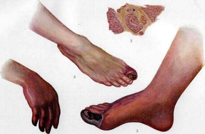
Rig, 2. Iodkpdinnn argspch lkuporgna spfshmn optiamsh»MIISP GrSMbON. SmshyA tpotttitt PVDMMMo! u-etiu. Pm. 3. IpokPSh of large pldshl gtopm. 9SHSl ^ Dry Miirpriu (YVDLYL frMMtIT shiishg KYSD RY* Y» VLYSPM tAiiifi.n.i . piys. ij JI hivta-fl Г<К1Г1п\ иф. fc_iiiLiiiMi 1 МГУ*) K /irnv/acwt. rii and condition of collaterals, in order to choose the lowest possible place in case of amputation where one can still count on good healing of the wound; this question gives only an approximate answer: for example, in case of pulsation of art. poplitea and art. tibialis posterior with non-pulsating art. dorsalis pedis, one can only say that art. tibialis anterior is blocked, but one cannot say exactly where. In the most recent time, the method of introducing lipoid-iodine and bromine into the artery with subsequent X-ray photographs is being developed. Collateral circulation is examined according to Moschkovitsch: the limb is raised so that it becomes pale, then it is quickly lowered and the appearance of skin hyperemia is observed; usually it reaches only a certain boundary, which will indicate the level of the skin incision for amputation. If the hyperemia is unclear, the limb can be immersed in hot water. The general prognosis in this disease is unfavorable: improvement is usually temporary, and gradually one limb after another becomes involved in the process. However, the prognosis for the life of patients must be considered in most cases not bad, especially if one is dealing with elderly patients with clearly expressed arteriosclerosis; the prognosis is worse for young people with rapidly progressing Buerger's disease, which sometimes carries to the grave within a few years. As for the restoration of working capacity of the diseased limb, the prognosis is more often unfavorable: after temporary improvement, a recurrence as a rule sets in, and the case ends in amputation; if the improvement turns out to be prolonged, the limb, living at the expense of collaterals, never returns to normal: it remains somewhat emaciated and does not have its former strength; patients, thus, always lose a certain degree of working capacity and become disabled. - Since the etiology remains unclear, there is no rational prevention of the disease. Patients should be protected from all harmful factors that cause or worsen arteriosclerosis; the limb in the ischemic period should be protected from injuries, cooling, and fatigue. For the treatment of spontaneous G., very many measures have been proposed; however, none of them is correct. In order to combat the formation of thrombi, treatment with leeches has been proposed, which is beneficial in the very earliest stages of thrombophlebitis; due to lack of clinical observations, it is premature to judge it. Japanese researchers, who have noted the significant and almost constant increase in blood viscosity in G., have proposed administering large amounts of physiological NaCl solution to patients daily or every other day (500.0 subcutaneously or into a vein). The treatment is carried out in repeated courses of several weeks each. Koyano (Kouano) reports 92 cases treated in this way, and all patients were discharged with improvement; no amputations were performed. The infusions indeed lower blood viscosity, calm pain, and sometimes improve circulation in the limb. In the USSR, this method is propagated by Protopopov, giving his own recipe for the infusion fluid (mineral serum). French authors infuse solutions of sodium citrate in order to act not only on viscosity but also on blood clotting; the introduction of fluid is also possible in the form of enemas or through a duodenal tube. This treatment is indicated for high blood viscosity, as well as in all cases where the patient's condition allows for prolonged treatment; its significance may also be that diuresis is increased and toxins are excreted. - A number of measures are aimed at expanding the collaterals; the simplest of these are hot baths or baths with variable temperature; success from them is observed only at the very beginning of the ischemic stage of the disease; more effective are hydrogen sulfide baths (for example, Matsestinsk baths) and then diathermy; these two methods always give success to a greater or lesser degree, calming pain and improving circulation to such an extent that ulcers often heal. This treatment is indicated not only in the ischemic stage, but also in the presence of superficial foci of necrosis, if time permits. For the same purpose, solutions of nitroglycerin, acetyl-choline are administered. Expansion of collaterals is pursued by the operation of V. I. Razumovsky - injection of 1.5-2 cubic cm of 80% alcohol into the sciatic nerve and the great subcutaneous nerve (n. saphenus) of the affected limb; the operation calms pain, causes redness of the limb. When vasoconstrictive phenomena predominate in the clinical picture (which happens more often in young people), operations on the sympathetic nervous system are successfully applied: the operation of Leriche, the operation of Prokin (see Angioneuroses-surgical treatment, as well as Desympatization of arteries). These operations promote the development of collaterals, since the limb immediately after the operation becomes warmer; pains also usually disappear; in many cases, healing of ulcers has been noted; however, in most cases the success of the operations turns out to be only temporary; after a year or two, a recurrence is usually observed, and only in a small number of cases was the result permanent. According to the collective statistics of Rubashev (1926), out of 118 Leriche operations for G., a good result was obtained: immediate in 59 cases, remote - only in 20 cases. Two operations are performed directly on the vessels: ligation of the vein of the same name or of a higher order in relation to the occluded artery (the first operation of Oppel), and arteriovenous anastomosis (operation of San.-Martin and Santrustegui). Ligation of the vein aims to bring the venous system into correspondence with the arterial (to create reduced circulation); it is indicated in cases where lowering the limb brings relief to the patient, which, as is known, happens often; the effect of the operation in these cases is always obtained, but, unfortunately, it is not permanent, and after a few months the pains and objective signs of ischemia return again. Arteriovenous anastomosis aims to send arterial blood through the venous bed; it is applied between the artery above the site of occlusion and the corresponding vein; on animals Carrel showed the full possibility of carrying out this idea; however, in surgical practice it did not take root; the anastomosis has to be applied to vessels with a changed wall, the venous valves represent a significant obstacle for blood to penetrate in the reverse direction, and usually thrombosis occurs at the site of the anastomosis; the clinical effect is very rarely good. In 1921, Oppel proposed his second operation for G. - epinephrectomy. It is a logical conclusion from his theory, which considers gangrene as a consequence of hyperfunction of the adrenal gland; according to the author, epinephrectomy is indicated in young persons (not older than 40 years); in the ischemic stage it aims to stop the disease, and in the presence of G. - to allow a relatively low amputation. Martynov considers the operation indicated in cases where blood examination for adrenaline would show an excess of it; however, in his clinic not a single such patient was found; apparently, these cases are not frequent. Then several cases of removal of a single adrenal gland with fatal outcome have been published; from this it is clear that before epinephrectomy it is necessary to be sure of the favorable condition of the other adrenal gland, and this is rather difficult. Despite all these difficulties in choosing suitable patients, epinephrectomy was nevertheless performed in a fairly large number of cases both by the author himself and by other Russian surgeons; in recent years Leriche (Strasbourg) has also applied it in 6 cases. According to the Russian collective statistics of Herzberg, out of 110 epinephrectomies, death, depending on the operation, was observed in 15% of cases; an immediately positive result was observed in most cases - 31 times out of 48 cases; pains disappeared immediately, in three cases ulcers healed quickly; pulsation appeared in 24.5% of cases; however, this success turned out to be temporary, and in 44.34% of cases gangrene reappeared after a few months, and it was necessary to resort to amputation. The remote result from one to three years is known in 54 cases; of these, ten patients can be considered cured (working capacity was restored), two are considered in a somewhat improved state, in the others a recurrence appeared. Based on the same assumption of hyper-adrenalinemia in G., Vaquez recommends the injection of insulin. The oldest, but still the most common operation for G. is amputation; it is indicated in cases of failure of therapeutic and palliative measures, as well as in cases where necrosis is rapidly progressing, causing general poisoning; the peculiarity of amputation in these cases is the necessity to amputate very high where one is sure of good circulation, where arteries still pulsate and reactive hyperemia of Moschkovitch is achieved.
The method of amputation should be the simplest (for example, circular), since plastic procedures are doomed to failure.
M. Egorov.
Decubital gangrene, see Bedsores. Gangrene symmetrical, see Raynaud's disease.
Related articles
Mentioned in
Cite this page
“Gangrene.” Soviet Medical Encyclopedia. English translation of Bolshaya Meditsinskaya Entsiklopediya, 1st ed. (Moscow, 1928–1936), ed. N. A. Semashko. https://sovietmedicalencyclopedia.pages.dev/article/gangrene/