Necrosis
Historical document, translated for reference. It reflects medical knowledge of the 1920s–30s and is not medical advice.
Summary
Necrosis refers to the local or partial death of a part of an organism while the whole organism remains alive. It can occur rapidly without preceding degenerative changes or develop gradually through degenerative processes, with morphological changes including coagulation, liquefaction, and specific nuclear changes.
Encyclopedia article (1928–1936)
NECROSIS (from Greek nekros - dead, mortification, a term used to denote so-called local or partial death, i.e., the death of some part of an organism (cells, groups of cells, a tissue area, or an organ) while the life of the whole continues. Sometimes death of this area occurs rapidly, so to speak in complete health or at any rate without significant preliminary damage, but much more often it is preceded by gradually developing and intensifying degenerative processes, associated with one form or another of progressive destruction of tissue elements, which ultimately leads to their death (for example, protein, fatty, vacuolar degenerations, ending in protein, fatty decay or complete melting of the corresponding area). Such a process of gradual tissue dying is designated as necrobiosis (see). 49S In cases of N. occurring without sharply expressed preceding degenerative processes, as for example with mechanical effects (concussion of the brain), asphyxiation of tissue due to closure of the feeding artery, under the sharp action of radiant energy, certain poisons, etc., in the first period after cessation of life processes in the tissue, no visible structural disturbances may be detectable, or they may affect only certain special cellular organelles (for example the chondriosome apparatus), so that with ordinary histological treatment the tissue will appear normal. The same is observed in tissue killed and fixed by some chemical reagent (alcohol, sublimate, formalin, etc.). However, after a certain, usually short, period of time in the dead tissue remaining among living tissues, more or less marked morphological changes develop, and the most conclusive evidence for the occurrence of mortification are changes in the nuclei, expressed in the form of pyknosis, karyolysis and karyorrhexis (see) (fig. 1). As for the protoplasm and intercellular substances, in most cases the first morphological expression of N. in them is coagulation of the protein constituents. Such N. is designated as coagulative or dry. In this case, the general outlines of the tissue elements, depending on the cause of N., the nature of the tissue, its preceding state, etc., may for a certain time be preserved rather well, as for example in ischemic infarcts of the kidney, myocardium, or in the so-called mummification (see), or, conversely, the tissue elements quickly undergo complete destruction, causing the transformation of the mortified area into an amorphous mass of fine-grained decay, as for example in toxic focal N. of the spleen, liver, mucous membranes, lymph glands and other organs, observed in many acute infectious diseases (scarlet fever, typhoid fever, diphtheria, dysentery, smallpox, etc.). The so-called curdy, caseous, or cheesy N., most often developing in tissues and various pathological products in tuberculosis and syphilis, more rarely in some other infections (lymphogranulomatosis, measles) and sometimes in tumors (sarcomas), also belongs to the category just described. Its distinctive feature is the presence in the dead mass of considerable quantities of fatty decay, thanks to which it in appearance and consistency bears great resemblance to cottage cheese or soft cheese. Besides coagulation, necrosis can also proceed with phenomena of colliquation, i.e., melting of the tissue (moist, colliquative necrosis). This kind of process sometimes begins from the very start of N., more often however after preliminary coagulation. In the first case, its causes are either the preceding conditions of existence of this area (as for example markedly expressed edema or vacuolar degeneration of tissue ending in its complete melting) or the special physico-chemical properties of the corresponding elements, which is revealed for example very brightly in the central nervous system, where necrotic foci immediately soften (the so-called foci of softening) and subsequently transform into cysts. As for liquefaction after preliminary coagulation, it is observed in the most diverse necrotic products and is explained partly as a manifestation of autolysis (see) and partly as the result of the action of proteolytic enzymes entering the dead focus with leukocytes or with lymph. This includes liquefaction of tuberculous curdy masses with formation of the so-called tuberculous pus, melting of infarcts, formation of cysts with liquid contents in place of foci of N. of the pancreas, etc. The morphological picture of tissue death occurring in the outcome of necrobiosis is determined to a considerable degree by those degenerative
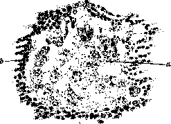
Figure 1. Necrosis: a-phenomena of karyorrhexis; b-normal lymphocytes.
changes which have developed in the tissue preceding the death of the given area; c-glomerulus of the kidney.
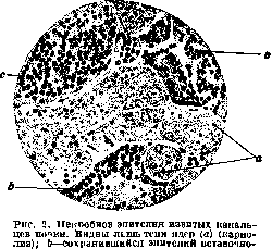
department; c-glomerulus.
by the changes that caused them (see Cloudy swelling, Waxy, Vacuolar fatty degeneration, etc.), but as numerous observations show, the moment of death can only be judged by the state of the nucleus (Figure 2). The presence of the latter indicates the continuation of life processes, and consequently the possibility of reverse development of pathological changes and restoration of normal structure, whereas the death of the nucleus, morphologically manifested as karyolysis or karyorrhexis, undoubtedly indicates the cessation of life of the cell. A special form of N. is the so-called gangrene (see), which in turn can be dry or wet. Regarding the microbes that play a role in the development of wet gangrene—see Gangrene. Regarding N. of fatty tissue—see Fatty necroses. The causes of N. are extremely diverse. Accordingly, they distinguish 1) mechanical, or traumatic necroses (as a result of a blow, rupture, crushing, concussion, compression, etc.); 2) N. as a result of the action of physical agents (high and low temperature, electricity, various types of radiant energy); 3) N. as a result of the action of chemical and infectious-toxic agents: strong acids, alkalis, arsenic, phosphorus, salts of heavy metals, alkaloids, poisons secreted by various animal and plant parasites, as well as those formed by the body itself (in jaundice, uremia, eclampsia, etc.); 4) N. as a result of disorders of blood and lymph circulation (local anemia, venous stasis, "sharp edema"); 5) N. as a result of nervous influences of vasomotor and trophic nature (for example, in perforating ulcers of the foot, symmetrical gangrene of the extremities, multiple gangrene of the skin, syringomyelia, tabes dorsalis, etc.). In the origin of all the listed types of N., the presence of predisposing factors in the form of (preceding the main cause) action of other harmful agents, the nature of the tissue (in terms of its sensitivity to harmful influences), individual peculiarities of the organism, its general state, etc., is of great importance. For example, in the development of decubital necroses and ulcers (see Decubitus), not only pressure on the tissue and its vessels plays a role, but also the usually present weakness of blood circulation in such patients, and often also disorders of trophoneurotic order. When the feeding vessel is occluded, nervous tissue, due to its sensitivity, dies earlier than all other tissues; in internal organs, the parenchyma dies significantly earlier than the stroma, etc. In persons suffering from any idiosyncrasy (see), the corresponding substance can cause N. when applied in a dose that does not cause any noticeable damage in others (for example, N. of the skin when using weak solutions of carbolic acid). In deep old people and patients with prolonged exhausting diseases (e.g., in chronic severe lesions of the intestine, prolonged disorders of cardiac activity, diabetes, some malignant tumors), necroses can occur under the influence of very minor and not always determinable factors (minor disorders of blood circulation, mild inflammatory processes, etc.)—so-called marantic necroses. The usual consequence of necrosis is the development of a reactive delimiting (demarcation) inflammation around the necrotic focus, as a result of which either organization of the dead area or its delimitation from the surrounding tissues occurs by the development of one or another kind of capsule [sequestration of bone, formation of cysts with connective tissue or glial (in the central nervous system) walls, etc.]. Sometimes the reactive zone in turn undergoes necrosis, which leads to an increase in the volume of the primary necrotic focus. The latter is often observed, for example, in cases of wet gangrene in nomas or in cases of progressive gangrene of the extremities on the basis of ascending lesions of the corresponding artery (thromboangiitis). If N. does not progress, and on the other hand, neither complete organization nor dissolution of the dead parts occurs (as often happens, for example, in encapsulated tuberculous caseous foci), then usually these parts are impregnated with lime salts (see Calcification). This type of process can sometimes develop extremely rapidly (even earlier than reactive phenomena in the surrounding tissue), which is especially often observed in isolated N. of individual small tissue elements, such as ganglion nerve cells, renal epithelium, muscle fibers, capillaries, etc.--Regarding the biochemical bodies that arise in tissue N., the so-called necrohormones—see Wound hormones. There are also indications of the formation of strongly acting substances of the histamine type (see) in necrotic areas.
Related articles
Mentioned in
Cite this page
“Necrosis.” Soviet Medical Encyclopedia. English translation of Bolshaya Meditsinskaya Entsiklopediya, 1st ed. (Moscow, 1928–1936), ed. N. A. Semashko. https://sovietmedicalencyclopedia.pages.dev/article/necrosis/