Radiotherapy
Historical document, translated for reference. It reflects medical knowledge of the 1920s–30s and is not medical advice.
Summary
Radiotherapy is the treatment of diseases using radium and other radioactive substances. This article details the properties and biological effects of various radioactive materials used in medical treatments during the 1920s-1930s.
Encyclopedia article (1928–1936)
RADIOTHERAPY (curie therapy), treatment with radium and other radioactive substances. Of these, radium, radium emanation-radon, mesothorium, and much less frequently radiothorium and thorium X are used for treatment. Pure metallic radium is unstable in air; therefore it is released in the form of salts-chloride, bromide, carbonate, and sulfate. In 1 mg RaCl2 there is 0.7612 mg RaEl; in 1 mg RaBr2-0.5857 mg RaEl; in 1 mg RaCO3-0.7902 mg RaEl; in 1 mg RaSO4-0.7020 mg RaEl. In Paris and Vienna are stored international standards of radium from preparations of high purity. Since the value of radium depends on the intensity of its radiation, its evaluation is done not by weight, but by radiation. For practical purposes, there is no need for especially pure preparations of radium. For solutions, it is desirable to have radium with barium impurity; this makes it possible to minimize the loss of metal during various manipulations or when it precipitates from solutions. Radium emits a large amount of alpha rays and in smaller amounts beta and gamma rays. The rays that come from a tube containing Ra are not emitted by Ra itself, but by the products of its transformation, the radioactive precipitate, which gives the most active rays. The emanation of radium, or radon, was discovered in 1900 by Dorn. It is the direct product of radium decay due to alpha ray emission and has all the characteristic properties of a gas; as a noble gas, radium emanation does not form chemical compounds. Radon has a half-life of 3.825 days, i.e., its ability to radiation decreases by half during this time. In the case where radon is removed from its source of formation, radium at the same time forms radon again. For a certain period of time in a radium preparation enclosed in a closed vessel, radium decays in the same quantity as it is formed; radium under these conditions is in a state of radioactive equilibrium. Radon dissolves in water; its solubility decreases with increasing temperature, and its solubility in salt solutions and in blood is even less. The amount of radium emanation that is in radioactive equilibrium in 1 g of radium is called a "curie". This amount serves as the unit of measurement for radium emanation. For practical purposes, 1/1000 curie-millicurie and 1/1,000,000-microcurie are used. The amounts of emanation contained in air and natural medicinal waters, however, are many times less than one microcurie. Therefore, the unit "Mache" was introduced, equal to the content of 3.6×10⁻¹⁰ curie in 1 liter of water. The radiation from emanation of alpha and gamma rays occurs, as in the radium preparation, from decay products-radioactive precipitate. The latter consists of rapidly decaying radium A, B and C, and radium D, E and F, in which the decay process proceeds more slowly. With alpha radiation of thorium, mesothorium is formed, which is an isotope of radium. When obtaining mesothorium, it is not possible to separate radium from it. Therefore, in preparations available for medical purposes, mesothorium contains about 20-25% radium. In radium preparations after being placed in a closed vessel, activity reaches a maximum after a month and thereafter remains practically constant, since its half-life is 1,600 years. The radioactivity of mesothorium, however, after its extraction slowly increases and reaches a maximum after 3 years, after which it begins to fall; but after ten years it is still higher than at the beginning. By the end of the second decade, the radioactivity of mesothorium falls to half. Mesothorium is cheaper than radium. In view of the fact that its radioactivity decreases after ten years, manufacturers, in order to be able to sell it, replace it with fresh after a certain period. Radiothorium is obtained from mesothorium and has a half-life of 1.9 years. From it arises thorium X with a half-life of 3.64 days. Of all the radioactive substances used, radium is most often used for treatment, since its existence time is a practically constant quantity. The degree of biological effect of radioactive substances on a living organism depends on the dose of radiation applied and is not related to which preparation was used. Each living cell under the influence of radioactive substances, provided that the dose absorbed by it is sufficient, can be killed. There is no selectivity in the sense that there are living cells on which radioactive substances do not act. The character and strength of the reaction of cellular elements to irradiation with radioactive substances, besides the size of the dose, also depend on specific sensitivity-radiosensitivity, which is different for each type of cell. Histological changes consist of the usual chain of degenerative processes leading to cell necrosis. Based on old observations, mainly by Hertwig, it was believed that the necrobiotic process first begins in the nucleus, then in the protoplasm, after which cell death occurs. Recently, thanks to fine cytological methods of processing, it has been possible to prove that the protoplasm is most sensitive to radioactive substances; degenerative changes appear in it when the structure of the irradiated nucleus still shows no deviations from the normal (Weil, Frenkel). Under certain technical conditions when using very small doses, under the influence of radioactive substances, in some cases an increase in the functional activity of an organ occurs (increase in growth, secretion, increased metabolism). It is not sufficiently studied whether this reaction can also occur due to regenerative changes in cells or from their irritation. The school of Holtzreck believes and apparently with full reason that here too we are dealing with the manifestation of the destructive effect of radioactive substances on living tissue. In treatment, not one isolated cell is irradiated, but an organ consisting of a group of cells; due to the interaction of individual cells and individual groups among themselves, their different radiosensitivity and unevenness of irradiation in terms of dosage, conditions are created for the exclusion of some cells from the whole organ. The latter circumstance can lead to the destruction of the inhibitory apparatus, which leads to a temporary increase in the function of the organ. This theory is disputed by many. Of normal tissues, the elements of the lymphatic system and sex glands are most sensitive. Experiments on animals have shown a significant effect of radio-irradiation on blood and hematopoietic organs: the spleen decreases, follicles atrophy; after a transient leukocytosis, the number of leukocytes decreases. In humans, small doses cause a transient leukocytosis. With large doses, prolonged leukopenia with neutrophilia and a shift to the right, i.e., the appearance of hypersegmented elements, occurs. Red blood cells are least sensitive to radioactive substances. In pathological processes-in secondary and pernicious anemia-injection of thorium X causes a short-term increase in the number of red blood cells. With toxic doses of radioactive substances, the erythroblastic apparatus is also damaged; the bone marrow turns into a homogeneous mass colored in dark cherry color. On microscopic examination, severe phenomena of destruction of white blood cells, capillaries and bone marrow elements are found. In terms of radiosensitivity, the epithelium of the sex glands is close to the blood-forming organs. In women, follicles gradually disappear, menstruation stops; in men, azoospermia occurs. Libido usually does not suffer. With small and medium doses, the ability to fertilize can be restored. Much less sensitive to the radiation of radioactive substances are the liver, pancreas, prostate and thyroid glands. With intense irradiation of the salivary glands, their function decreases and a severe sensation of dryness in the mouth appears. Cells of the central nervous system are least sensitive; histological changes in these cells from irradiation occur only with very high doses. The mechanism and functional effect of the anesthetic effect of irradiation with radioactive substances on peripheral nerves in various pathological phenomena accompanied by pain is completely unexplored. Muscles are not very radiosensitive, but with repeated deep radiotherapy, processes leading to scar changes and their shortening also occur in them. With direct through-the-skin irradiation of the uterus, a process of regression also occurs in it. In bones and cartilage, changes are possible only with very high doses. More often, degenerative changes in the periosteum are encountered, as secondary, due to the destruction of small blood vessels. Changes in the skin under the influence of irradiation with radioactive substances are divided into three stages: the first - hyperemia, erythema, swelling and slight infiltration of the skin, often accompanied by mild local pains. At the same time, hair loss may occur. The second stage is characterized by the formation of blisters. The third-deep ulcers with damage to the epidermis and cutis. All the described damage to the skin occurs after a certain period after the application of irradiation with radioactive emanation, lasting from one to 4-5 weeks. In some individuals, a slight reddening of the skin occurs immediately after irradiation-early reaction, which soon passes.
First and degree skin changes pass in a relatively short time, leaving behind pigmentation, and with second degree also atrophy and dryness of the skin. Ulcers with third degree are painful, sometimes requiring many months and even years for their healing, but in their nature and outcomes they are more benign than ulcers obtained from X-ray therapy, and cause patients less suffering. Late skin necroses sometimes occur where there is insufficient subcutaneous fatty layer, and the skin is tightly adjacent to the bone. Sometimes a uniform thickening appears in areas with rich fatty padding. These changes and telangiectases appear after many months and years of a hidden favorable period. The effect of exposure to radioactive substances on metabolism after skin irradiation with large doses is manifested by an increase in the amount of uric acid, purine substances, and phosphoric acid in the urine. When emanation is used for treatment, an increase in gas exchange and a decrease in blood pressure are observed. For introducing radioactive substances into the body, the following methods are used: 1) Inhalation of radium emanation, for which specially constructed apparatus is used. The patient inhales radium emanation mixed with atmospheric air. 2) In the form of baths - the skin absorbs radium emanation dissolved in natural medicinal waters or artificial ones, like other gases. The contact of radium emanation and its decay products with the skin plays no less an essential role in the general treatment with radioactive substances than inhalation. 3) By internal administration as drinking water - in the form of natural and artificial medicinal waters. There are special apparatus for dissolving emanation (emanators). 4) By injection - subcutaneously, intravenously, or intramuscularly. For general effect, soluble salts of radium, radium emanation, and thorium-X are used. To create a long-term reserve of radiation in a limited area of the patient's body, insoluble salts are injected. This method of injecting insoluble salts is rarely used at present. The entire system of application of radioactive substances, with very rare exceptions, is used primarily for internal diseases. In dermatology, surgery, gynecology, etc., radioactive substances are used in special apparatus of which there are several types: 1) In the form of small quadrangular metal plates, on which radium is distributed evenly. On 1 cm2 of such a plate, 5 mg, 2.5, or 1.25 RaEl are placed. The radium is attached with a ray-permeable varnish. These plates are sometimes made flexible so that they can be applied to any part of the body. Mostly flat applicators are used in skin therapy. 2) In the form of cylindrical tubes, first used by Dominci, 7a to 4 cm long, 3-4 mm in diameter. Each applicator contains from 5 to 100 mg RaEl. The radium is placed in a glass tube, which is covered with a metal casing (preferably platinum) 0.2-0.3 mm thick. The radium can be placed directly in a platinum tube. The tubes can be joined in length and inserted into various body cavities, or, when placed parallel, used for illuminating large surfaces. 3) Applicators in the form of platinum needles containing 1 to 10 mg RaEl. These needles are injected into diseased tissue; one end has a point, the other has an eye for threading a string, by which at the end of the treatment session the needle is pulled out of the body. 4) Glass capillaries filled with emanation - bare needles. Since these capillaries cause strong necrosis, they are inserted into metal, platinum, or gold needles. 5) Applicators for teletherapy. In this case, large amounts of radium up to 5 g are used at a great distance from the object being irradiated, in order to increase the percentage of deep irradiation and to achieve maximum uniform irradiation. There are a limited number of such apparatus in the world, constructed on their own, mostly hanging stands. Depending on the goals pursued in the treatment with irradiation by radioactive substances, either the entire sum of emitted alpha, beta, and gamma rays or a separate group from this bundle is used. As is known, when rays pass through dense media, part of them is absorbed. On the basis of this law, in radiotherapy certain substances are used to absorb certain rays, so-called filters. Alpha rays are completely absorbed by a sheet of black paper, rubber, or three to four sheets of aluminum 0.04 mm thick. For filtering beta and gamma rays, substances of high atomic and specific weight are necessary. For complete absorption of beta rays, a filter of 2-3 mm of lead is necessary. Gamma rays penetrate even through a layer of lead 15 cm thick. Usually in medical practice, filters of platinum, gold up to 2 mm thick, lead from 0.1 to 5-6 cm, brass 1-2 mm, aluminum up to 3 mm, etc. are used. There is a whole system of devices, from ordinary adhesive plaster to complex apparatus, for fixing radioactive substances on those parts of the body where they need to be applied. The impetus for the development of radiotherapy was the discovery of emanation. After it was proven that in healing sources known for favorable treatment results there is significantly more emanation than in ordinary water, experiments with the use of radioactive substances on humans begin. The first experiments date back to 1905 (Dautwitz, Lowenthal, etc.). Systematic research by the school of His made it possible to develop the technique, dosage, indications, and contraindications for the use of radioactive substances in a large number of internal diseases. Radium emanation is inhaled by the lungs like any gas, is partially absorbed by the bloodstream, and with the next breathing movements is exhaled back. To create a reserve of radioactive gas in the blood, it is necessary to ensure continuous delivery throughout the entire treatment session, which is achieved by the patient staying in a specially constructed enclosed room - an emanatorium. The minimum dose for each session is 5 Mache, the maximum is 50 Mache. Per month - 20-30 sessions. Dissolved radium emanation, taken internally, passes from the gastrointestinal tract into the portal vein system, liver, and right heart. Together with the blood, radium emanation penetrates all parts of the body and causes various biological changes in it. Partially it is exhaled, partly reabsorbed by the alveoli and again enters the bloodstream. As soon as the delivery of emanation from the gastrointestinal tract ceases, it soon disappears from the blood. A small part of the emanation is excreted with feces, urine, saliva, and sweat. Injected solutions of radioactive substances - radium, radiorium, and thorium-X - are retained in the body much longer than radium emanation and are excreted slowly, over weeks. Solid components are excreted with feces, urine; gaseous ones are exhaled by the lungs. Residual radioactive substances retained are found in the hematopoietic organs - bone marrow and spleen. There are observations of the beneficial effect of radiotherapy in gonorrheal arthritis. In acute rheumatism, radioactive substances are used only in cases where other methods give no effect. First of all, with favorable treatment, pain, swelling, joint swelling decrease, temperature drops, leukocytosis. If after several sessions no improvement occurs, further treatment is useless. In chronic arthritis with atrophy of periarticular tissues and swelling of the synovial membrane, emanation reduces pain and exudate. Gudzent obtained improvement in 78% of 262 cases. The results are approximately the same in gouty processes. Lazarus believes that in these cases a complete cure cannot be expected, but the analgesic effect of radiotherapy makes it possible to resort to movement therapy and thus improve the final outcome. Large doses are used both for internal administration and for inhalation. Falta gives 1,000,000 Mache as drinking water and about 600 Mache per day for inhalation. Gout patients are very sensitive to radiotherapeutic interventions. Therefore, they are contraindicated in acute attacks. In tabes dorsalis, neuralgia, radiotherapy is sometimes used in stubborn cases as a painkiller. Application of radium, mesothorium is much more effective than internal administration of radioactive substances or inhalation. Among internal diseases, radiotherapy gives the most satisfactory results in myeloid and lymphatic leukemia; spleen, liver, glands, and mediastinal tumors decrease, and blood composition improves. The treatment process proceeds as in X-ray therapy. The preference that some authors (Lazarus) give to radiotherapy over X-ray therapy is not justified; besides that radiotherapy does not give better results, it is inaccessible to many patients in the relatively common case of leukemia. But in cases where X-ray therapy at the beginning of treatment gives no results or after multiple sessions ceases to be effective, radiotherapy is recommended. Sometimes it is still possible to achieve a satisfactory condition in such cases, which can last for many months or even years.
Radiotherapy is contraindicated in acute cases of leukemia. The dose in chronic leukemia must be small, and treatment must be carried out under strict control of the blood composition and the general condition of the patient. The decrease in the number of leukocytes should not be rapid; the number of erythrocytes and Hb should not decrease. The dose should be such that leukoplastic hyperplasia is suppressed without impairment of erythrocyte formation. A radium preparation of 14-100 mg is applied to the area of the spleen, liver, or glands. Its distribution over the surface of the organ should be uniform. A filter of 1-1.5 mm of copper or lead is used at a distance of 1-2 cm from the patient's body. No more than 1,000 mg RaEl is given per session; it is better to give it every other day and not more than 10,000 mg RaEl per course of treatment. Injections of thorium-X are not safe, as at large doses damage occurs not only to hyperplastic tissues but also to a decrease in blood pressure; the general condition worsens, loss of appetite, diarrhea, and weight loss occur. Therefore, the administration of thorium-X must be carried out with extreme caution. It is absolutely contraindicated in cases of a tendency to bleeding, in septic processes, in poor general condition of the patient, in hypotension, and in exacerbations of the disease. Falta proposed combined treatment with thorium-X and X-rays. With fairly good results, curietherapy is applied in Vaquez's disease; the long bones are irradiated, with a dose up to HED. Radiotherapy is also used in pernicious anemia, although the results to date are unsatisfactory. Lazarus describes cases of favorable outcome from the treatment of radium in agranulocytic angina; the tibia is irradiated with small doses of radium or mesothorium. Radiotherapy is also used in Basedow's disease, where the indications are similar to those for X-ray therapy. Radium or mesothorium is applied to the thyroid gland; the doses are small. It is necessary to carefully monitor the patient's subjective condition and the metabolism of substances. The widest field of application of radiotherapy is in surgical diseases and in particular in malignant tumors. As is known, the effect of treatment depends to a large extent on the individual radiosensitivity of cells of different histological structure. Only the amount of radioactive energy that is absorbed per unit volume of the diseased focus is effective. Only this amount manifests itself in various biological consequences. The task of delivering the appropriate amount of radioactive energy to the diseased focus, i.e., dosing, is very difficult and has not yet been finally resolved. The following problem is not yet fully clear: 1) whether to give a certain dose in one session - single massive irradiation; 2) whether to distribute the same dose over a period of 8-10 days - prolonged irradiation; 3) whether to perform irradiation in separate sessions with an interval of one to two weeks - fractionated intensive treatment. There are experimental and clinical data to prove the correctness of each of these methods. Eymer, for example, in cancer of the uterus, places 75 mg of Ra in its cavity for 72 hours and at the same time 25 mg in the vagina for 48 hours, totaling 6,600 mg. With such a single high dose, he achieved recovery in 55.6% of surgical cases and in 11% of inoperable cancer of the cervix. The Americans give even larger doses - at one time about 13,000 mg/hour. Regaud, on the contrary, placed several radium preparations of 6.66 and 13.33 mg RaEl in the uterine cavity and vagina for 120 hours and gives in almost the same total dose as Eymer. A number of authors use fractionated intensive irradiation: for example, they give 2-3 sessions of 1,500 mg RaEl each. Nemenov gives 3-4 sessions with intervals of 8 days. At the Moscow Cancer Institute (Frenkel), treatment of cancer of the uterus is also carried out by the method of fractionated intensive irradiation combined with X-ray therapy. All authors, with different techniques, achieve approximately the same results: out of 10 inoperable cases, one recovers; in operable cases, every second one recovers. It is necessary to be careful not to give too small a dose. Frenkel considers fractionated irradiation to be the most effective and safe. The advantage of radium treatment over X-rays lies in the fact that with radiotherapy it is possible to concentrate in the minimum volume of the diseased focus the maximum amount of rays. This factor can be further enhanced by cross-irradiation. General poisoning phenomena in radium treatment are significantly weaker than in X-ray treatment. This is explained by the fact that in X-ray therapy the necessary dose is given in a short time, whereas with radium the diseased focus is subjected to radiation therapy slowly, for a longer time. Therefore, where the diseased focus is accessible in size, radiotherapy is preferable. Healthy tissues lying over the diseased focus to be treated undergo less damage than in X-ray therapy, since in the latter the tissues intermediate between the skin and the tumor absorb at least 15%, whereas in radiotherapy, due to the greater hardness of the radiation, only 5%.--The deeper the diseased focus is located in the body, the further the radiation source should be placed to increase the percentage of absorption in depth, and the thicker the filter should be to allow only the hardest rays to pass. In accordance with these requirements, three methods of treatment are used: 1) contact irradiation, 2) irradiation at close range, and 3) telecurietherapy--irradiation at a distance. In contact irradiation, the applicator is placed directly on the skin, on the diseased focus, or at a distance of no more than 1 cm. It is used where the tumor is not deeper than 2 cm from the skin--predominantly in diseases of the skin, mucous membranes, hemangiomas, etc. In irradiation at close range, from 2 to 4 cm from the skin, radium tubes are evenly distributed on the skin. This method of irradiation--distribution of radium in small sources--has the advantage that cross-irradiation is obtained in depth--below the skin. Thus, powerful irradiation of the deep focus is created with relatively little absorption of rays by the skin. Applicators are laid on special masks made of a mixture of wax and vaseline. Masks can be made of lead, leather, etc. Irradiation by radioactive sources follows the law of decrease in intensity of irradiation inversely proportional to the square of the distance between the light source and the object being irradiated. Taking this factor into account, the radioactive source is placed at the necessary distance. When distributing radium, the dose must be chosen so that each cm3 of the tumor receives no less than the cytolethal dose for the cancer cell--from 150 to 200 mg/hours RaEl. From Table 1 it can be seen what influence the position of the irradiation source has in relation to the skin and tumor, in terms of its effect on the percentage of deep absorption of rays. Table 1. Distance in cm Intensity in % Relative decrease in intensity in % 1 2 3 4 5 100 25 11.1 6.25 4 75 56 41 35 The deeper the rays penetrate, the smaller their relative decrease in intensity; the further the radiation source is from the focus, the greater the percentage of deep irradiation. The most sharp decrease in irradiation intensity occurs in the first two centimeters. This law is of great importance in radiotherapy: it makes it possible to increase the percentage of radioactive energy necessary in the depths of the body if the cancerous focus is located there. Irradiation at a distance--telecurietherapy--differs in that the radioactive source is at a distance of 5-10 cm from the skin and lead filters up to 5 mm thick are used. By this method, it is possible to significantly increase the percentage of deep irradiation and create uniform (homogeneous) irradiation of the entire tumor. Regaud uses an apparatus on a well-balanced stand, made of iron (weighing 80 kg) and 6 mm lead. In the box are placed 80 tubes of 50 mg of radium each; the apparatus is placed at a distance of 10 cm from the patient's skin. In cancer of the uterus, about 3-4 hours daily is given for 2-3 weeks. Dermatitis occurs with 4 g RaEl on a certain area of skin at a distance of 10 cm after 13 hours of treatment. After 9 hours, only erythema is obtained. Berwen uses only 3 g with a 5 mm lead filter and 5 cm distance of the apparatus from the skin. German authors dispute the importance of telecurietherapy in the treatment of malignant tumors, as this treatment requires large expenses and is accessible only to a limited circle of patients. According to Lazarus, the same successes can be achieved with X-ray therapy and the usual method of treatment with radium at close range.
According to the latest data from clinics where curietherapy is widely used (Paris, New York, Stockholm), the objections of German authors are not sufficiently substantiated, and telecurietherapy deserves great attention in the fight against cancer. In addition to the methods described, the intratumoral introduction of needles with radioemission or radium has become widespread. Americans were the first to use glass
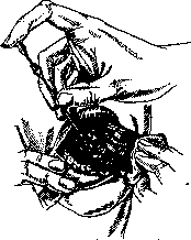
tubes about 3mm long and 0.3mm in diameter, made with radon emmanation, without a filter. These so-called bare tubes are introduced into the tumor with a thin trocar (fig. 1). Each tube usually contains 1 millicurie of emmanation. The needles are not removed, sometimes surrounded by a capsule in the tumor, and remain there permanently or come out on their own. For many years this method has been practiced at Memorial Hospital (New York). Its advantage is that it uses both alpha, beta, and gamma rays, and the tumor is irradiated for a prolonged period.

Figure 2. Cancer of the tongue with enlargement of regional glands before treatment and during treatment with fixation of the needles introduced into the tumor.
and the trauma caused by the introduction of glass tubes is insignificant. The treatment itself is accompanied by a strong reaction—sharp pains, elevated temperature, intense necrosis of tissues around each tube, and bleeding. The necrosis must be attributed exclusively to the influence of alpha and beta rays. For this reason, it was proposed to absorb alpha rays during radon emmanation treatment by covering the glass needles with a filter of pure gold 0.2-0.3mm thick or placing them in needles of platinum-iridium. With this method of treatment, the results are no worse than with bare needles, but the reaction and necrosis are significantly less—by 60%. Most authors prefer for intratumoral treatment to use needles of platinum-iridium filled with radium (fig. 2). These needles almost completely absorb alpha rays. With this method of intratumoral treatment using exclusively beta and gamma rays, it is possible to achieve homogeneous irradiation of the tumor and achieve maximum prevention of necrosis around the needles. Healthy cells remain undamaged and retain their ability to react to radiation treatment. Each needle contains from 2.5 to 10-15 mg RaEl and is introduced into the tumor for 3-4 to 12 hours. Rego, based on his experimental and clinical data, proved that with the same dose, treatment applied for a longer time is more effective than treatment performed in a short time. His method of applying intratumoral treatment currently gives the best clinical results. In some cases, combined treatment can be applied—contact or close-distance irradiation together with the introduction of needles into the tumor» The absolute unit of measurement 'g', which exists in roentgenotherapy, is not applied in curie therapy. In practical work, the dose of radium is determined by the amount of the radium element multiplied by the number of hours of application of the preparation. The effectiveness of biological action
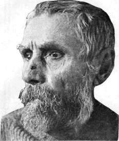
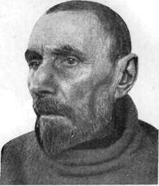
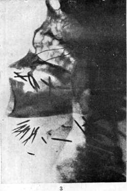
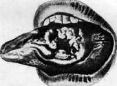
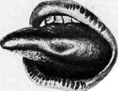
Figure 1. Epithelioma of the nose before treatment. Figure 2. The same case after treatment with radium. Figure 3.
Roentgenogram of the head after insertion of radium needles into the tongue tumor and regional glands. Figure 4. Palate before treatment. Figure 5. The same case after treatment with radium. as is known, also depends on the distance between the applicator and the skin and on the nature of the radiation, i.e., the filter. Both of these indicators should also be indicated when determining the nature of irradiation. Lam studied the biological relationship between radium dosage and X-ray doses. The skin dose (HED) in irradiation with radium rays is obtained with 600 g. The same effect is obtained with 450 mg[hours RaEl at a distance of 1 cm from the skin and a brass filter 1mm thick. The unit of measurement for emmanation is the curie. In practical work, the thousandth part of a curie is used—the millicurie. Due to the rapid decay of radon emmanation, the amount remaining in a tube or needle taken for treatment decreases each day: from 100 millicuries, 83.43 remains after one day, 69.6 after 2 days, etc. The amount missing from 100 millicuries determines the applied dose—that amount which has decayed. The French call this amount of decay the term 'millcurie detruit' (mcd). This number can be converted into milligram-hours if multiplied by 133, since the average lifespan of one millicurie of emmanation is 133 hours. The clinical result from R. of malignant tumors depends on the location of the tumor, its histological structure, the degree of spread of the tumor, its relationship to surrounding tissues, and the general condition of the patient. The most favorable for treatment is cancer of the facial skin. Treatment here is carried out both by contact application of radium and needles with emmanation, and by introducing needles, bare and covered with filters and filled with radium. The result is better for superficial cancers than for deeply penetrating, infiltrated ones, and reaches 85% recovery, [see separate table (pp. 191-192), figs. 1 and 2]. *
tab. 2. Authors Duration of observation Number of cured and followed-up cases Rego........... Fopsell (superf.) . . adorsel (infiltrr.) Oncological institute in Leningrad ........ . . Neienov and Grosman . . Institute for treatment of tumors in Moscow ....... 1- 3 years. 8-13 years. 8-13 » 80 82 64 58,5 1-2»/g g. 46 immediate 19 31,' result percentage of immediate cure - 63.5%, 5 years-50% Some cases do not respond to methods of radiation treatment, in particular R. Often after complete 'cure' from R, (in superficial cancers up to 100% of all cases, in infiltrated ones up to 30%) recurrences occur. In cancer of the lower lip, the results from surgical treatment are satisfactory, but recently, under the influence of data obtained in Western European and American clinics, the use of radium in cancer of the lower lip is expanding. Treatment also, as in cancer of the skin, by application of radium or intratumoral. Special attention should be paid to regional glands. They are removed and radium is placed in the wound. Rego during observation for 7 years had 61.6% cures, Quick-68%, Forsell - after 5 years on 208 patients (125 superficial and 83 deep)
obtained cure-in 121 patients. In cancers of the oral cavity and tongue, it is proposed (Berven) to perform telecurie therapy before the operation, which significantly improves the outcome. Clinically, cancers of the oral cavity proceed extremely severely and are accompanied very soon after appearance by metastases in regional glands. Surgical treatment is unsatisfactory: Patch's combined statistics on 306 operated cases gives 11% cure with 15% postoperative mortality; Menegaux indicates 10-14% long-term cure after operations. Such are the results for most surgeons. The results from radium treatment are much better [see separate table (pp. 191-192), figs. 3-5]. Rego in five years from 311 cases with operable 20%, inoperable and borderline 80% obtained from R.-intratumoral with needles - after 3 years 20.8% recovery and immediate cure in 46.3% of the entire material. Berven on 86 cases (operable 48%, inoperable 35%, recurrences 17%) has 38% recoveries and immediately after treatment 53% with absence of symptoms of
the disease. Patients were subjected partly to telecurie- ^gastrostomy in cancer of the pi-therapy, partly intra- *
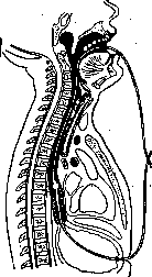
esophagus. For tri-tumor therapy with needles and contact treatment. Treatment with radon emanation needles is extremely painful for patients and sometimes accompanied by severe complications that can lead to a fatal outcome. N. N. Petrov had 5 fatal outcomes out of 74 cases. Despite the possibility of complications, curietherapy is currently the method that gives the best results for oral cavity cancer compared to the surgical method and radiotherapy, which is only permissible for the basal form of cancer. However, surgery is recommended for cancer of the tip of the tongue and its edges. In many cases, the best results are achieved with combined treatment - surgery and curietherapy. Particularly good results were achieved by Berwen for tumors of the pharyngeal ring with tele- and intra-tumor curietherapy. In treating cancer of the larynx, it is necessary to consider the possibility of cartilage damage and the onset of laryngeal edema, which often requires emergency tracheotomy. The question of whether to use X-rays or radium is far from resolved. But in both cases, intensive irradiation is required. Americans propose using telecurietherapy with large doses. According to some authors, intra-tumor introduction of needles with emanation is not contraindicated. In addition to direct irradiation of the tumor, it is necessary to irradiate regional glands with radium at close range. When the case is operable, the glands are removed surgically. In the commonly occurring cancer of the esophagus, the only means that gives temporary improvement in the patient's condition is the introduction of a probe filled with radium at the end into the esophagus and contact irradiation. Many such probes have been proposed. The best method is probing after applying a gastrostomy according to the Kurtzahn method (Fig. 3). Cases of complete recovery from esophageal cancer after radiotherapy have been described, but these reports are not trustworthy. Kurtzahn with his treatment method in 24 cases almost in all patients obtained a decrease in difficulty in swallowing and an increase in the patient's weight by 7-8 kg. The longest observation period of improvement was 1 year 8 months. Jungling managed in only 8 out of 36 cases to prolong the patient's life for more than one year. Improvement is possible only in fresh cases. Treatment with radium for cancer of the lungs, stomach, and small intestine is completely hopeless. Satisfactory results are obtained with radiotherapy for cancer of the rectum. A condition for a good outcome is the preliminary application of an anus praeternaturalis. Radium is introduced in a tube into the rectum and fixed to the tumor. Radiopuncture or telecurietherapy is also possible. In the treatment of malignant tumors of the central nervous system, more or less significant results are obtained from radiotherapy of pituitary tumors. Malignant diseases of the female reproductive organs stand completely separately in this group, where recovery is achieved more often, especially for cancer of the uterus. Treatment method: combined radiotherapy and simultaneously radiotherapy; radiotreatment - introduction of radium into the uterine cavity and vagina, intra-tumor introduction of needles (Fig. 4) and telecurietherapy. The most frequently used method is the introduction of radium in tubes into the uterine cavity and vagina. The dose is from 3,000 to 7,000 mg/hours RaE.l. The results are almost the same for all authors; they depend mainly on the stage at which treatment is started. Most authors divide their material into three groups: operable, borderline, and inoperable. For cancer of the cervix, Heymann over 10 years had 1,157 patients whom he treated with radium, placing the preparation in the uterine cavity and vagina; 734 patients were inoperable, 309 operable, and 114 on the border of operability. Of these patients, after five years of observation, 15.7% of the inoperable cases were healthy, and 40.8% of the operable cases. For cancer of the body of the uterus (80 cases), recovery in 42.5% after 5 years. The attached table presents the results of combined treatment with radium and X-rays. Table 3. Absolute cure in the operable group (in %) Number of cases Authors Total cure in all cases (in %) (operable) (in %) Observation periods Lahm .... 23.5 50.0 1915-23 Gal..... 10.5 40.0 1919-25 » Voltz .... 21.4 41.5 1913-23 » Wickham and Laborde . 22.0 80.0 1921-26 » Clauberg . . .18.7 - 1917-22 » Nahmacher . 17.8 - 1919-23 » Feldweg. . . 16.8 - 1913-21 » No worse results have been obtained in the Leningrad X-ray and Moscow Cancer Institutes. Treatment with radium for inoperable cancer of the breast and inoperable recurrences gives significant improvements in the patient's condition; it often makes it possible to prolong the patient's life. There are many cases of complete recovery. Very good results have been achieved with telecurietherapy (Berwen). Radiotherapy is also used for cancer of the prostate and bladder with temporary success. The same result is also obtained for cancer of the upper jaw and inoperable cancers of other organs; in such cases, radiotherapy acts as a painkiller. - Among the methods of treating malignant tumors, radio-surgery deserves great attention. It consists in using a series of surgical techniques to make deeply located tumors accessible to curietherapy. The goal of radio-surgical intervention is as follows: first, to produce homogeneous radioirradiation in areas where this cannot be done with ordinary techniques, second, to irradiate the entire affected area, and third, to obtain precise orientation regarding the extent of the tumor and the degree of involvement of nearby tissues in the process. Radio-surgical operations are most often performed on the rectum, pharynx, larynx, uterus, and prostate. Among benign tumors, radiotherapy is most often used for uterine fibroids in cases where radiotherapy does not give results and where surgery is contraindicated. For skin diseases, good results are obtained for keloids, warts, angiomas; it is also recommended for precancerous keratoses.
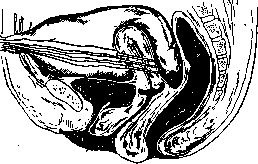
Figure 4. Introduction of radium needles into the body of the uterus by surgical means.
st benign tumors, radiotherapy is most often used for uterine fibroids in cases where radiotherapy does not give results and where surgery is contraindicated. For skin diseases, good results are obtained for keloids, warts, angiomas; it is also recommended for precancerous keratoses.
Related articles
Mentioned in
Cite this page
“Radiotherapy.” Soviet Medical Encyclopedia. English translation of Bolshaya Meditsinskaya Entsiklopediya, 1st ed. (Moscow, 1928–1936), ed. N. A. Semashko. https://sovietmedicalencyclopedia.pages.dev/article/radiotherapy/