Trichophytia
Historical document, translated for reference. It reflects medical knowledge of the 1920s–30s and is not medical advice.
Summary
Trichophytia is one of the most common fungal infections in humans, particularly in children, affecting the skin, hair, and nails. It is caused by fungi of the genus Trichophyton and classified into different types based on their characteristics and location.
Encyclopedia article (1928–1936)
TRICHOHPHYTIA, trichophytia [syn. herpes tonsurans (ringworm), dermatomycosis trichophytica, mycosis tonsurans, tinea tonsurans, porrigo furfurans]. One of the most common in humans, and in children the most common fungal disease of the skin, affecting both the skin itself and its appendages—hair and nails. T. belongs to the so-called epidermomycoses (Darier). The causative agents of T. are vegetable fungi of the genus Trichophyton (see separate table, figure 4). Most dermatologists accept the classification of these fungi proposed by Sabouraud (Sabouraud, 1910), who distinguishes trichophytons by the appearance of colonies when growing on a special medium with maltose, by the location of the fungus in relation to the hair, by the size of the spores, and by their predominant location on humans or animals. The following types of Trichophyton are distinguished: 1) Type Endothrix purum; located inside the hair, human forms; these include Trichophyton violaceum, crateriforme, acuminatum and others. 2) Type Neoendothrix; located both inside and outside the hair, occurs in humans and animals (mixed form); these include Trichophyton cerebriforme, plicatile and others. 3) Type Ectothrix; affects the hair mainly from the outside, animal forms; here are distinguished: a) Ectothrix microides (microsporoides) with small spores (Trichophyton gypseum, niveum) and b) Ectothrix megasporum with larger spores (Trichophyton faviforme, equinum, rosaceum and others). Trichophyton of humans mostly causes superficial lesions, Trichophyton of animals—deep (deep trichophytia). Trichophytia is distributed in all parts of the world. Among skin patients who applied in 1919-27 to the Odessa Dermatovenereological Institute, there were 12.5% of patients with T. A German among Uzbek schoolchildren of Tashkent found 10% of patients with T. About half of the cases of T. of the hairy skin of the head fall on the age from 6 to 10 years; twice less frequently than this age, children from 1 to 5 years and from 11 to 15 years are affected. Individual species of Trichophyton occur very unevenly in different countries and parts of the world. In the USSR, the most common causative agent of T. is Trichophyton violaceum, to a much lesser extent Trichophyton crateriforme and gypseum are distributed, other species are rarely encountered. Trichophyton violaceum is also common in Italy, France, Romania, Hungary, on the Balkan and Iberian peninsulas, in Japan and others. At the same time in Germany this species is rarely isolated, in Berlin for example Trichophyton cerebriforme predominates. During large movements of masses of people, the introduction of any species of Trichophyton into new areas for it is possible. Thus, Trichophyton rosaceum, which had not been previously encountered in Germany, became the cause of a large epidemic of T. there in 1918, being brought by French and Belgian prisoners of war. The main sources of spread of T. in humans are patients, things that come into contact with them, and animals affected by T. A particular danger is posed by fresh cases of lesions of the smooth skin of open areas of the body, as well as the period of hair loss after X-ray epilation. Of the things that come into contact with patients, hats, scarves, bedding, combs, brushes, scissors and others can play a significant role in the spread of infection. Transmission of infection occurs especially easily in places where children gather: in orphanages, kindergartens, schools, playgrounds and others (school epidemics of T.). In the presence of a sick child in the family, other children as well as the mother can easily become infected from him. Patients among the serving personnel of children's institutions can be the source of many infections of those served.
812 of children. Without adherence to necessary sanitary measures in barber shops, transmission of infection is also possible. Among animals, horses, cattle, pigs, dogs, cats, birds, etc. contract T. Horse T., as well as T. in cows and calves, often occurs in an epizootic form, which is favored by warm, damp, and poorly cleanable premises, as well as rainy, damp time of year. Transmission of fungal infection from animal to human occurs most easily during molting, as well as during agricultural work when there is more frequent and close contact of people with working livestock. Sick dogs and cats are a frequent cause of human infection, mainly of children, under domestic conditions, often causing family epidemics of T. Numerous recent studies have shown that a significant number of cases of T. are not merely local, purely skin diseases, but general diseases. Deep T. always leaves immunity (J. Jadas-sohn). Epstein and Grünmandel (Epstein, Griin-mandel) experimentally demonstrated the existence of a pronounced local immunity in superficial T. in humans, both at the sites of the foci themselves and at their periphery. Chernogubov and Muskatblit were able to establish on extensive material that of children who had T., about 97% had T. only once despite the great possibilities of reinfection. On re-inoculation, the guinea pig reacts faster, more intensely, and for a shorter time than with primary vaccination, i.e., it shows a state of relative allergic immunity. Upon irritation (medicinal, by X-rays during epilation, etc.) of primary foci of T. in humans, especially in deep T., widespread or less extensive secondary allergic rashes of various clinical characters sometimes appear on the skin, first noted by Jadassohn and then by analogy with tuberculids named trichophytids (Br. Bloch). The hematogenous-toxic theory, which currently has few supporters, explains the occurrence of trichophytids by the transport of fungal toxins into the skin by the bloodstream from the primary focus. The results of work on hematogenous infection by dermatomycetes, obtaining blood cultures, finding in individual cases elements of the fungus in trichophytid eruptions all increasingly incline researchers today to accept the hematogenous-microbial, rather than hematogenous-toxic, pathogenesis of trichophytids. For diagnostic and therapeutic purposes in T., trichophytin is used, which is an extract from the corresponding fungal bodies. Preparation of liquid trichophytin: 2-4 month-old fungal cultures, grown on sugar broth, are filtered through a Chamberlan candle or the cultures are ground with silica, after which the fungal suspension in broth is filtered under high pressure. Recently, dry trichophytin has been proposed. It is prepared as both polyvalent (from a mixture of several types of fungus) and monovalent trichophytin. Trichophytin is used mainly in the form of intradermal injections, single doses of 0.1-0.2 of diluted (1:100-1:10) or pure trichophytin, intervals between injections are set depending on the reaction; local reaction to trichophytin is always positive in deep T. and in trichophytids, it is often positive also in superficial T.; general and focal reactions occur much less frequently. The diagnostic value of trichophytin is small due to the often obtained non-specific positive results, for therapeutic purposes trichophytin is used mainly in deep T. Trichophyton, like other dermatomycetes, is characterized by kerato(dermo)tropism: when introduced into the guinea pig's organism subcutaneously, intravenously, or into internal organs, with a positive result T. occurs only in the superficial layers of skin-provided it has been previously irritated. The pathological anatomical picture of T. varies depending on the clinical form of the disease. In superficial T. of smooth skin, changes are limited to loosening of the horny layer, parakeratosis, acanthosis, and serous inflammation in the Malpighian layer up to the formation of vesicles; in the papillary and subpapillary layer, edema, dilation of vessels, and polymorphous perivascular infiltration. Between the horny plates, mycelium and spores of the fungus are found. In superficial T. of the hairy part of the head, changes from the follicles and hairs are added; the latter are filled with threads of mycelium and spores, and in the follicular and perifollicular tissue, more or less inflammatory changes are noted (see figure). In deep T., the entire dermis and subcutis are penetrated by a powerful infiltrate with a large number of eosinophils, inflammatory phenomena are especially intense in the follicles and in the perifollicular tissue (abscesses).-Clinical classification of trichophytia lesions: 1. Superficial T.: a) superficial T. of the hairy skin of the head, b) superficial T. of smooth skin, c) chronic T. of smooth skin. 2. Deep T. 3. T. of nails. Superficial T. of the hairy skin of the head (trichophytia superficialis capillitii, superficial follicular trichophytia (see separate table, fig. 3) occurs in the following forms: erythema-vesicular, squamous, and impetiginous. In the first form, multiple, scattered pink plaques, the size of a 10-15 kopeck silver coin, appear, protruding above the surface of normal ch skin and studded with small vesicles. In the squamous form, on small areas, bran-like, whitish-gray scaling appears, with almost no clinically noticeable inflammatory phenomena. On affected areas, some hairs are altered, they are thickened, juicy, and break off easily not far from the place of exit from the hair follicles (leaving 'stumps' sticking out) or at the level of the skin (leaving as if black dots in the follicles). Sometimes affected areas are covered with yellowish crusts, which can simulate vulgar impetigo-the impetiginous form of T. On the beard and mustache, the squamous form occurs mainly. The course of all these forms is chronic.
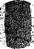
Hair in trichophytia superfic. capillitii.
Superficial trichophytia of smooth skin (trichophytia superficialis cutis glabrae) appears as sharply defined, round, slightly elevated inflammatory disks, in which in most cases two zones can be distinguished: a peripheral spot-vesicle-crust-like rim and a regressive scaling center (see separate table, figure 1). In some cases, vesicles do not form and the entire lesion appears as a scaling, sharply defined spot with regression in the center; sometimes the changes are uniform throughout the plaque, in such cases there is a similarity with seborrheic eczema. More often the open parts of the body are affected: face, neck, hands and forearms; the number of foci varies, sometimes they merge, forming bizarre patterns on extensive areas of skin. Subjectively—itching. The course is acute. In recent years, the not infrequent cases of so-called atypical chronic trichophytia of smooth skin (trichophytia chronica cutis glabrae) have been studied, which occurs mainly in young women and is localized mainly on the legs, thighs and buttocks. This form differs from ordinary trichophytia of smooth skin by the absence of sharp demarcation and peripheral rim; the lesion appears as merging dark-violet or rosy-blueish, slightly scaling spots, resembling disks of seborrheic eczema or psoriasis. This very therapy-resistant and sluggishly progressing form of trichophytia is usually caused by Trichophyton violaceum and develops on a predisposed soil, more often in women with a labile peripheral vascular system and endocrine disorders. Often the nails and hairy scalp are simultaneously affected; there are known cases of trichophytia affecting almost the entire skin, mucous membranes, lymph glands and bones (Chernogubov and Pelevina). Special mention is deserved by chronic trichophytia of the palms (trichophytia chronica palmarum), which in most cases is combined with trichophytia of the nails. The lesion here is expressed only in diffuse or limited scaling, inflammatory phenomena are clinically either not noticeable at all or barely expressed. Isolated trichophytia of the palms and nails is relatively not infrequent, always runs for a very long time, is very resistant to therapy. In all forms of chronic trichophytia of smooth skin, trichophytin reactions are as a rule negative, which is why one can speak here of anergic forms of trichophytia. In cases of infection with Trichophyton of animal origin (more often in rural residents), the process usually takes an acute and deeper character, deep trichophytia (trichophytia profunda) develops, which occurs both on the hairy parts of the head and face, and on smooth skin (see separate table, figure 2). When localized on the hairy scalp, deep trichophytia is called kerion Celsi, when localized in the area of the beard and mustaches—sycosis parasitaria (parasitic sycosis). Acutely inflammatory disks of various sizes, protruding above the level of normal skin and sharply defined, arise, densely studded with small holes, from which, upon pressure, like from a honeycomb, pus is released. The hairs on the affected areas are partially absent, while the remaining ones are easily removed. Subjectively—painfulness. The number of foci rarely exceeds 5-6. Reactions with trichophytin are always positive (allergy). Unlike superficial trichophytia of the hairy scalp, deep trichophytia resolves quite quickly, sometimes even spontaneously, and often leaves behind scar atrophy. - Trichophytia of the nails (onychomycosis trichophytica, trichophytia unguium)—see Onychia. While trichophytia of the hairy scalp and superficial trichophytia of smooth skin are more often in children, chronic trichophytia of smooth skin, trichophytia of the nails, and deep trichophytia of smooth skin are more often found in adults. Superficial trichophytia of the hairy scalp is often complicated by pyoderma, staphylococcal impetigo arises. Another complication (more often of deep trichophytia) is the development of trichophytid, which develops on allergic skin by the carriage of individual elements of the fungus from the primary focus into the bloodstream. For the development of trichophytid, three moments are necessary: a primary focus of trichophytia, irritation of it, and an allergic state of the skin. The appearance of trichophytid is often preceded by prodromal phenomena: malaise, headache, vomiting, increased temperature, etc., sometimes the regional lymph glands enlarge (more rarely suppurate). With the appearance of the rash, these phenomena subside. The clinical picture of trichophytids is diverse. Along with lichenoid trichophytid (see Lichen) there are (more rarely) measles-like, scarlatina-like, eczematoid, nodular and other rashes. Within 5-20 days, trichophytids regress spontaneously. Sensations from the rashes are minimal. In the rashes of trichophytids, fungi as a rule cannot be found, although there are individual exceptions to this, as well as the successful isolation of fungi from the bloodstream during the acme of trichophytid. In case of suspicion of trichophytic nature of the skin lesion, it is necessary to perform a bacterioscopic examination for fungi of altered and broken hairs and scales. In the absence of a laboratory on site, the material for examination can be sent to the laboratory in a test tube. In suspicious cases, a single negative result is not conclusive, after 3-4 days a repeated examination is performed, and during this time all kinds of ointments and head washing are prohibited. At the same time, in such cases, sowing on Sabouraud's medium can be performed. Bacterioscopic examination for fungi is performed as follows: 1-2 drops of a 10% solution of caustic soda (for softening and clarifying the horn) are applied to the hairs and scales placed on a slide, a cover slip is placed on top, the preparation is heated for 1-2 minutes over a flame and examined under high magnification. If the fungus is present in the preparation, its elements are found: a uniform in thickness, more or less long mycelium consisting of quadrangular, oval and round spores, and the latter in an isolated state from each other. Superficial trichophytia of the hairy scalp differs clinically from microsporia (see.) by its smaller size, indistinct demarcation and various shapes of foci, as well as by the presence of 'black dots' (hairs broken off at skin level). From favus (see.), trichophytia differs mainly by the absence of favus-like shields (scutula) and the outcome in atrophy. The squamous form of trichophytia of the hairy scalp sometimes presents some similarity with dry seborrhea, from which it differs by its focal arrangement, changes in the hairs and the presence of fungi. The impetiginous form differs from vulgar impetigo by less pronounced inflammatory phenomena, the absence of individual pustules and the presence of fungi. When complicated by pyoderma, the clinical picture of trichophytia is obscured, and in the absence of fungi, a final diagnosis can be made only after the elimination of the complication. The spot-squamous form of trichophytia of smooth skin sometimes resembles disks of seborrheic eczema, from which it differs by a brighter inflammatory coloration, the correctly round shape of the foci, an acute course and the presence of fungi. Kerion Celsi differs from pyoderma by the sharp demarcation of protruding disks, slight inflammatory phenomena around the latter, a honeycomb-like appearance and the presence of fungi; regarding the latter, it must be remembered that due to the acuteness of the process and suppuration, finding fungi in deep trichophytia may be difficult; fungi should be sought in the peripheral, not yet disintegrated areas. Sycosis parasitaria differs from sycosis non parasitaria (see Sycosis) by the presence of large, deep infiltrated, sharply defined and very painful nodular tumors. The prognosis for deep trichophytia and superficial trichophytia of smooth skin is always favorable. As for superficial trichophytia of the hairy scalp, the prognosis is also good if appropriate treatment can be carried out. It is significantly worse in chronic trichophytia of smooth skin and in cases of multiple nail lesions. Treatment of superficial trichophytia of the hairy scalp (and microsporia): the fungus settles inside the hair or outside it, forming on a certain space as it were a sheath for it, which makes the application of epilation (removal) of diseased hairs necessary. The basic principle of treatment consists in epilation followed by the use of disinfectants. The best method of epilation is X-ray rays, which cause not only the falling out of hairs but also a temporary suppression of the function of the hair follicle [usually new hairs appear on average on the 20th day, after X-ray epilation—through 65-70 days (Chernogubov)]. A longer period of absence of new hair growth allows for better subsequent medicinal disinfectant treatment, which to a greater extent guarantees against relapse. Most authors consider it necessary to epilate the entire head, even in cases where there are only 1 or 2 small foci of lesion. Irradiation of the entire head takes several days depending on the method (three-field, four-field, etc.). Epilation with X-ray rays should be carried out only by experienced radiologists, since very precise dosing is necessary (see Radiotherapy).
Administering a smaller dose leads to subsequent relapse because not all diseased hairs fall out, while administering a larger dose can lead to mild or severe X-ray dermatitis or, in the best case, to permanent alopecia. X-ray epilation can be performed on children starting from 2 years of age. Hair begins to fall out on the 11-14th day after irradiation, by the 25-30th day only individual hairs remain, which must be epilated manually. Vellus hair should also be removed, as fungus can also be present in them. Before hair loss begins, the affected areas are rubbed with a 1-2% salicylic or salicylic-sulfur ointment, and the head is washed 2-3 times. During the period from the onset of hair loss until the completion of epilation, daily washing of the head and removal of easily removable hairs is performed. After epilation is completed, disinfecting ointments and liquids are applied for a long time: tincture of iodine, Wilkinson's ointment, ointments with tar, salicylic acid, β-naphthol, pyrogallic acid, etc. It is recommended to prescribe Ung. Wilkinsoni at night and tincture of iodine (2-10%) in the morning. Such treatment (with breaks of 1-2 days after 5-7 days of application) is continued for 1-1½ months, and both during and after treatment, control examinations for fungi should be performed. A child is considered cured if, upon repeated microscopic examinations of new hairs, fungal elements are not found. For some reason, if X-ray epilation cannot be used, manual epilation has to be resorted to, which requires great patience from the patient and time from the person performing it. With the help of epilation forceps, hairs are removed completely not only on the affected areas but also in a space of 1 cm around them. Thallium acetate, proposed by Buschke for epilation purposes, often causes unpleasant side effects (see Thallium). Thallium is given internally at a dose of 0.008 per 1 kg of the patient's weight; it is taken in one dose on an empty stomach in sweet water. Hair loss begins after 12-20 days and ends in a few days, but new hair starts to grow after 8-22 days; such a short 'bald' period is one of the major disadvantages of this method. At present, thallium cannot be recommended for widespread use. For single foci and when X-ray epilation cannot be used, the Sachs-Sobolev method can be used: the affected area is epilated with forceps and rubbed 3 times in a row (every hour) with 10% tincture of iodine; after the 3rd rubbing, a 10% white mercury ointment is immediately applied; the rest of the head is rubbed with tincture of iodine or wiped with 1% sublimate alcohol once a day. After 2 hours, the ointment dressing is removed, the skin shows a picture of bullous dermatitis; after puncturing the blisters, a 10% ichthyol ointment is applied. In the following days, the same mercury ointment is used. After 5-6 days, when the reaction subsides, the head is washed with soap and the entire procedure is repeated; this is done 3-4 times. For widespread lesions, this method cannot be recommended. Treatment of superficial T. of smooth skin: for 5-7 days, the affected areas are rubbed 2 times a day with 10% tincture of iodine or once a day with tincture of iodine and the other time with Wilkinson's ointment. If a medicinal dermatitis develops during treatment, indifferent ointment is prescribed instead of disinfectants; after the irritation subsides, if necessary, peeling therapy is repeated. In cases of chronic T. of smooth skin, along with local treatment, general treatment should also be used - systematic intradermal injections of trichophytin. Treatment of deep T.: epilation is not required, as here the fungus, due to the intense inflammatory reaction, dies off relatively quickly even spontaneously. For severe inflammatory phenomena, cold compresses of aluminum acetate (2 tablespoons per glass of water), of resorcin (1-2%) or others are used; for less inflammation - alcoholic (30°) or sublimate (1:1,000) compresses. If the inflammatory phenomena are not so pronounced, a 5-10% white mercury ointment Ung. Wilkinsoni or simply a 5-10% tar ointment can be applied immediately, but one must take into account the possibility of allergic rashes - trichophytids - appearing due to irritation of the primary foci. In sluggishly progressing cases, specific (injections of trichophytin) as well as non-specific (injections of milk, autogenous blood, etc.) Reiztherapie are indicated. Treatment of T. of nails - see Onychia. Prevention of T. and microsporia. One of the main measures of public prevention of T. is the timely detection, isolation and treatment of patients. A child suspected of having T. should not attend schools or other places where children gather; until the diagnosis is clarified, children suspected of having T. in orphanages should be placed in special fungal isolation wards or rooms. A thorough examination of all children entering schools, orphanages, playgrounds and kindergartens is necessary, as well as periodic examinations at least once every three months. No less important is the examination upon admission and subsequent observation of the serving personnel of these institutions. Children in orphanages and kindergartens should have separate beds, towels, napkins, etc.; hair washing should not be done in common basins, but should use taps with warm water. If a schoolchild becomes ill, all children in the group in which he studies should be examined; if there are children in the family, the patient should constantly wear a cap on his head, preferably paper, which should be changed frequently, and the used one should be burned. Hats of patients should be destroyed, bedding, towels, etc. should be boiled in water with soda, pillows, blankets and outer clothing should be disinfected in a chamber (preferably in sulfur vapors). Children can be admitted to school only after treatment is completed, i.e., from the time new hairs appear, in which no fungus is found: on average - 2 months after X-ray irradiation; subsequently, clinical and microscopic control is necessary for another month. If only smooth skin is affected in children, they should be isolated until clinical and microscopic recovery (on average for 6-7 days), if only covered parts are affected - until clinical symptoms disappear (for 3-4 days). In the fight against T., great importance is attached to the supervision of the sanitary-hygienic condition of barber shops, with special attention to the disinfection of instruments, linen and periodic examinations of barbers. Fight against T. in animals: regular veterinary supervision, hygienic livestock yards, isolation and treatment of sick animals with disinfection of premises, harness, etc. From the above, it follows that sanitary-educational work plays a colossal role in the fight against fungal diseases.
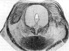
Fig. 1. Trichophytia corporis. Fig. 2. Trichophytia profunda capillitii. Fig. 3. Trichophytia superficialis capillitii. Fig. 4. Trichophyt tonsurans. Fig. 5. Nucleus n. trochlearis, transverse section of the brainstem. (Figs. 2-4 according to Friboes, fig. 5 according to Marburg.)
L. MASHKLLEYSON. TRICEPS-REFLEX, a tendon reflex consisting in extension of the forearm when a hammer strikes the tendon of m. triceps brachii. It is best elicited in such a way that the subject places his arm bent at an angle on the examiner's left hand, who strikes with the right hand just above the olecranon. The subject is asked to relax the muscles of the arm as much as possible beforehand. The T.-r. is less constant and varies more in its manifestation than the tendon reflexes of the lower extremities, often being weakly expressed even in normal conditions. For this reason, it is significantly inferior to the latter in diagnostic value. For this reason, when examining, it is necessary to attach importance mainly to a noticeable difference in the expression of T.-r. on both sides. The reflex arc of T.-r. passes through the C7 segment. -T.-r., like other tendon reflexes (see), shows changes in various processes of both peripheral and central nervous systems. It is weakened up to complete extinction in various pathological processes that disrupt the integrity of its reflex arc in any of its parts (lesion of the peripheral neuron). On the contrary, with lesions of the pyramidal system (central neuron), it is to a greater or lesser degree increased, as well as with a general increase in the excitability of the nervous system of a functional nature (neurasthenia, hysteria). In some cases, an inversion of T.-r. can be observed, which is expressed in flexion of the forearm instead of its extension when striking the tendon of m. triceps brachii. Inversion of T.-r. can occur, for example, with a lesion of the peripheral motor neuron of T.-r. or the extensor muscles of the forearm with a simultaneous lesion of the pyramidal system, as a result of which the irritation is transmitted to the flexors of the forearm instead of the extensors.
Related articles
Mentioned in
Cite this page
“Trichophytia.” Soviet Medical Encyclopedia. English translation of Bolshaya Meditsinskaya Entsiklopediya, 1st ed. (Moscow, 1928–1936), ed. N. A. Semashko. https://sovietmedicalencyclopedia.pages.dev/article/trichophytia/