Fixation
Historical document, translated for reference. It reflects medical knowledge of the 1920s–30s and is not medical advice.
Summary
An overview of fixation in histology from the 1920s-1930s Soviet medical encyclopedia, detailing various fixatives like formalin, alcohol, and chromium mixtures, their preparation, and their specific applications in tissue processing.
Encyclopedia article (1928–1936)
FIXATION in histology is the first and very important step in the process of preparing histological specimens. To fix a tissue means to prepare it for subsequent processing methods. As a result of fixation, quite complex changes occur in various tissue elements, due to which during subsequent processing these elements acquire the property of reacting in a specific way when exposed to various stains and reagents. Some fixatives, for example, make it possible to particularly well preserve and reveal the structure of the nucleus, others promote the subsequent processing of protoplasm and its organelles, still others preserve lipoids, and so on. Histological technique does not know of a fixative that would simultaneously allow all details in a specimen to be revealed equally well. Therefore, if one wishes to carry out a deeper, comprehensive study of tissue, the same object (pieces from it) must be fixed by various methods, each of which makes it possible to conduct the tissue study in one direction or another. For the theoretical foundations of fixation, see Histological Technique. The choice of fixation method depends mainly on the purpose of the further processing of the material. Another indication for choosing the fixation method is the time available to the researcher to obtain the final result of specimen processing, since various fixation methods require different amounts of time. Finally, the size and nature of the object itself matter when choosing a fixative. Thus, for the purposes of studying topography, orientation in the process and its spread, it is appropriate to use only those fixatives that easily and quickly penetrate tissues, such as formalin, and therefore allow one to take relatively large pieces, sometimes even whole organs. Fixatives used for cytological study purposes, for example, those containing osmium, on the contrary, penetrate very shallowly and require very small objects. Alcoholic fixation, with the exception of only some special purposes, is completely unsuitable for objects very rich in water (e.g., embryonic tissue), because it shrinks them beyond recognition, and so on. Thus, in each individual case, a suitable fixative is chosen, taking into account both all its positive aspects and disadvantages. For all fixation methods, it is necessary that the fixation object be in a fresh state. However, routine patho-histological examination can be successfully carried out on pieces taken recently from a corpse, for example, 24--48 hours after death. For some, especially cytological studies (study of mitochondrial structures, identification of nuclear division figures, etc.), it is necessary that the pieces be taken immediately after death or even from a living organism (biopsy) and immediately placed in the fixative. The significance of temperature during fixation comes down to the fact that raising it accelerates, and lowering it slows down fixation. Usually, fixation is carried out at room temperature. A temperature of 37° (fixation in a thermostat) is in some cases highly desirable, especially with fixatives that slowly penetrate into the depth of the tissue, or if one wishes to accelerate the fixation itself. Of the fixatives in histological technique, only a few, such as formalin, alcohol, and acetone, have independent fixing significance; in most cases, various fixing mixtures are used, the constituent parts of which (potassium dichromate, sublimate, osmium, and many others) complement and mutually correct each other. Formalin is used in 10-25% solutions [commercial 40% formalin (see) is considered one hundred percent]. Formalin perfectly preserves the general structure of tissues and the basic morphological details of the specimen, preserves fats, lipoids, allows the fixation of very large objects, even whole organs, makes it possible to use the freezing method, and allows most stains. In patho-histological practice, formalin fixation is the most widespread. Of the fixing mixtures that include formalin, the most commonly used are Orth's fluid (see Müller's fluid), Zenker's fluid (see Zenker's fluid), Helly's fluid (see below and Formalin, formalin in histological technique). Formalin in museum technique—see Anatomical preparations. Certain disadvantages of formalin fixation (insufficient revelation of details of the nuclear and protoplasmic structure) can be partially corrected by additional fixation of frozen sections with chromium salts, as well as osmium and sublimate (Weil). For chroming, frozen sections are placed for a day in a 5% solution of potassium dichromate. Then the sections are rinsed in water and, if they are very yellow, placed for some time in salt water. Absolute ethyl alcohol is also used for fixation (alcohol denatured with formalin can also be taken). Alcoholic fixation, due to a number of its major disadvantages, is used very rarely (shrinkage, especially of water-rich tissues, extraction of many pigments, dissolution of lipoids, etc.). Alcoholic fixation is necessary when it is necessary to preserve substances in tissue that are soluble in aqueous solutions of other fixatives (glycogen, uric acid, etc.). Alcoholic fixation gives good results when examining tissue for the presence of microbes. Of the fixing mixtures that include alcohol, the most commonly used is Carnoy's fluid (absolute alcohol 6 parts, glacial acetic acid 1 part, and chloroform 3 parts). It is used for rapid fixation followed by embedding of the specimen. The duration of fixation is several hours, followed by absolute alcohol, changed every 12-24 hours, and embedding. Kulchitsky's fluid (95% alcohol 100 parts and glacial acetic acid). It is used in the study of zoological material and in embryology. Acetone fixes quickly, but shrinks tissues severely. It is used rarely, mainly for very urgent preparation of a specimen. Fixation (of small pieces) takes several hours. Embedding in paraffin. Of the huge number of fixing mixtures and their modifications, the most widespread are mixtures containing salts of chromic acid (potassium dichromate). 1) Müller's fluid (see Müller's fluid) is currently not used in its pure form, because it possesses a large number of major disadvantages, but it often enters as a main component into other mixtures (Zenker's fluid, Helly's, Orth's, etc.). 2) Orth's fluid: 90 cm3 of Müller's fluid and 10 cm3 of pure formalin. The addition of formalin is performed ex tempore. The duration of fixation is no more than 18-24 hours, otherwise the pieces become brittle. Washing in running water—24 hours. It is very good to transfer pieces after fixation with Orth's mixture into pure Müller's fluid for 2-3 days. Müller's fluid must be taken in large quantities and changed daily. After fixation, washing in running water—24 hours. Orth's fluid fixes all tissue elements well. In addition, it is used, just like other chromium mixtures, to reveal so-called chromaffin cells. 3) Regaud's fluid in its composition and fixing properties is close to Orth's fluid. Its composition: 21/2% solution of potassium dichromate 80 cm3, pure formalin 20 cm3 (formalin is added ex tempore). Fixation 18-24 hours. Washing in running water—24 hours. Mixtures containing potassium dichromate and sublimate should be placed in one of the first places among all fixing mixtures. Of greatest importance are: 1) Zenker's fluid and 2) Helly's fluid (see Zenker's fluid). The nuclear structure, karyokinetic figures, cell protoplasm, blood hemoglobin, and its derivatives are perfectly preserved by these fixatives, which is why they can be widely recommended for cytological studies, especially in the study of hematopoiesis. In Helly's fluid, tissues swell less than in Zenker's fixative. Very good results are obtained with the combined use of fixatives, namely, after fixation in Helly's fluid (6 hours), the pieces are transferred for 24 hours into Zenker's fixative. Both fixatives require a relatively small size of pieces, thorough washing in running water, and removal of sublimate precipitates by iodination (see Zenker's fluid). Storage of material fixed in all the chromium and chromium-sublimate mixtures listed above is carried out in 10% formalin or 75% alcohol. The disadvantages common to all these mixtures are: a certain painstaking nature of the methods (washing, iodination), low and sometimes complete unsuitability of the material for producing sections by the freezing method, as well as the difficulty, and often impossibility, of revealing lipoids. Fixatives containing osmic acid (or rather its anhydride OsO4) are widely used in cytological practice, because they make it possible to study the finest structure of the cell. When preparing osmium solutions (usually a 2% solution is prepared), the ampoule with osmium, as well as the glassware in which osmium is dissolved, must be chemically absolutely clean. The ampoule is broken inside a jar and closed with a ground stopper. When formulating fixing mixtures, the osmium solution is added ex tempore. Both fixation and storage of osmium solutions are carried out in dark glass jars with ground stoppers. The thickness of pieces during fixation in mixtures containing osmium should not exceed 1-2 mm.
Following fixation, thorough washing in running water is necessary; otherwise, the preparation stains poorly and darkens easily. Storage of fixed material is carried out in 80-90% alcohol. 1) Flemming's fluid is a mixture of 1% chromic acid (15 cm3), 2% osmic acid solution (OsO4) (4 cm3), and glacial acetic acid (from 10 drops to 1 cm3). Fixation lasts for 24 hours or more. Washing of the pieces in running water takes 24 hours. Flemming's fluid makes it possible to very electively fix the finest structure of both protoplasm and especially the nucleus (caryokinetic figures). For the purpose of chromosome counting, 1% urea is added ex tempore to Flemming's fluid. 2) Hermann's fluid excellently preserves the fine structures of both protoplasm and nuclei (see Hermann's fixing fluid). 3) Altmann's fluid. A mixture of equal parts of a 5% potassium dichromate solution and a 2% osmic acid solution. Fixation takes 24 hours. Washing in running water takes 6-12 hours and longer. Embedding in paraffin. Altmann's fluid is an excellent fixative for studying the finest structures of protoplasm—mitochondria, chondriosomes, bioblasts, etc. The same fixing properties are possessed by 4) Meves' mixture: 15 cm3 of 0.5% chromic acid solution, 3-4 cm3 of 2% osmic acid solution, and 4-6 drops of glacial acetic acid. Fixation lasts for 3-4 days (preferably in an incubator). Washing in running water (24 hours) and embedding in paraffin. 5) Benda's method. Fixation in Flemming's fluid for 8 days (in a dark place). Washing in running water (1 hour). Then the pieces are placed for a day in a mixture of 1% chromic acid (1 part) and purified wood vinegar (1 part) (the fluid should be changed), after which the pieces are transferred for a day to a 2% potassium dichromate solution. Then the pieces are rinsed several times in distilled water and washed in running water for 24 hours. Embedding in paraffin should be done as quickly as possible. Among less common mixtures containing osmium, one can mention: 1) Maximov's fluid, which is a mixture of Helly's fluid (prepared ex tempore), 100 cm3, and 2% osmic acid, 10 cm3. 2) Champy's fixative: a mixture of 1% chromic acid (7 cm3), 3% potassium dichromate solution (7 cm3), and 2% osmic acid (4 cm3). Fixation of the pieces lasts for 24 hours. Washing in running water takes 24 hours. It is recommended to subsequently transfer the pieces for a day into a mixture of wood vinegar (1 part) and 1% chromic acid (2 parts). Then the pieces are treated with 3% potassium dichromate for 4 days and washed in running water for a day. Among the modifications of this method, Minouchi's method deserves attention, giving excellent results in the study of nuclear structure and chromosome counting. Fixation in a mixture of 2% osmic acid (4 cm3), 1% chromic acid (8 cm3), and 3% potassium dichromate solution (8 cm3). Washing in running water and embedding in paraffin. Fixation of smears of various exudates, secretions, microbe cultures, blood, etc. Fixation of smears for the presence of microorganisms is carried out by passing the smear, previously dried on a glass slide, through the flame of an alcohol or gas burner three times (smear side up). For the simultaneous study of cellular elements as well, fixation should be performed with absolute methyl alcohol or a mixture of absolute alcohol and ether (see Nikiforov's methods). Fixation for finding spirochetes and fixation of smears from broth cultures are carried out in the same way. Fixation of smears for the purpose of studying cell composition is undesirable with alcohol; it is more expedient to stain the smear with hematoxylin-eosin, Sudan, or perform the oxidase reaction (secretions from the mammary glands, punctates of neoplasms, etc.), which is very conveniently done in formalin vapors. Very good results, especially for cytological research purposes, are given by fixation of smears (blood, cell-containing fluids, etc.) in osmium vapors. For this purpose, a 1% osmic acid solution is poured into the bottom of a stoppered vessel. The smears are placed on a glass stand and the jar is tightly closed. Fixation occurs rapidly. Good results are obtained by fixing bone marrow punctates according to the Duesberg-Weidenreich method (see Bone marrow, functional diagnostics). For the fixation of blood smears, see Blood, Nikiforov's methods.
A. Kostin.
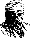
FILATOV, Vladimir Petrovich (born 1875), a prominent ophthalmologist. Upon graduating from the university in 1897, he took the position of an ordinarius at the eye clinic of Moscow University (under the direction of Prof. A. A. Kryukov). In 1899, he moved as an ordinarius to the Moscow Eye Hospital. In 1904, he passed the doctorate examination. In the same year, he moved to the position of ordinarius at the eye clinic of Novorossiysk University in Odessa, which was headed by Prof. S. S. Golovin. In 1908, he defended his dissertation for the degree of Doctor of Medicine on the topic "The Doctrine of Cellular Poisons in Ophthalmology" (Odessa, 1908). From 1909 he was a privatdocent, and from August 13, 1911, a professor at Novorossiysk University in the chair of ophthalmology with a clinic, a position he still holds at the Odessa Medical Institute. For several years he served as head of the research department of clinical and experimental medicine in Odessa and head of its ophthalmology section. Filatov is not only one of the largest researchers in his specialty in the USSR, but is also considered a major authority in general surgery, especially plastic surgery. The method of plastic surgery on a rounded pedicle proposed by him is now adopted not only in the USSR, but also abroad. In recent years, Filatov has acquired particularly wide fame through his successfully performed corneal transplantation operations (in 14 cases). 106 scientific works by Filatov are devoted to various issues of ophthalmology and its borderland fields.
Related articles
Mentioned in
Cite this page
“Fixation.” Soviet Medical Encyclopedia. English translation of Bolshaya Meditsinskaya Entsiklopediya, 1st ed. (Moscow, 1928–1936), ed. N. A. Semashko. https://sovietmedicalencyclopedia.pages.dev/article/fixation/