Histological Technique
Historical document, translated for reference. It reflects medical knowledge of the 1920s–30s and is not medical advice.
Summary
This article from the first edition of the Great Medical Encyclopedia covers the foundational methods of histological technique, including fixation, sectioning via microtome, paraffin and celloidin embedding, and staining of biological tissues for microscopic study.
Encyclopedia article (1928–1936)
HISTOLOGICAL TECHNIQUE. Contents: Methodology of histological research..... 242 Theoretical foundations of histological technique........... 246 Histochemistry................... 253 Stains used in histological technique......... 258 Histological technique—the technique of studying the microscopic structure of cells, tissues, and organs of plants and animals. The branch of histological technique dedicated to cell structure is usually designated as cytological technique. The simplest and in many cases extremely valuable method of histological technique is the study of native material, i.e., living or fresh, surviving histological elements or cells. This is possible, however, only in the case of very small and transparent objects, e.g., blood cells and others, as well as thin tissue films like the mesentery of lower vertebrates, etc. The method of teasing fresh tissues into separate elements is also frequently used, e.g., when studying muscle, nerve fibers, etc. In many cases, direct examination of living tissue can yield an extraordinary amount of information and serves as the basis for a correct evaluation of the results of standard methods of histological technique. Observation in the living state often contributes significantly to understanding the physiological significance of various structures, such as skeletal and contractile elements, etc. However, in most cases, direct study of native material is hindered either by opacity or by optical homogeneity of the studied objects. The latter is due to the fact that the refraction and absorption of light by various histological structures are extremely close; therefore, their optical differentiation is for the most part extremely difficult. However, by applying so-called fixatives, which cause the coagulation of protein and other constituents of protoplasm, one can achieve a state where various tissue elements form coagulates having different refractive indices. This makes it possible to reveal many histo- and cytological elements, e.g., nuclei, nucleoli, fibers, by very simple means, e.g., the addition of acetic or osmium acid, etc. However, the optical differentiation obtained in this way is far from perfect, and in most cases, the finest morphological details can be revealed only with the help of various stains. Staining is possible only in the case of dead tissues, since living cells do not stain at all, and can only accumulate dyes in the vacuoles of the protoplasm (see Vital staining). Therefore, cells must be preliminarily preserved or fixed, i.e., killed, but in such a way as not to disrupt natural morphological relationships as far as possible and not to produce gross artifacts (see). Histological objects are examined under the microscope mainly in transmitted light, less often in polarized light (reflected light is practically not used). For some special purposes, dark-field microscopy is used. For examination in transmitted light, objects must be sufficiently transparent. Therefore, the task of histological technique is the preparation of very thin specimens. Thus, histological technique reduces to the fixation of cells or tissues, the preparation of sections, and their staining. When studying individual histological elements, free or obtained by teasing, one can dispense with whole mounts, bypassing the preparation of sections. Methodology of histological research. Fixation. The purpose of fixation is the rapid killing of tissue by protein coagulation (and partly of lipoids) while preserving the natural morphology of the structure as much as possible. Originally, the significance of fixation was seen in the hardening of soft tissues so that thin sections could be made from them. However, many hardening substances are known that, although they coagulate protein, are nevertheless unsuitable as fixatives because they cause major structural changes. Fixatives are primarily salts of heavy metals: Hg, Cr, Os, Pt; among acids—picric, acetic, trichloroacetic, then alcohol, acetone, formol, and some other substances. Only the last three are used as independent fixatives, while in most cases the most diverse mixtures are used, since in this way the best results are achieved. The most widespread are the fluids of Flemming, Zenker, and Bouin. After remaining in the fixative for some time, from several minutes to several days (each fixative has its own optimum time), small pieces of tissue must in most cases be washed free of the fixing fluid. This is achieved by washing in water or alcohol.—This completes the first operation and proceeds to the preparation of sections. But if the pieces contain hard tissues, such as bone, chitin, or pathological lime deposits, they are preliminarily softened; thus, calcareous elements are dissolved by acid (see Decalcification), chitin is softened by diaphanol (chlorine dioxide dissolved in acetic acid) or cedar oil. The simplest method of preparing sections is cutting pieces by hand with an ordinary shaving razor. This method has been very widely applied in botany and is still used. In botanical objects, such sectioning without further processing (and even without fixation) is possible because plant tissues are relatively very dense; in the case of animal objects, this method is used extremely rarely. At the dawn of the development of sectioning technique, cutting in the liver was used, where a piece was clamped between two plates of fresh liver, and the cut was made through the entire mass. At present, microtomes (see) are used to prepare sections, allowing very thin sections to be made and their thickness to be regulated. The simplest method is cutting frozen pieces. A piece of tissue fixed in formol or other fixatives is frozen (with ether or liquid carbon dioxide) and thus freezes onto the stage of a special microtome. Frozen tissues are hard enough to allow the microtome blade to cut them into very thin sections (down to 5 µ). Sections are immediately transferred to water, where they thaw. Disadvantages of this method: 1) sections of some tissues crumble and disintegrate upon thawing; 2) it is impossible to work with very small objects; 3) it is almost impossible to prepare a large series of sections, since part of them always perish.—Much more widespread is the cutting of tissue pieces embedded in paraffin or celloidin. When embedding in these substances, the piece must be preliminarily impregnated with a liquid that, on the one hand, would mix with water, and on the other hand, would dissolve paraffin and celloidin. In the case of paraffin, various hydrocarbons and their derivatives serve this purpose: benzene, xylene, toluene, chloroform, etc., as well as various oils. Usually, the piece is transferred first to 50% alcohol, then to 70-80-100% alcohol; next follow mixtures of 100% alcohol with increasing amounts of xylene, then pure xylene -> xylene saturated with paraffin (at 37°), and finally pure paraffin (at 60°), which is usually changed 3-4 times. Paraffin embedding is performed in an incubator. When the piece is thoroughly impregnated, it is quickly cooled, because upon slow cooling paraffin crystallizes. Before the universal adoption of paraffin, embedding pieces in soap was used. When embedding in celloidin, the object is likewise passed through alcohols of increasing strength, then transferred to a mixture of alcohol and ether (aa) and finally impregnated with alcohol-ether solutions of celloidin of ascending concentration up to 8%. Then it is removed, placed on a piece of wood or ebonite, and transferred to a vessel with chloroform vapors; in this process, the celloidin hardens. Next, the piece attached to the wooden block and embedded in celloidin is immersed in 70% alcohol, where it reaches the consistency of soft cartilage. The embedded piece is called a block. In this state, objects can be preserved indefinitely without any harm to the structures. Both of these embeddings require a lot of time, because in most media the piece must lie for a day or more. With the help of a microtome, any piece can be sliced into sections from 1 µ to 20 µ without losing a single one. From paraffin blocks, ribbons of sections can be easily obtained due to the fact that during cutting they adhere to each other by their edges. Sections are transferred to slightly warmed water, on which they flatten out easily (they must not melt). After this, sections are placed on glass slides or cover slips and dried at 37°, during which they adhere firmly to the glass. Before staining, the paraffin must be removed, for which the glass slides with adhered sections are immersed in xylene (or another substance) in which the paraffin dissolves; then the xylene is washed out in 100% alcohol, and through alcohols of descending strength, the sections are brought down to water (if staining with an aqueous dye is intended). In the case of celloidin embedding, the prepared sections go into 70% alcohol and then into water; they are stained in small dishes. The celloidin itself in most cases does not interfere with staining, and if necessary, it can be easily removed with a mixture of alcohol and ether. Staining. Its purpose is the clear differentiation of various structures. The outcome of staining strongly depends on the preceding fixation. The best stainability is achieved after alcohol, formol, then sublimate and trichloroacetic acid, whereas after osmium salts, the best of the known fixatives, staining is frequently difficult.
However, staining can be significantly facilitated by treating the section with hydrogen peroxide, thus removing all tissue-bound osmium, or by keeping the sections in sublimate. For successful staining, the complete removal of all traces of the fixative is usually necessary. An important factor in staining is also the treatment of the tissue with alcohol (to remove fats); during paraffin and celloidin embedding, this is achieved in the process of embedding, whereas when cutting on a freezing microtome, it is highly recommended to place the sections in alcohol for some time before staining. In staining, the chemical structure of the dye, as well as the method of its application, is of paramount importance. Not every colored compound is a dye, but if chromophore groups are present in it (see Aniline, aniline dyes), the charge—in other words, the reaction of the dye (basic or acidic)—is of the greatest importance; this also determines the method of its application. Some dyes stain the substrate directly. Other dyes are incapable of direct action and require prior mordanting of the substrate with a substance, most often colorless, which on the one hand binds to the substrate and on the other to the dye itself. Thus, colored lakes are obtained. In this process, the color of the dye itself is often greatly changed. Various salts of aluminum, chromium, iron, etc., are used as mordants; their alum compounds are most commonly used (see Hematoxylin). A distinction is made between progressive and regressive staining. Progressive staining is called such when, after the dye bath, the preparation already has the final degree of staining. For example, the basic dye Methyl green in an acidified solution gives a very clear staining of the nuclei, without overstaining them at all and without staining the protoplasm. Progressive stains can also be obtained with acid dyes; they serve mainly for revealing connective tissue fibers (this is, for example, Orcein according to Unna, Fuchsin S-picric acid according to van Gieson). - Regressive staining always proceeds in two stages: 1) intensive and uniform overstaining of the entire object and 2) differentiation of the preparation, which consists in removing excess dye from the section. Experience shows that various tissue and cellular structures do not stain at equal speeds; therefore, it is possible to stain any single structure in isolation. An example of regressive staining is Heidenhain's iron hematoxylin stain. Depending on the degree of differentiation, this method can produce staining of nuclei, fibrils, cell walls, mitochondria, etc. - By other methods, isolated staining of elastic fibers, myelin sheaths of nerve fibers, fibrin, etc., can be obtained. Extremely widespread is the staining of preparations in many colors (so-called double or triple staining). The simplest case is the diffuse counterstaining of a progressively or regressively stained preparation with some acid dye. For example, after hematoxylin staining, when the nuclei have an elective blue or black color, the protoplasm is counterstained with acid dyes: eosin, chromotrope, etc., or the van Gieson mixture (picric acid and fuchsin). Multi-color preparations can be obtained by applying (progressively or regressively) mixtures of many dyes. Homogeneous mixtures contain either only acid dyes, for example, Unna's mixture 'WEP' (Wasserblau, Eosin, Phloxin), which stains cell walls blue, nucleoli red, and chromatin dark red progressively, or only basic ones, for example, his Methyl green-Pyronin mixture, used regressively; the result of staining is green chromatin, red nucleoli, and red basophilic protoplasm. - Heterogeneous mixtures are composed of basic and acid dyes. Examples include the Biondi-Ehrlich-Heidenhain mixture, consisting of one basic and two acid dyes (Methyl green, Fuchsin S, Orange G), and Giemsa's stain, composed of two basic and one acid dyes (Methylene blue, Azure I, Eosin). In the mixture, the dyes do not remain indifferent to each other, but form compounds that easily precipitate out of solution. Such mixtures of acid and basic dyes are sometimes called neutral mixtures. Staining can be performed not only on sections, but also on whole pieces of tissue before embedding them in the medium for sectioning. Thus, ready-stained sections are obtained. Staining of pieces can, of course, be combined with additional counterstaining of sections. Whole-mount staining methods are used mainly in zoological and embryological work. The most favored dyes in this respect are borax and alum carmines and hemalaun. The described combination of various acid and basic dyes, as well as mordants, exhausts the main methods of histological staining. Regarding special staining methods, see below - histochemistry. Metal impregnation methods are not stains in the strict sense of the word, although they serve the same purposes of histological and cytological differentiation. The principle of impregnation is based on the fact that various organoids retain salts of certain heavy metals to a very different degree. In practice, the procedure is as follows: pieces are placed in solutions of these salts, and after a certain period, the salts are reduced to the metal. An example of impregnation is the silvering of the Golgi apparatus (see Golgi method) according to Ramón y Cajal. Impregnation methods have been developed mainly for nervous tissue (see Nerve cells, Bielschowsky method); they are very numerous and often very complex, but in principle remain the same. The stained or impregnated section must be mounted in a clearing medium in order to impregnate the preparation with a medium having a refractive index close to glass and thus eliminate light scattering. For this purpose, glycerin, glycerin-gelatin, gum arabic, levulose, and various oils are used; mounting in Canada balsam is the most widespread. The sequence of mounting a preparation in Canada balsam is as follows: after staining, the preparation is passed through alcohols of increasing strength into absolute alcohol and finally into pure xylene; then a drop of balsam is applied to the preparation, and the preparation is covered with a cover slip. The thickness of the glass must not exceed 150 µm, otherwise, it will be impossible to examine the object with microscope objectives of short focal length. If the preparation is mounted in a water-soluble medium, no intermediate media are needed. Gum arabic or glycerin-gelatin is applied to the sections immediately after staining and washing the preparation. Theoretical foundations of histological technique. Fixation. To fix a tissue without changing it at all is, of course, an impossible task. Therefore, one must strive to fix it in such a way that structural changes can be taken into account. When studying the fine structures of the cell, this is very difficult to achieve, and therefore control by vital observation is desirable. In fixation, which usually leads to complete and irreversible coagulation of the protoplasm, one must always reckon with artifacts because this causes a change in the physical state of the cell colloids, whereby their dispersion changes, and the decomposition of lyophilic mixed colloids sets in. The morphological character of the protoplasmic coagulum is determined on the one hand by its colloidal-chemical properties and on the other by the properties of the fixative used. Thus, the microscopic structure of fixed protoplasm is essentially an artifact and reflects its morphology in the living state only to a very conditional degree. Consequently, only a comparative study of the action of various fixatives on a given protoplasm and its structure in the living, unchanged state can give an idea of its true structure. The inevitability of such artifacts must be reckoned with not only when evaluating the finest cytological observations, but also in purely histological questions, since under the influence of fixatives and subsequent processing in media, tissues can swell or shrink. Thus, on the basis of precise measurements, it has been established, for example, that after fixation in Müller's fluid, the spleen swells by 19%, after passage through alcohols, its size is increased by 10% compared to the initial value, and after embedding in paraffin, it turns out to have shrunk by 21% compared to the initial value (W. Berg). Many similar examples can be given. Therefore, the control of one fixative by another fixative of a different composition is highly desirable. - The first group of fixatives consists of those that do not enter into a chemical compound with the tissue, such as alcohols and acetone, which little change the nature of proteins. Here coagulation occurs as a result of dehydration of colloids. Thermal coagulation—fixation with boiling water and drying of blood smears, bacteria, etc., on a burner flame or on a copper plate at 120° (according to Ehrlich)—can also be classified here. However, these agents are used very rarely, as they produce significant artifacts (used mainly in hematology and bacteriology). Much more common is fixation by chemical coagulation, when the fixative enters into a bond with the tissue substance. These compounds have been little studied from the chemical side, since all fixatives were introduced and developed purely empirically.
Theoretically, all substances capable of coagulating proteins can be used for fixation, but practically only a few prove suitable, since the majority yield overly coarse coagulates that destroy biostructures. For example, copper salts have found no application whatsoever as fixatives, despite the fact that they coagulate proteins very rapidly and energetically. Thus, the first requirement demanded of a fixative is that it produce a fine, uniform, and compact coagulate. It was found empirically that the best fixing substances are: osmium tetroxide, platinum chloride, chromic acid, and potassium dichromate (all in acidified solution). A second advantage of these substances is their property of also fixing fats and lipoids. This is especially important, since the amount of lipid-like substances in the animal organism averages 5% with 12-15% protein. Finally, the third requirement demanded of a fixative is its rapid action—the rapid killing of the tissue and penetration into its deep layers, since otherwise a slow dying-off occurs in the depths of the piece being fixed, leading to disintegration. The speed of penetration of a fixative into tissue, being a function of the rate of free diffusion, is nonetheless not strictly proportional to the latter. Upon mixing with proteins, most fixatives are bound by them, and in this case diffusion is inextricably linked with processes of changes in the physical state and chemical nature of the substance being fixed. Consequently, the fixative exerts a sort of "membranogenic" action, which extremely impedes the further penetration of the fixative into the tissue. In the opinion of some, of particular importance in this regard are the lipoids liberated at the boundary, and the more easily the fixative dissolves them, the faster its penetration proceeds. From this standpoint, an explanation is found for the circumstance that acetic acid enters into the composition of most fixatives as an obligatory component, easily destroying and therefore removing lipoids and penetrating tissues very rapidly. Acetic acid as it were leads along other substances that otherwise would penetrate into tissues only with great difficulty. Of course, the circumstance that acetic acid itself easily precipitates proteins is also of significance, and therefore, even if the fixing agents of mixtures have a purely partial action, substances that penetrate living protoplasm with difficulty will diffuse much more readily into the precipitated colloid. Below is given a table showing the comparative rate of penetration of fixatives into spleen tissue after 12 hours of exposure at 20° (according to Tellesdnitsky): 0.3%
»
picric acid ....
»
osmic
»
....
»
chromic
»
....
»
»
»
potassium dichromate .
»
»
»
nitric
»
Empirically, it has long been concluded that mixtures of fixatives usually act much better than any single substance. Approaching this question from the standpoint of penetration speed, one arrives at the same conclusion: Flemming's fluid (1% CrO₃ - 15 hours, 2% OsO₄ - 4 hours, acetic acid - 1 hour) - 4.8 mm; Tellesnitsky's fluid (3% K₂Cr₂O₇ - 100 hours, acetic acid - 5 hours) - 5.5 mm. From the chemical side, it is interesting that OsO₄ and K₂Cr₂O₇ - the two best fixatives - themselves precipitate proteins very little, but in the presence of acetic or other acids, they act very energetically. In Zenker's fluid (5% HgCl₂, 3% K₂Cr₂O₇, 1% Na₂SO₄, 1-5% acetic acid), Glauber's salt plays a similar role (it can be replaced with table salt), which in itself is by no means a fixative. Its significance can be understood by analogy with the process of tanning leather with chrome salts. It turns out that when tanning leather with the same salt (K₂Cr₂O₇), the addition of the above-mentioned salts improves their quality (strength); a total chemical analysis shows a percentage increase in bound chromium. Along with converting some compounds into an insoluble state, the fixative can dissolve other substances and thereby remove them from the tissues. Thus, for example, to this day no method has been found for preserving glucose in a pressed preparation. In general, with all aqueous fixatives, when dealing with animal tissues, it is necessary to take into account the loss of carbohydrates. To preserve glycogen, fixatives containing at least 50% alcohol are used, but then fats and lipoids, which have much greater importance in terms of preserving structure, are removed. Acetic acid, chloroform, and others are also fat- and lipoid-dissolving fixatives. For this reason, the best fixatives are mixtures of chrome-osmium salts, which convert both proteins and lipoids into insoluble compounds. Finally, proteins can also undergo partial dissolution. A fixative containing too much acetic acid will dissolve albumins, which are abundant in protoplasm. Their coagulation is best ensured by chrome salts. For the same reason, alkaline fixatives are not used at all in histological technique due to their dissolving effect on all proteins. Thus, histological technique does not know of an ideal fixative. To obtain a more or less complete picture of the structure of a given tissue or cell, the same material must be fixed in parallel with various fixatives and then the obtained results compared. The theory of fixation is very little developed. We know that fixation is a complex process in which both external and internal factors participate. Among the former, the coagulating properties of fixatives and the speed of their diffusion should be mentioned. Among the latter, the nature of the object being fixed has the main importance. Therefore, fixatives that give very good results on some objects are almost unsuitable for others. However, it is unknown why one fixing liquid is good and another is bad. In general form, this question can be answered that it all depends on the method of formation and the nature of the coagulum. It is never possible to predict theoretically whether fixation by any liquid will be 'good' or 'bad.' This is determined empirically. Staining. Theories of staining have very great importance, since in a number of cases the morphological interpretation of the preparation depends on them. At present, there is no generally recognized theory of staining, since the final resolution of these questions is still far off. All the proposed theories can be divided into two groups: chemical and physical. The chemical theory of the staining process considers that during staining a real chemical process occurs, which in the simplest case can be expressed by the following equations. I. Methylene blue hydrochloride + tissue = salt-like compound of the dye with the tissue + NaCl. II. Tetrabromofluorescein potassium (eosin) + tissue = salt-like compound of the tissue with the dye + KCl. As proof of this equation, cases are cited where tissue is stained by leuco-compounds (the base) in the dye's own color, when consequently a colored 'tissue salt' is obtained, while the anion is removed in the form of sodium or some other salt. The authors of this theory are Ehrlich, then Unna, Heidenhain, and to some extent Pappenheim. They consider that in staining, electrochemical polar affinity has primary importance. From this arose the terminology proposed by Ehrlich: oxyphilia, basophilia, and neutrophilia. Due to the amphoteric nature of proteins, most tissues can be stained with both acid and basic dyes. However, only in rare cases do the basic and acid groups balance each other to such an extent that the tissue takes up both dyes to an equal degree from a heterogeneous mixture. We know little about the structure of neutrophilic tissues; in most cases, staining occurs with either a basic or an acid dye, and then one speaks of their baso- or oxyphilia. Besides electropolarity, as another factor of chemical affinity, which according to Unna has primary importance in staining, is the oxidizability of tissues: Unna believes that some structures have oxidizing, others reducing properties (some structures are indifferent); the former have affinity for reducing substances, the latter for oxidizing ones. Different dyes are also not indifferent to reduction and oxidation. Thus, Methyl green is very sensitive to so-called reducing places, which decolorize it (therefore it stains only oxygen-rich places). Besides such general principles of the chemical theory of staining, specific inexplicable affinities between tissues and dyes have also been proposed, for example between collagen fibers and Fuchsin S. Parallel to chemical hypotheses, there also exist purely physical theories of staining. Witt's theory (1890), which now has only historical interest, considers staining as solid solutions. Staining in Witt's understanding occurs because the coefficient of solubility of the dye in the tissue is greater than in water or alcohol. Auerbach's theory (1891) and others are based on adsorption. This theory therefore assumes that the force penetrating the dye into the tissue is diffusion, the force holding the dye is adsorption, analogous to the adsorption by charcoal. The diffusion of the dye into the tissue takes place between its particles, in the so-called 'intramitochondrial spaces.' Therefore, if the dye particles are small, they diffuse easily; if large, they may not pass at all. As a general rule, dyes of the left part of the spectrum are finely dispersed, dyes of the right part are coarsely dispersed. Therefore, narrow-porous tissues will be 'xanthophilic' (i.e., stained with red and yellow dyes), and wide-porous tissues will be 'cyanophilic' (i.e., stained with blue and violet dyes). These terms are still occasionally used today, but have no absolute value. All recent authors attribute primary importance to adsorption processes. However, 35 years ago Auerbach could not yet take into account the importance of charge in the staining process and therefore he compared staining with adsorption by an indifferent adsorbent (charcoal). Michaelis (1920) proposed to consider staining as exchange adsorption. This builds a bridge between physical and chemical theories, and in favor of the latter. Michaelis assumes that the staining process is adsorption and proves this by quantitative studies on the retention of dye at its various concentrations. At the same time, curves typical for adsorption were obtained. The adsorbents in tissues are protein and cellulose, i.e., electrolyte-like adsorbents, which can be compared with kaolin, talc, iron hydroxide, and silicic acid. In Michaelis's view, in these cases there is exchange adsorption. This must be understood as follows: the adsorbent is never absolutely pure, but always has various ions adsorbed on it; thus, kaolin, for example, always contains calcium ions; if one filters methylene blue hydrochloride through it, calcium chloride will be found in the colorless filtrate. This means that the methylene blue ion has taken the place of calcium on the adsorbent, while the calcium has reacted with the free chlorine group. A similar thing will happen when filtering through iron hydroxide, which easily adsorbs anions (e.g. Cl) of ammonium eosin: the eosin ion will remain on the adsorbent, while ammonium chloride can be detected in the filtrate. Michaelis tries to prove these laws by a series of model experiments. He showed, for example, that the purer the adsorbent, i.e., the fewer adsorbed ions it contains, the worse it adsorbs. The main conclusion from his work will therefore be that the adsorption of dye by protein occurs according to the type of salt-forming bonds. In general, this direction must be considered as a return to Ehrlich's views, but only with a deeper understanding of the processes occurring. Michaelis approached the theory of staining as a physical chemist. Mollendorf (1924) reconsidered this question from the histologist's point of view. He does not judge about the forces holding the dyes in the tissues and is mainly concerned with the morphology of this process. Mollendorf distinguishes 2 types of staining: impregnating and precipitative.
Impregnation stains were studied by him mainly on muscles. It turned out that when they are stained, 1) there is no fundamental difference between acid and basic dyes, 2) no connection between the structure of the dye and the staining results; but a close dependence is observed between staining and the dispersion of the dye. On this basis, Mendorf divided all dyes into 3 groups. I. Molecularly-dispersed dyes (such as Orange G, Auramin O)--they stain the entire muscle quickly and evenly and are also easily removed from the preparation. II. Highly colloidal dyes (such as Congo Red, Baslerblau)--with progressive staining, they first stain the Z disk in the muscle and later and worst of all the Q disk; with regressive staining-the opposite: the Z disk is decolorized first and the Q disk last. III. Semi-colloidal dyes-their majority (Eosin, Methylene blue, etc.)-occupy an intermediate position. The theoretical explanation of impregnation staining is very simple and coincides with Auerbach's theory. It is known that the muscle stripes have different densities; for this reason, colloidal dyes first penetrate the least dense Z, then J->m->Qh and finally Q. When staining a overstained preparation, we will of course see the opposite-the less dense the structure, the faster it gives up the dye. Therefore, all homogeneous acid mixtures are always composed of dyes of different dispersion, e.g., the van Gieson mixture, which consists of colloidal acid fuchsin (Fuchsin S) and molecularly-dispersed picric acid. Loose collagen fibers are stained red by it, other, denser ones-yellow. Thus, impregnation staining depends on diffusion, which is the faster, on the one hand, the less the dispersion of the dye, and on the other hand, the less the density of the tissue. Thus, it is proved that in the mechanism of staining in this case, physical forces have primary importance, while the chemical nature, as Mendorf notes, is not captured, even if it exists. Of course, regarding the forces that hold the dye, these observations say nothing. It should be added that Mendorf confirmed the dependence between stainability and the rate of diffusion with numerous model experiments on the diffusion of dye into gelatin of different concentrations. Impregnating the substrate, the dye reveals its structures, as if getting stuck in them. Precipitation staining is a different phenomenon: it turns out that basic dyes not only impregnate tissues but also precipitate in the form of a crust around many structures (of an acidic nature), creating the impression of a shell. In other words, the dye sits not only inside the structure but more often on it. If one follows the process of precipitation staining, e.g., on a cartilage section, first the deposition of individual small grains is seen over its entire surface, then the number of grains quickly increases, and soon so many of them accumulate that they form a continuous layer. The morphological picture in such staining is therefore a typical artifact; therefore, if the essence of the process is not known, an incorrect idea of the structure will be obtained (e.g., the shells described by various authors around chromosomes). From this point of view, the phenomenon of metachromasia [when the dye stains a structure not in its own color, e.g., blue (Toluidin-blau) stains cartilage in a red-violet color] receives a very simple interpretation. Some authors explained metachromasia (see) by the formation of free bases of dyes (Pappenheim, Hansen), and others-by their polymerization (Giemsa, Michaelis). From Mendorf's point of view, metachromasia is a special case of precipitation staining, in which the change in dispersion, i.e., the enlargement of particles during the precipitation of the colloid, is associated with a change in color. Mordant dyes, according to Mendorf, are based on the same two principles. Precipitation staining is characteristic only of basic dyes. This is explained by the fact that in organisms we do not know substances with strongly basic properties that could energetically precipitate acid dyes, just as substances of an acidic nature-nucleoproteins, cartilage, etc.-precipitate basic dyes. However, with the help of mordants, it is possible to achieve precipitation staining of "acidic" structures with acid dyes, e.g., hematoxylin and carmine in alum solutions. This is explained by the fact that the alums used in histological technique always have a clearly expressed basic character and easily form complex compounds with acid dyes, which have a clearly expressed basic character. In other words, in this case, staining occurs not with the original dye but with a new compound-alum+dye, having an opposite charge. The main conclusion from these works is that staining is determined by physicochemical, not chemical, forces. Therefore, Mendorf believes that the terms "oxyphilia" and "basophilia" should be abandoned. "Oxyphilia" is characteristic of all structures, since acid dyes can stain everything; thus, oxyphilia will be an expression of the impregnation of the structure with acid dyes. The concept of basophilia is replaced by the concept of precipitation staining with basic dyes. It is very likely that the charge of the structures also strongly influences the outcome of staining (Pieschinger; 1927-28). Through model experiments, Pieschinger showed that the accumulation of dye occurs according to Gibbs' rule; this proves that staining is an adsorption process. Furthermore, he shows that the intensity of staining depends on the magnitude of the charge, which is determined by the concentration of hydrogen ions in the solution. When the substrate is at its isoelectric point (see), no staining will occur, since adsorption will be zero. Transferring these observations to the staining of histological preparations, Pieschinger considers it possible to determine the isoelectric point of different structures. Technically, this is done by staining identical preparations with the same dye in buffer solutions with different pH values. The value of the hydrogen ion concentration at which staining of a particular structure does not occur is considered its isoelectric point; for nuclei it lies at pH = 3.3, for muscle disks J-at pH = 4.8 and for ZnQnpn pH = 6.4. Based on the magnitude of the charge of different structures, their different stainability can be explained very simply. It should be noted that, based on these concepts, it is impossible to draw a line between precipitation and impregnation staining; therefore, Pieschinger does not recognize such a division. Thus, there is no generally accepted theory of staining at present. Histochemistry. Histochemistry is called the microscopico-chemical analysis of cells and tissues. Unlike microchemical analysis, which is ordinary chemical analysis carried out only with an extremely small amount of substance (of course, in it too the microscope is often used, but only to observe the results of crystallization), the task of histochemistry is to establish the chemical nature of various cellular inclusions and structures. Histochemistry is still very poorly developed; this is explained by the extreme difficulty of the technique. At present, only two methods are used: 1) the method of color reactions and 2) the method of specific solvents. It is clear that the range of applicable reactions is limited only to those which, on the one hand, give colored products that do not go into solution, and on the other hand, do not destroy the morphological structures beyond recognition. The methods of dissolution themselves give comparatively little and are used mainly as a control for color reactions. Almost all known reactions are performed after fixation. Only agents that little change the nature of the substance (alcohol, acetone, boiling water) can be used as fixatives; mixtures containing salts of heavy metals are mostly unsuitable, since difficult-to-react compounds are obtained thereby. Almost all reactions are performed on sections. At present, histochemical questions are developed mainly by the school of Unna. Unna is an advocate of the chemical theory of stains, and from the fact that all structures can be stained with both basic and acid dyes, he concludes that they are built from different proteins. By the sequential application of various solvents, Unna tries to separate the cell proteins and characterize them individually. Thus, his method consists in combining solvents and stains-hence the name of his technique "chromolysis." This technique is developed very thoroughly. Unna imagines that each cell structure is built from layers, or, as he calls them, stories (Stockwerk, see scheme). In his study of the cell, Unna starts from staining it with a homogeneous mixture of two basic dyes: Methyl-grun-Pyronin. In many cells, the cytoplasm is stained red by pyronin; but if the preparation was previously placed for a day in distilled water, then the staining no longer succeeds. From this, Unna concludes that there was a layer of acid protein (granoplasm=cytosa) in the cytoplasm, which is easily soluble in water, and, based on these two properties (basophilia and solubility), considers it possible to identify it with acid albumose. The intense red staining of the nucleolus by pyronin, Unna explains by the presence of globulin there. This is proved by the fact that staining will not occur if the preparation is previously treated with a specific solvent for globulins (a 2-3% solution of neutral salts).
The green staining with methyl green is caused (according to Unna) by nucleoprotein; it is easily removed by a weak alkali (Na2CO3). After treating the preparation with water, 2% NaCl, and soda, the acidic (i.e., basophilic) proteins have been removed from the cell - this can be judged
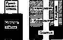
// Schema of cell structure according to Unna: I-protoplasm; II-nucleus; III-nucleolus. However, Methylgreen-Pyronin does not stain it. But no structure has disappeared, therefore the basic (i.e., oxyphilic) proteins remain; Unna no longer identifies them with any definite chemical concepts and calls them by conditional terms. For the basic protein of the protoplasm he uses the term 'spongoplasm' (it is stained only by acid dyes); in the nucleus he distinguishes two floors: the upper-mesoplasma-is stained by hemalum (acid dye+mordant) and is removed only by 15% hydrochloric acid; the lower-plasma (basic substance)-is stained only by acid dyes. In this connection, Unna's concepts of oxygen-forming and oxygen-consuming places in the cell are very important. To determine these places, fresh sections of frozen tissues are treated with the oxygen reagent-Rongalitweiss, which gives a bluish color in excess of oxygen. Rongalitweiss is methylene blue, which is reduced by rongalite (a condensation product of formaldehyde with the sodium salt of sulfurous acid). In excess of O2 methylene blue is reduced. It turned out that all acid places coincide with places containing oxygen (i.e., are stained blue by this reagent), but among them chromatin is particularly distinguished, which receives oxygen from the lymph and activates it with the help of the iron contained in it (O=O is converted to the form O-O-). The activated oxygen is transferred to the surrounding places with the help of chromatin peroxidase (it is revealed by the benzidine test). Other acid places stained by Rongalitweiss: granoplasm, globulin of the nucleolus, cartilage, etc.-only accumulate the oxygen received from chromatin, but they themselves cannot activate it. In all other places (muscle fibers, nerve processes, spongoplasm, keratinized cells, etc.) oxygen is consumed chemically. These so-called reduction places are revealed on preparations with KMnO4, which is an oxidizing agent. Giving up oxygen, it decomposes with the formation of insoluble dark-colored manganese oxides, which are deposited in places of their formation. Many authors-Zacharias, Schwartz, Meyer, Pratje, etc.-used the dissolution technique. They gave descriptions of the processes of dissolution of the nucleus, but, unlike Unna, the named authors relied only on the morphology of the structures, however, they did not consider the stains as chemical reactions and used them only to more clearly see the changes that had occurred. The main disadvantage of this technique from the point of view of its chemical evaluation is that in most cases swelling processes are observed and true dissolution rarely occurs. Swelling as such depends far from only on the chemical structure of the substance, and therefore the swelling capacity of even very different substances may turn out to be quite the same. In relation to the nucleus, the only valuable histochemical conclusion is the establishment that the nucleolus is built mainly of globulins. Of all substances, the greatest attention was paid to nucleoprotein, since its discovery has very great importance for many cytological questions. The approach to the discovery of nucleoprotein (they identified it with the concept of chromatin) was different: 1) Zacharias and Schwartz (1899-1901) gave tables of specific solvents in their opinion, mainly alkalis; 2) later they applied dissolution by enzymes, using the property of nucleoprotein to be digested in trypsin and not to change in pepsin; van-Herwerden (1910) proposed to use nuclease as the most specific solvent, however, this method did not receive wide distribution, since so far it has not been possible to isolate this enzyme in a sufficiently pure form; 3) Unna (1912-21) discovered nucleoprotein with the help of chromolysis, i.e., on the basis of staining it with methyl green before and after various solvents, at the same time he proceeds from the electric polar properties of this dye and its sensitivity to reduction; 4) finally, recently Feulgen (1924) proposed an excellent histochemical reaction for nucleic acid, the so-called nucleal reaction. In view of the fact that nucleic acid does not occur in a free state, but only in combination with proteins, then by discovering it, nucleoprotein is thereby discovered. The reaction is based on the property of thymonucleic acids after moderate acid hydrolysis to split off purine bases and free aldehyde bonds, as a result of which thymine acid is obtained. If one acts on it with fuchsin-sulfurous acid (a colorless compound), then as a result of its attachment at the places of aldehyde groups, a new compound of dark lilac color is obtained. It can already be considered established that nowhere except in nuclear chromatin is nucleic acid contained. There are very few histochemical reactions for protein, and all of them are essentially reactions for amino acids. Such reactions are: the Millon reaction (reveals tyrosine), the Ehrlich diazo reaction (tyrosine and histidine), the Romieu reaction (tryptophan) and the xanthoprotein reaction (tryptophan and tyrosine). The ninhydrin test stands apart, indicating generally free amino groups (amino acids in the alpha position) and introduced into histochemistry by Berg. A positive reaction result indicates processes of protein destruction in tissues. Thus, Berg described a very interesting process of protein destruction in the muscles of starving salamanders. Fats in a free state are revealed relatively easily; all fat-like substances bound with protein are inaccessible to observation. The best method for detecting fats is staining with neutral azo dyes: Sudan III, Scharlach R, dissolved to 40-50% in alcohol. The staining is explained purely physically-by extraction of the dye by fats, since its coefficient of solubility in fats is many times greater than in alcohol (distribution coefficient). The same property is the basis for the staining of fats with chlorophyll and many others. All fats are stained by these methods, but there are also methods for differentiation of various fats from each other. It is believed that on chrome preparations (this achieves insolubility of fats in alcohol and xylene, therefore, embedding in paraffin is possible) Sudan stains neutral fats red, and lipoids orange-yellow. Staining preparations with Nilblausulfat gives the following effect: neutral fats-red color, fatty acids and soaps-blue, phosphatides-blue, cholesterol esters-violet. The theory of this method is unclear. Fatty acids are also revealed by the method of Fischler; it is based on their ability to be mordanted with copper salts and to give with hemalum insoluble black lacquers; neutral fats are incapable of this. The oldest method for determining fat is its blackening with osmic acid, based on its ability to be reduced by fats to metal or its oxides. It is believed that unsaturated fatty acids reduce osmic acid already at 15°, saturated ones at 37°; lipoids, in order to be blackened, require in addition a long time. There is a good method for phosphatides by Stühler: by treating the tissue with cadmium bromide, insoluble cadmium salts of phosphatides are obtained, then cadmium is revealed by the formation of hemalum lacquer. Cholesterol is well recognized with the help of a micropolarizer according to its double refraction or with the help of a somewhat modified Schulze's Lieberman test. The reaction is based on the property of oxysterol to give with a mixture of acetic and sulfuric acids an intense blue color. Carbohydrates are revealed only in a polymerized state or in combination with proteins, i.e., in the form of glucoproteins. So far there is not a single method that would allow sugar to be preserved in the preparation. The methods for revealing carbohydrates are first of all empirically found stains; Best's ammonium carmine stains glycogen; safranin-tannin, according to Fischer-various glucoproteins, mainly mucus; mucicarmine and mucihemalum, according to Mayer-mucins; Kongorot, according to Bechhold-amyloid; thionin stains give metachromasia of all carbohydrates. However, since these are stains and not true reactions, they always require control by the dissolution technique. As solvents, distilled water, ptyalin and diastase are used. It is always necessary to have iodine reactions as control (Lugol's solution, iodine-sulfuric acid and chlorine-zinc-iodine). When combined with iodine, different carbohydrates give colors of different shades: from brown to green, blue and violet; due to this, a certain qualitative differentiation of them is possible. Until recent years, the staining of carbohydrates with iodine was evaluated as a physical phenomenon, however, the works of Euler, Bergmann and others show that the chemical interpretation is much more correct. On the other hand, empirical observations show that besides carbohydrates, iodine stains only a few substances of plant origin. Apparently, it is possible to stain some lipoids with iodine, but they can be easily excluded by dissolving in alcohol-ether. All this makes the reactions with iodine extremely valuable.-They have been proposed for a long time, mainly
By Maccallum, various methods for the detection of inorganic substances. Methods have been developed for iron, zinc, calcium, potassium, gold, bismuth, chlorine, phosphoric acid, iodine, sulfates, and carbonates. All these methods are based on the most characteristic reactions of qualitative analysis. Thus, iron is detected by the reaction with Berlin blue (see); this reaction is the most accurate, all others do not always give satisfactory results. Nevertheless, they sometimes prove to be very valuable. The field of histochemistry also includes the so-called reactions for oxidase and peroxidase. Nitro- dyes used in H. t. 2 z t a ch as S я й ^ SO , The group of rosaniline (tri-amido-derivatives of triphenylmethane) 3. The group of phthalein I Malachitegreen Brilliantgreen (Fuchsin R Fuchsin S Methylviolet Methylgreen Iodgreen Anilinblau Wasserblau /Fluorescein I (Uranin) ' Eosin I Erythrosin I Phloxin A very diverse group of dyes both in their properties and in their methods of preparation. This group includes the best basic progressive nuclear dyes. The acidic dyes of the rosaniline group used in H. t. have well-defined colloidal properties and are used in homogeneous mixtures for staining fibers. The phthalein group is represented exclusively by acidic dyes; they are used for restaining protoplasm and stain evenly and very intensely. {Thionin Methylenblau Toluidinblau Azur I Neutralrot {Safranin G Janusgreen Magdalarot Derivatives of quinone-imide of safranines: safranines ( Nigrosin (Indulin 3B indulines The dyes from the thiazine group used in H. t. are very similar to each other. Their most important property is the ability to show very clear red metachromasia; their orthochromatic color is blue. All of them are widely used for the preparation of complex heterogeneous mixtures (such as: Giemsa, May-Grünwald and many others), which have great polychromy. The azine group is very diverse in its properties. Indigo - indigo carmine - sodium indigo sulfonate. This dye is used as an acidic colloidal dye for staining fibers, as well as for intravital introduction into the body. Organic dyes of little- ( carmine of known structure \ hematoxylin Carmine is a product of cochineal, which is obtained from dried females of the insect Coccus cacti (Mexico). It is used only in combination with mordants, i.e., in the form of a lacquer; it gives the most permanent non-fading colors. Hematoxylin dye is acidic-obtained from the wood of the tree Haematoxylon campechianum (Mexico, Jamaica, etc.). By itself it does not stain; with mordants it gives excellent dark-colored stains.
Related articles
Mentioned in
Cite this page
“Histological Technique.” Soviet Medical Encyclopedia. English translation of Bolshaya Meditsinskaya Entsiklopediya, 1st ed. (Moscow, 1928–1936), ed. N. A. Semashko. https://sovietmedicalencyclopedia.pages.dev/article/histological-technique/