Vacuole
Historical document, translated for reference. It reflects medical knowledge of the 1920s–30s and is not medical advice.
Summary
This 1930s encyclopedia article defines vacuoles as fluid-filled spherical spaces within plant and animal cell plasma, detailing their diverse types in protozoa such as digestive and pulsating vacuoles, and also covers vacuolar degeneration.
Encyclopedia article (1928–1936)
VACUOLE (from Latin vacuus—empty, hollow), spherical spaces in the plasma of plant and animal cells, filled with liquid of various chemical compositions. The sizes and number of vacuoles vary greatly both in different cells and within the same cell in its different physiological states. Vacuoles may have various contents. They are especially often formed as vacuoles in cells re--.
Figure 2. Pulsating vacuole of an infusorian...
according to Nasonov: B—vacuole; H—afferent channels with their swellings, or ampullae;
Figure 1. Amoeba (A. proteus) with two digestive vacuoles (DV) and one simple pulsating vacuole (V); A—nucleus.
C—connecting tubules between the channels and the vacuole; against the background of the vacuole, the excretory opening is visible.
excretates and secretions, food is absorbed, reserve substances are accumulated, etc. The most diverse in composition and function are the vacuoles in the protozoan cell. In Protozoa, the following main types of vacuoles are distinguished: 1. Digestive vacuoles. They are formed in amoebae, flagellates, infusoria, etc., around swallowed food particles, circulate through the body of the protozoan, and serve as the site of food digestion (see Figure 1). Soluble food components diffuse from the vacuole into the plasma, and the vacuole itself with the indigestible waste remaining in it is excreted outward at any point of the body or through a special opening (cytoproct).
2. Pulsating vacuoles are found in all Protozoa except Sporozoa, predominantly in freshwater species (see Figure 2). They have a constant location and periodically fill and contract, expelling their contents through a special pore out of the body. Sometimes they have the appearance of a simple vesicle, while at other times (in infusoria) they are very complexly arranged. Such vacuoles consist of the vacuole proper, into which a ring of fine channels flows—afferent channels that collect fluid into the vacuole—and an excretory tubule with an opening on the body surface (see Figure 3). The rate of pulsation varies in different species, reaching 5–6 times per minute; it depends on temperature, accelerating with an increase in the latter.
The function of pulsating vacuoles is apparently multifaceted. Firstly, they serve for the excretion of metabolic waste (lately there is data on the excretion of urea through them); secondly, they perhaps play a role in respiration, removing oxygen-depleted water from the body, which is replaced by fresh water. But the main significance of vacuoles is currently seen in the fact that they serve the animal in the form
R-lk.
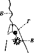
Figure 3. Anterior end of the flagellate Euglena: B—flagellum; V—pulsating vacuole surrounded by a ring of spherical afferent channels; G—eyespot; R—reservoir into which the vacuole empties...
V. Dogel.
VACUOLAR DEGENERATION (from Latin vacuus—empty), or hydropic degeneration (also called "vacuolization"), belongs to the group of protein degenerations, of which it is closest to the so-called cloudy swelling, or granular degeneration. The latter, like vacuolar degeneration, is characterized by an increase in the amount of water in the cell, but there this water is probably bound with pre-existing granular substances of the protoplasm, causing them to swell; in vacuolar degeneration, the moisture perceived by the cell remains in it more or less isolated in the form of droplets of various sizes, which as it were form the "empties" (vacuoles) of the protoplasm [see separate table (pp. 351–352), Fig. 4]. There may be one or several such vacuoles; often there are so many of them that they lie closely adjacent to one another, filling the entire cell and giving it a reticular appearance.
Their contents consist of a fluid very poor in protein and are demarcated from the surrounding protoplasm by a lipoid layer. This layer, however, may be absent, and then the protoplasm is more or less uniformly impregnated with fluid, which with its large accumulation often leads to complete liquefaction of the cell body, leaving only a narrow marginal rim. On a histological preparation of such a cell, only the contour and the nucleus lying as if in a void are visible. The latter is also capable of undergoing hydropic degeneration, expressed either in the appearance of individual vacuoles within it or in the complete dissolution of the nuclear substance, turning the nucleus into a thin-walled vesicle, sometimes containing a nucleolus suspended in the fluid.
Vacuolar degeneration is observed most often upon the impregnation of tissue with edematous fluid of both inflammatory and non-inflammatory origin, as well as as a result of various toxic and some other harmful effects on the cell (for example, in internal organs during infectious diseases, in tumor cells under the action of X-rays and radium, etc.). In inflammation, the penetration of moisture into the cell often occurs very quickly, obviously under the influence of equally rapidly occurring physicochemical changes in the cell body itself. In non-inflammatory edema, this process usually occurs much more slowly. There are almost no cellular elements in the body that cannot undergo vacuolar degeneration. It is most frequently observed in the covering epithelium, connective tissue cells, in various types of tumors, in the parenchyma of the liver and kidneys, striated muscles (skeletal and cardiac), leukocytes, nerve cells of both the central and peripheral nervous system, and in myelin fibers. The process is capable of regression, but in sharply expressed cases can end in cell necrosis due to its ultimate dissolution and disintegration.
Related articles
Mentioned in
Cite this page
“Vacuole.” Soviet Medical Encyclopedia. English translation of Bolshaya Meditsinskaya Entsiklopediya, 1st ed. (Moscow, 1928–1936), ed. N. A. Semashko. https://sovietmedicalencyclopedia.pages.dev/article/vacuole/