Infusoria
Historical document, translated for reference. It reflects medical knowledge of the 1920s–30s and is not medical advice.
Summary
Infusoria, or Ciliata, are a diverse class of protozoa characterized by the presence of cilia during their vegetative life. They exhibit complex morphology, diverse reproductive methods, and include both free-living and parasitic forms, some of which are pathogenic to humans and animals.
Encyclopedia article (1928–1936)
INFUSORIA (Infusoria, s. Ciliata, s. Cilio-phora), an extensive class of the type Protozoa. Its representatives are characterized by the presence of cilia on their bodies during their vegetative life. The exception is the group of sucking infusoria (Suctoria), which have cilia only in early developmental stages but lose them in the adult state. The form and body coverings of Infusoria are extremely diverse. They are covered with a dense and complex, mostly elastic pellicle, having a more or less complex external sculpture. Inside the pellicle, contractile elements, myonemes, as well as supportive skeletal formations, are often located. From it extend the cilia, and in many forms, also cirri. Frequently, cilia are arranged in more or less regular longitudinal rows; in some cases, they cover the entire body uniformly, in others they form characteristic clusters on certain areas of the body. - Nucleus. The vast majority of Infusoria are characterized by a dimorphic nuclear apparatus, which in them consists of macro- (Ma) and micronuclei (Mi). The numerical ratios of the latter vary among different Infusoria, but for each given form they are constant. In the simplest case, 1 Ma corresponds to 1 Mi. But there are infusoria with 1 Ma and many Mi. There are also Infusoria with many Ma and many Mi. The role of the latter is clarified in the processes of reproduction, when they form chromosomes and become sexual, generative nuclei. Macronuclei are assigned trophic functions as somatic nuclei. They divide mostly amitotically. Only in some Infusoria does the division of Ma also approach mitosis. However, in the course of individual development of Infusoria, a certain connection between Ma and Mi is revealed, since during conjugation the old Ma die, and new Ma in exconjugants are restored from derivatives of parental Mi. Only in one group, Opalinata, is there no nuclear dimorphism. In various species of this group, the number of nuclei varies - not less than 2 and up to several dozen, but all nuclei are identical and constructed according to the type of micronucleus. The protoplasm of Infusoria contains various inclusions, the number, form, and distribution of which characterize individual groups of Infusoria. Such inclusions are glycogen, glucoproteins, fats, lipoids, crystalline formations, etc. Some Infusoria contain characteristic stinging elements--trichocysts, which react to external irritations by ejecting their contents. Mitochondria and formations homologous with the Golgi apparatus have also been described in Infusoria. For some Infusoria, the presence of zoochlorellae--symbiotic green algae--is characteristic. - Nutrition. In this respect, Infusoria are divided into two groups. Some ingest formed food by means of special oral apparatus (mouth, pharynx) and digest it in the so-called digestive vacuoles, others lack oral apparatus, do not form food vacuoles, and feed osmotically (Opalina). - Respiration and osmoregulation occur by means of the so-called contractile vacuoles, the number and form of which are characteristic of different species. Parasitic Infusoria, and among free-living forms--marine forms living under conditions of high osmotic pressure, do not have contractile vacuoles. In these forms, respiration apparently occurs by diffusion of gases through the pellicle. - Reproduction. The majority of Infusoria reproduce both asexually and sexually. The asexual method consists of division, which in most Infusoria occurs in the mobile state, and in some forms in the encysted state. The sexual process proceeds in most forms in the form of conjugation, during which the macronuclei of conjugants die, and the micronuclei form sexual nuclei. Conjugants exchange parts of Mi, which mutually fuse, forming fertilized syncaria, after which the conjugants separate, and the syncaria of exconjugants divide, and from them both micro- and macronuclei are restored. In some Infusoria (Vorticellidae), the sexual process has the character of anisogamous hologamy. For some Infusoria, the possibility of so-called endomixis, i.e., reconstruction of the nuclear apparatus outside of connection with the sexual process, has been proven. Thus, Mi is the source of formation of all components of the complex nuclear apparatus of Infusoria and serves for its constant renewal, while Ma has a limited period of existence, after which it dies and must be restored at the expense of Mi. The rhythm of divisions, as well as the alternation of asexual and sexual cycles, depend to a large extent on external conditions: temperature, composition of the medium, etc. Under certain conditions, it is possible to completely turn off conjugation, but in these cases endomixis periodically occurs. In one group of large multi-nucleate Infusoria (Opalinata) (fig. 1), parasitic in the intestines of tailless amphibians, the sexual process occurs between small single-nucleate gametes, which are formed from maternal individuals by a series of successive divisions. Gametes pair up (isogamy) and form zygotes, which grow and become vegetative individuals. In this group of Infusoria, the alternation of sexual and asexual generations is connected with the metamorphosis of the host: asexual reproduction occurs in sexually mature frogs, and the sexual process--in their larval forms--tadpoles. Usually, however, the alternation of generations in parasitic Infusoria occurs in the same host. - Cysts. The majority of known Infusoria form cysts (fig. 1a), with some Infusoria forming protective cysts and reproductive cysts. The cyst wall is usually very complex, and for some forms their multi-layered nature has been proven, with two outer shells (ectocyst+endocyst) performing mechanical protective functions, and the inner, semi-permeable intinocyst serving for physicochemical protection. In reproductive cysts, vegetative division of the maternal body into several (4) daughter individuals occurs. Cysts of parasitic Infusoria apparently open only in the intestine of the corresponding host. Ecology. Infusoria are inhabitants of the most diverse fresh and saltwater bodies; in salt lakes they maintain viability at a NaCl concentration of up to 30%. In a biological respect, different Infusoria very accurately characterize the degree of pollution of different waters. Parasitic Infusoria are very widespread and occur as commensals and parasites in representatives of almost all classes of both invertebrates and vertebrates. The most common place of residence of Infusoria in the organism is the intestine and its appendages; thus, in many mollusks they are found in the intestine and in the liver. Parasitic Infusoria have also been described in the kidneys (Chromidina in cephalopod mollusks). In addition to endoparasitic Infusoria, ectoparasitic ones are also known; thus, some representatives of the genus Trichodina (fig. 18) parasitize in the urinary bladder of newts, while others--on the skin of flatworms (Planaria). Glaucoma pyriformis (fig. 8) is interesting in that, being a free-living form, it can attack small planktonic animals, e.g., fish fry, larvae of mosquitoes (Aedes, Chironomus, Theobaldia), multiply in their body and cause their death (McArthur, 1922; Epstein, 1925). Ichthyo-phthirius multifiliis parasitizes on the skin of fish, causing extensive necroses. - Pathogenicity. Among parasitic Infusoria, some are undoubtedly pathogenic, for example Balantidium coli, the causative agent of chronic ulcerative colitis in humans. At the same time, for pigs, the same Infusoria, or at least one close to it, Balantidium suis, is not pathogenic. When finding Infusoria in human excrement, it is always necessary to keep in mind the possibility of the development in the excreted feces of cysts of free-living Infusoria, swallowed by him together with drinking water and having no pathological significance. Thus, the following accidental findings of Infusoria in human excrement have been described: Chilodon dentatus, s. uncinatus (fig. 5)--in dysenteric stool (Guiart; 1903) and in schistosomiasis (Manson et Sambon; 1913); Colpoda Steini (fig. 6) was found in Germany (Schulze; 1899) and in the USSR (Yakimov and Kolpakov; 1921); Uronema caudatum (fig. 7)--in dysenteric material in China (Martini; 1910) and in the USSR (Yakimov; 1921). - Cultures. Pure cultures of Infusoria, i.e., free from bacterial impurities, have not been obtained with certainty. For cultivating free-living infusoria, various empirical conditions of joint cultivation with different bacteria, algae, etc., have been proposed. For parasitic infusoria, different protein media have been proposed (see Balantidium coli). - The study of infusoria is proceeding in the direction of morphological research of their structure, processes of their division, encystment, rhythm of their reproduction, their reactions to physicochemical conditions of the environment, their action on the tissues of the host. Recently, Infusoria are gradually also becoming objects of immunological research; thus, the phenomenon of anaphylaxis was studied on them (Levinson). Apparently Infusoria are also suitable for testing the action of toxins, e.g., diphtheria (Tunnicliff). Systematics of Infusoria. Ciliata, s. Ciliophora are one of the classes of the type Protozoa and are characterized by the presence of cilia throughout life or at least some stages of their development. Group I. 1st subclass-Opalinata. Large mouthless Infusoria, two-, four-, or multi-nucleate. All nuclei are homogeneous. Parasitize in the intestines of amphibians. One species--in the intestine of fish. Typical species: Opalina ranarum--a non-pathogenic parasite of the frog (fig. 1). Group II. 1st subclass-Eueiliata.
Most representatives are equipped with a mouth opening. The nuclear apparatus is dimorphic. Free-living and parasites. -2nd subclass-Aspirigera; some representatives lack a mouth; those that have it are devoid of an adoral zone of cilia. 1st order-Holotrichida, covered with cilia entirely or partially. The mouth is absent. 1st suborder-Astomatea. Devoid of a mouth. Parasites of the intestine or body cavity of various invertebrates. Typical genus: Maupasella-in the intestine of the earthworm (fig. 2). 2nd suborder-Stomatea. Have a mouth. Free-living or parasites. Typical genera: Paramaecium-the slipper, a free-living form (fig. 3); Isotricha (fig. 4)-parasitizes in the stomach of ruminants; Chilodon cucullulus (fig. 6), Colpoda Steini (fig. 6), Uronema (fig. 7); the last 3 forms are free-living, but are occasionally found in human feces. Glaucoma pyriformis-a free-living form, but can temporarily parasitize small aquatic animals (fig. 8).-3rd subclass-Spirigera, the mouth is equipped with an adoral zone of cilia, which in all is turned to the left, with the exception of the 4th order. 1st order- Ж!Ж
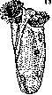

J-Opalina ranarum. la-cyst, 2-Maupasella nova. 3-Paramaecium caudatum. 4-Isotricha intestinalis. 5-Chilodon cucullulus. 6-Colpoda Steini. 7-Uronema. 8-Glaucoma. 9-Balantidium coli. 10-Balantidium suis. 11-Nyctotherus faba. 12-Nyctolherus cordiformis 13-Diplodinium ecaudatum. 14-Cycloposthium bipalmatum. 15-Troglodytella abrassarti. 16-Euplotes patella. 17-Carchesium polypinum. 18-Trichodina. 19-Allantosoma intestinalis. Heterotrichida. The entire body is uniformly covered with cilia. Typical genus: Balantidium, parasitizes in the intestine of various animals, for example Bal. coli-a pathogenic parasite of humans (fig. 9), Bal. suis-a non-pathogenic parasite of pigs (fig. 10); Nyctotherus faba (fig. 11) is described in human feces; pathogenicity is doubtful; Nyctotherus cordiformis (fig. 12)-a non-pathogenic parasite of frogs.-2nd order-Oligotrichida. Cilia are present only on certain parts of the body. Non-pathogenic parasites of the stomach of ruminants, the cecum of horses; found in the intestine of chimpanzees and gorillas. Typical genus: Diplodinium-in the intestine of ruminants; Diplodinium ecaudatum-in the stomach of ruminants (fig. 13); Cycloposthium bipalmatum-in the cecum of horses (fig. 14); Troglodytella abrassarti-in the large intestine of chimpanzees (fig. 15).-3rd order-Hypotrichida. The body is flattened. Cilia form an uneven covering and in places are transformed into spines (cirri). Free-living. Typical genus: Euplotes; Euplotes patella (fig. 16). - 4th order - Peritrichida. The body is conical. The adoral spiral of cilia is turned to the right. Cilia are limited to it alone. The body is mostly attached to the substrate by a contractile stalk. Many forms are colonial. Free-living or parasites. Typical genera: Vorticella, Carchesium, Trichodina-suckers, Carchesium polypinum (fig. 17), Trichodina pediculus-parasite of the skin of worms or of the skin and urinary bladder of amphibians and fish (fig. 18).-Group III. Suctoria. Cilia are present only in young forms. They lack a mouth. They are equipped with sucking tubes, and many also with a stalk. They reproduce by budding, division. Conjugation is observed. Most are free-living predators. Ecto- and endoparasites of aquatic animals, including protozoa. Example: Allantosoma intestinalis-parasitizes on Infusoria living in the intestine of horses (fig. 19).
Related articles
Mentioned in
Cite this page
“Infusoria.” Soviet Medical Encyclopedia. English translation of Bolshaya Meditsinskaya Entsiklopediya, 1st ed. (Moscow, 1928–1936), ed. N. A. Semashko. https://sovietmedicalencyclopedia.pages.dev/article/infusoria/