Laboratory Animals
Historical document, translated for reference. It reflects medical knowledge of the 1920s–30s and is not medical advice.
Summary
This article from the 1928-1936 Soviet Medical Encyclopedia describes laboratory animals used in various types of laboratories for scientific and practical purposes, including their classification, common and rare species, purposes of use, and methods of maintenance and breeding.
Encyclopedia article (1928–1936)
Laboratory animals, animals that serve in various types of laboratories for scientific and practical purposes. Laboratory animals may be those that are easily obtained, well-kept or bred in laboratory conditions, and in addition are suitable by their properties for certain experiments and tests. The composition of the group of laboratory animals by species is not constant. As the experimental direction in biology and medicine grows, in connection with the special tasks arising from new questions and with the discovery of new qualities of some animals, the latter are also drawn into the sphere of laboratory application, as for example Drosophila since the classical experiments of Morgan.-Practically, ordinary and rare laboratory animals are distinguished. The first includes species that have very wide application, for example rabbit, white mice and rats, guinea pig, dog, frog, etc. The second group includes species of animals less commonly used to solve special problems (for example turtle, fish, newts), as well as those having limited distribution in laboratories (for example monkeys in temperate zones). Under laboratory animals are usually understood vertebrate animals, but various invertebrates are also practically laboratory animals, as for example infusoria for pharmacological experiments, Anopheles mosquitoes for experimental infection of patients suffering from progressive paralysis with malaria, Drosophila fruit fly, etc. List of laboratory animals. Type Protozoa (protozoa). Infusoria - Paramaecium caudatum (slipper infusorian).-Type Arthropoda (arthropods). Class Insecta (insects). Diptera: Anopheles maculipennis (common malaria mosquito). Drosophila (fruit fly). Aphaniptera. Fleas, various species. Rhynchota (beaked bugs): Cimex lectularius (bed bug). Pseudorhynchota. Lice of the genus Pediculus. Lepidoptera (butterflies), Bombyx mori (silkworm), Lymantria dispar (gypsy moth). - Type Vertebrata (vertebrates). Class Pisces (fish). Various species of fish: perch, pike, etc. Class Amphibia (amphibians): Rana temporaria (common frog), Rana esculenta (edible frog), Siredon pisciformis (axolotl). Various species of newts. Class Reptilia (reptiles). Various species of turtles: Testudo graeca. Class Aves (birds): Gallus domestica (chicken), Columba livia (pigeon), Dryospiza canaria (canary). Class Mammalia (mammals). Order Rodentia (rodents): Mus musculus (white mouse - albino house mouse), Mus decumanus (white rat - albino rat), Cavia cobaya (guinea pig), Lepus cuniculus (rabbit). Order Ungulata (ungulates): Bos taurus (ox), Ovis aries (sheep), Capra hircus (goat), Sus scrofa (pig), Equus caballus (horse). Order Carnivora (carnivores): Felis domestica (domestic cat), Canis familiaris (domestic dog). Order Insectivora (insectivores): Erinaceus europaeus (hedgehog). Order Primates (primates): monkeys - guenons Cercopithecus, macaques (Macacus rhesus, Macacus sinicus, etc.), Inuus ecaudatus, etc. Purposes of using laboratory animals. 4 directions in the use of laboratory animals are distinguished. 1. Experiments for scientific research purposes, covering general biology, genetics, psychology, physiology, endocrinology, general pathology, pathological anatomy, parasitology, medical zoology, hygiene, bacteriology, the doctrine of infectious diseases, serology, toxicology, the doctrine of poisonous animals, pharmacology with the doctrine of medicinal substances, disinsection, etc. 2. Experiments for diagnostic (applied) purposes in private pathology, bacteriology, serology, parasitology, forensic medicine, toxicology. 3. Use for production purposes: production of vaccines, antitoxic sera, virus fixe, various components for immunobiological reactions, sera for determining blood group affiliation; for obtaining natural gastric juice, extracting various enzymes and other components of the body, for extracting or chemically extracting the active principles of endocrine organs, poisons of some laboratory animals, etc. 4. For special clinical purposes (transplantation of various internal secretion organs, bones, etc.). Maintenance of laboratory animals. Cultivation of protozoa, namely infusoria, is carried out by infusing finely chopped hay in water in a large jar kept warm. Infusoria appear in 2-3 weeks. If infusoria do not appear, a little pond or swamp water is added; in such cultures, the maximum abundance of infusoria occurs at the end of the 2nd week. The maintenance and breeding of insects as laboratory animals has mainly the purpose of studying the transmission by them of pathogens of various parasitic and infectious diseases, as well as the use of insects as a means of infecting humans with certain febrile diseases (malaria) for therapeutic purposes and for studying disinfection means.-A. Anopheles maculipennis-common malaria mosquito; in winter it can be obtained in winter quarters, which serve as basements, animal rooms, cellars, sheds, etc. For finding winter quarters, it is useful to have connections with local malaria stations. Fertilized females hibernate. When transferred to warmth, the mosquitoes warm up, become active and readily drink human blood. Mosquitoes are kept in lamp glasses, the ends of which are covered with gauze held by a rubber ring, or in various wooden (wire, etc.) cages with mesh walls (Fig. 1). When feeding on gametocyte carriers and kept warm, it is easy to obtain Anopheles with sporozoites in the salivary glands even in winter (Simanin). By feeding such mosquitoes on patients with progressive paralysis, malaria can be induced in the latter. When keeping mosquitoes at a lower temperature, they can be preserved with infecting sporozoites for up to 3 months. With repeated warming, such mosquitoes can repeatedly drink blood and infect people (James). For feeding on humans, a lamp glass or glass with mosquitoes is applied to the skin of the hand or arm, through which mosquitoes can sting even through a gauze mesh. Considering that Anopheles does not always suck blood equally readily and quickly, it is more convenient to use boxes with mesh walls and two fabric sleeves, through which the hand is inserted into the box where the mosquitoes are located. In summer, mosquitoes are reared from caught larvae and pupae. They are caught in various bodies of water with a net, plate, cuvette, etc., scooping up the surface layer of water where the aquatic stages of Anopheles transformation usually remain. The collected material is distributed into aquariums (jam jars, glasses, glass vessels, etc.), into which water from the corresponding body of water is poured. The aquarium is covered with a cap of mesh or gauze, through which the emerging mosquitoes are retained. In special large cages, which are a combination of an aquarium with a terrarium, Anopheles can copulate and lay eggs, but such breeding in ordinary laboratory conditions is almost impossible, while the yellow fever mosquito (Aedes argenteus) breeds easily in laboratory conditions. Hatched and unfertilized Anopheles generally drink human blood poorly, but experimental infection of them with malaria is still possible (Veshezerov). B. Fleas can be successfully bred in the laboratory. Flea larvae are selected from the bedding of mammal nests or shaken out of fur, where they can be present in large numbers in neglected dogs. For mass selection of fleas from the debris of ground squirrel nests, the Ioffe thermoelector is used. The best food for flea larvae in the laboratory is dry blood ground into a fine powder. Together with sawdust or sand, it is poured in a layer of 1-2 mm on the bottom of a Koch dish, which is covered with a lid. Dry blood is highly hygroscopic; therefore, a cage charged with it should be dry. One can set up a 'flea factory' in a liter material bottle, the stopper of which is pierced with a piece of glass tubing (Fig. 2). Sawdust to the depth of two fingers is poured on the bottom and adult fleas are placed. For feeding them, a cage made of perforated sieve is lowered into the bottle, into which a white mouse is locked. After an hour the mouse is released and the fleas are removed from it and returned to their place in the bottle. Fleas lay eggs here, from which larvae hatch, feeding on the excrement of adult fleas. For mass keeping and breeding of fleas, larger 'factories' are used (Fig. 3). For individual storage, fleas are placed in test tubes with pieces of filter paper. The test tubes are tied with gauze and stored in a cellar or in boxes with moistening. To feed such fleas, the gauze is removed and the test tube is placed over the body of a host tied to a stand. After feeding is completed, a strip of cardboard is slid under the test tube, covering the opening of the test tube, the latter is turned over and tied with gauze again. Most fleas can feed on the blood of various hosts, which facilitates the laboratory maintenance of these parasites. C. The bed bug is also a polyphagous parasite. Bugs are kept in Koch dishes between layers of filter paper. At a temperature between 25-30°, bugs must be fed every 2-3 days. At a temperature below 12° they do not drink blood. A bug culture can be preserved for a long time by placing them in a cool, dark place (cellar, ice house, etc.). Bugs can be fed on a mouse locked in a cage, described above (as for keeping fleas). D.
Human lice (Pediculus capitis and Pediculus vestimenti). For mass collection of material, take the underwear or clothing of a heavily infested person, as well as cut hair. The obtained items are placed on a large sheet of glass (fig. 4), on which a protective ring of wet clay has been applied. The sheet is positioned with its narrow side toward the window. On the inner side of the protective ring, a narrow strip of paper used for catching flies is placed. The entire apparatus is placed in a warm room at a temperature not below 15°. Hungry lice crawl toward the light source and are caught on the sticky paper, from which they are collected with tweezers, holding them by a leg. It is safer to work with bare arms to the elbow, as lice easily catch on sleeves. Nits are collected by cutting out pieces of belts or seams, especially rich in them, or by tearing apart the fibers of the material itself. Rearing lice in the laboratory is more difficult than other ectoparasites, as their almost exclusive food is human blood. For keeping lice, they are placed in special boxes with round nests, the bottom and lid of which are covered with thin gauze. To feed the lice, such boxes are tied to the forearm or thigh (fig. 5) overnight or worn for long periods. When working with infected lice, persons who have had typhus or relapsing fever are used as 'feeders.' Rocha-Lima and Sikora fed infected lice on pigs and guinea pigs, dressing them in special blankets with fixed receivers for lice. Pieces of material are placed inside the boxes with lice for laying nits on them. The nits are incubated in an incubator in Petri dishes with some moisture. The duration of incubation depends on the temperature (see Lice). V. Vertebrates. Freshwater fish are kept in aquariums of glass, zinc, or cement with running water or with air blown in, depending on the size of the fish and capacity of the aquarium. A supply of frogs is kept in large wooden, painted metal, or cement boxes with sloping bottoms and wooden flooring; water is poured on the bottom so that the frogs have space to get out of it. The box is covered with a mesh lid from above. The supply of frogs is examined from time to time to remove dead ones and to change the water. Frogs are kept in winter without ANY FOOD.
V. Pavlovsky.
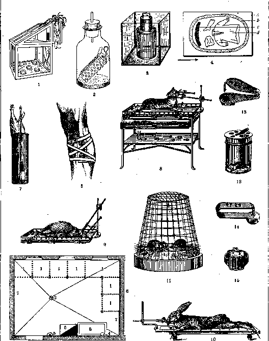
Figure 1. Mesh enclosure for malaria mosquitoes. Figure 2. Enclosure for cultivating fleas. Figure 3. Flea farm: in the inner cylinder of mesh a rodent is placed; at the bottom of the box in sawdust live fleas that move to the animal for feeding. Figure 4. Apparatus for mass collection of lice: a-glass; b-protective ring; c-strip of sticky paper; d-louse-infested clothing; arrow shows the direction of light fall. Figure 5. Wearing a box for feeding lice on a person. Figure 6. Plan of a dog kennel at the physiol. institute of 1st Moscow State University: 1-cages; 2-drain hole for water; 3-bath; 4-boiler; 5-sink with water faucet; 6-Dutch stove; 7-wall lining with Metlakh tiles. Figure 7. Metal cylinder for guinea pigs (thermometry, injections). Figure 8. Debrand's operating table for a cat. Figure 9. Operating table for a chicken. Figure 10. Mallassez's operating table for a rabbit. Figure 11. Metal cage for mice. Figure 12. Glass cylinder for mice. Figure 13. Ear tags for rabbits. Figure 14. Im Nu ear mark.
Figure 15. Leg ring for birds. 282 Warm-blooded animals, mostly mammals L. a. Housing, feed, care, etc.--1) When keeping L. a., the basic rules of hygiene must be strictly observed regarding the size of the premises, its ventilation, light, heat, cleanliness, adequate feeding of animals, separation of sick from healthy, etc. 2) The living conditions in the nursery should be as close as possible to the living conditions of the animal in nature (especially important for wild-living animals, for example monkeys). 3) When arranging the premises and establishing the routine, it is necessary to consider for what purpose the animals are kept (breeders, reserve for experiments, animals for studying conditioned reflexes, for operations, etc.).--Housing for L. a. The premises for experimental animals should be separated and, if possible, remote from the premises for reserve animals and breeders. The basic type of housing is a cage, in which animals are kept singly (carnivores, especially dogs, for studying conditioned reflexes, operated, experimental and control) or in small groups together (rodents, especially guinea pigs, rats, mice). In the latter case, precautions must be taken to avoid fights between males (rabbits, guinea pigs), early mating (rabbits), the male eating the offspring [rabbits, rats, mice (not always)]. Cages for the same animals are sometimes connected together into one common enclosure, aviary, etc. in the open air. In this case, measures must be taken against the possibility of escape (deeply buried outer wall in the rabbit enclosure), against rodent pests, against rain, dampness, and cold [arrangement of special nests with warm bedding (straw, hay), located at some distance from the ground]. The size of the cage, generally speaking, depends on the size and number of animals in it. For dogs, the best size is considered 3-4 m2 per dog, for rabbits-a space of 75 cm depth, 75 cm width, and 50 cm height per rabbit (the depth of a rabbit cage should not exceed 80 cm to avoid inconvenience when removing animals). The basic principles for arranging the cage floor: a) the most complete removal of feces and urine; b) material that does not deteriorate from constant contact with urine and feces; c) minimal heat loss through the floor. In connection with this, either a sloping floor of concrete, galvanized iron, etc., with a hole in the lowest part, on which a layer of frequently changed sawdust is poured, or a double floor is arranged in cages: the lower one, sloping with a hole or sliding out in the form of a flat box covered with galvanized iron, and the upper one-grill-like of metal mesh or bars, on which the animals are placed. The mesh size of the grill floor should be large enough to pass animal feces, but not so large that animal legs get stuck. The walls and ceiling of the cages should be durable and have poor heat conductivity. In well-heated premises, cages can be entirely metal; with insufficient heating and in the open air, wooden cages covered with iron are preferable. In rodent cages, protruding wooden parts inside the cage should also be covered with iron. The construction of cage walls from metal bars or mesh facilitates ventilation, cleanliness, and lighting of the cage, but requires good heating of the room and precautions against rats and mice. Constant keeping of laboratory rats and mice in glass jars is not rational due to poor ventilation. When connecting several cages into one building-nursery, the cage walls are usually made of ordinary building materials (concrete, brick). The door, occupying part of the front wall, and sometimes the entire wall, is usually made of metal mesh or bars on a strong frame. The door should open and close conveniently and quickly (preferably automatically), freely pass the animal, and be securely hung. For dogs, the arrangement on the back wall of the cage of a hinged frame with stretched and easily removable canvas is especially recommended as bedding. For rabbits and guinea pigs in cages of enclosed rooms, special bedding is not needed except at the time when the female should have young. On bedding when kept in the open air-see above. In cages for rats and mice, it is desirable (and for birds-mandatory) to arrange a nesting place (a small wooden box with a ceiling and an opening on the side) and material for it: hay, straw, twigs, wool, cotton wool. For climbing animals (monkeys, cats, rats, mice), as well as for birds, special equipment (trunks, bars, platforms, perches) must be arranged. When keeping a significant number

Figure 16. Dog kennel (plan-see figure 6).
it is very convenient for animals to house similar cells together in one common facility and to equip it with a special kitchen, bathroom, storage room, isolation ward, etc. (figs. 6 and 16). The food for laboratory animals, in connection with the special purposes for which they are usually kept (physiological experiments, operations) and the special conditions of their life (insufficient exercise or its complete absence), must be fully nutritious, easily digestible, but not too abundant. The food for monkeys is predominantly plant-based: fruits, berries (except grapes), nuts, and grains - rice and corn. Among nitrogenous substances, eggs are recommended. White bread and crackers are also given. The main components of a dog's diet are: 1) meat with bones (not fatty, beef or horse) or meat powder making up at least 1/4 of the weight of the entire food ration. Raw meat is incomparably more nutritious and digestible than cooked meat, but it is inconvenient due to its tendency to spoil easily and the possibility of parasite infection. Bones are given depending on the development of the dental apparatus (bird bones should be avoided); 2) carbohydrate substances: various grains, especially oats, in the form of porridge. Bread, especially dark bread, is not tolerated equally well by all dogs. It is useful to give sugar before experiments measuring muscle strength, endurance, before operations under general anesthesia, etc.; 3) table salt added to the food, and 4) clean water. The total amount of food is calculated per 1 kg of animal based on heat production = 45.3 large calories per 1 kg at rest and room temperature. On average, with a weight of up to 12 kg, the total weight of food = 1/4-1/2 kg; with a body weight from 12 to 25 kg, the weight of food = 3/4-1 1/2 kg; and with a body weight over 25 kg, the weight of food = 1 3/4-2 kg. Feeding should preferably be done once a day, at precisely set hours, giving only as much food as can be eaten at one time. The food should not be too hot or too cold. The food for cats differs from that of dogs by the significant predominance of meat (raw), the amount of which is best brought to 100% of the ration. The most nutritious food substance for rabbits is oats (from 200 to 400 g per day per animal). It is also useful to give root vegetables, fresh vegetables (especially for nursing females), and hay; in summer - green plant food (grass, nettles, etc.). The introduction of green fodder should be gradual to avoid diarrhea. For the same reason, it is not recommended to keep rabbits on green fodder for extended periods; it should not be given to young rabbits and nursing females. When feeding root vegetables, fresh vegetables, and green fodder, there is no need to give water separately. The food for guinea pigs differs little from that of rabbits, but due to the guinea pig's tendency to develop scurvy, it is mandatory to give fresh vegetables and root vegetables in winter and green fodder in summer. For rats and mice, various grains (buckwheat, oats, millet - a mixture is best) and, to avoid avitaminosis, milk with white bread are used. It is useful to give rats pieces of raw meat from time to time. The dietary ration for pigeons and other birds consists of various grains and water. White bread soaked in milk is also given. Basic rules for caring for laboratory animals: 1. The temperature in the rooms for laboratory animals should not be below 20° for monkeys and other tropical animals; from +20° to +16° for Japanese mice and about 16° for other laboratory animals. Guinea pigs, rats, and mice can be outdoors for several months a year, while rabbits and pigeons can be outdoors for most of the year. 2. Ventilation, dryness, and cleanliness of the premises are equally necessary for all laboratory animals. The young are particularly sensitive in this regard, and among adults, monkeys. Cages for dogs and cats are cleaned every day, or in extreme cases every other day, and disinfected (water with creolin and lysol) 2-3 times a year; dogs are washed (water, green soap, creolin or lysol) monthly. Rabbit cages should be cleaned 3-4 times in summer and 2-3 times a month in winter; 2-3 times a year, cages are scrubbed with disinfectant solution (water with creolin or lysol, lime water). The urine receptacles under cages are emptied daily. The bedding in rat and mouse cages is changed 2-3 times a week; cages are cleaned once a week; bedding in nests is changed once a week (handle the young with care!). 3. Even, not too strong light is necessary in all cases except during childbirth. 4. Sick, experimental, and operated animals should be kept separately and require different care in different cases. 5. Animals that are to undergo heavy experiments or surgery should be placed in exceptional conditions both in terms of nutrition and detailed care. 6. Animals that are to undergo surgery on digestive organs should not receive anything except water the day before the operation. 7. Handling of laboratory animals should be as gentle as possible. 8. For each animal in the facility, there should be a special entry in a special book noting the time of birth, parents, sex, special markings, purpose, experiments and operations performed, diseases contracted, instances of mating, births, etc.
n. Okuiev. Use of L. animals. The cat mates twice a year—from February to early April and in early June. The duration of pregnancy is 54-56 days. It gives birth to 5-6 blind kittens, which gain sight on the 9th day. The normal duration of feeding by the mother's milk is 5 weeks. Sexual maturity is reached by the end of the first year. The lifespan is up to 15-20 years. It is used to study the effects of O.V. and various pharmacological substances, as well as in experimental parasitology, with kittens being especially used for the diagnosis of amebic dysentery (suspicious material is injected into the rectum and the anus is sealed with a tampon for 2-3 days; if cysts of Entamoeba histolytica are present, the kitten develops typical amebic dysentery), in immunology (tissue immunity) and others.—The dog is very widely used as a laboratory animal for various operations, the study of metabolism and other physiological processes, in endocrinology, pharmacology, experimental pathology, neurology and parasitology.—The ram (sheep) is used for serological and bacteriological purposes. In particular, washed ram erythrocytes are used as one of the ingredients in the Wassermann reaction. It is not recommended to use the same ram for a long time, as its erythrocytes become very fragile and hemolyze when physiological solution is added. The scrotum of a ram is convenient for feeding ticks.—The goat serves as an experimental animal, particularly for studying the causative agent of Malta fever (Micrococcus melitensis), which people contract from goat's milk. Goat erythrocytes can be used in serological reactions instead of ram erythrocytes.—The horse is an immunizing animal that is widely used as a "factory" for producing various antitoxic and other sera by the method of active immunization. The blood of a horse normally contains a certain amount of diphtheria antitoxin.—The cow (calves) finds special application for obtaining smallpox debris by vaccinating the animal with smallpox virus. Fresh organs of the cow (as well as the goat, sheep), taken from the slaughterhouse, are used for pharmacological experiments and in endocrinology (the method of isolated organs).—The rabbit serves as a classic animal for the purposes of experimental pathology, neurology, physiology, pharmacology (isolated heart and ear), toxicology, tissue culture (rabbit embryo), immunization, serology, bacteriology and genetics.—The guinea pig is also widely used in serology (fresh blood serum as complement), bacteriology (for example, infection with various bacteria), in experimental parasitology (for example, studying the migration of ascarids), in the study of invasive and infectious diseases (e.g., typhus and relapsing fever), for diagnostic purposes (e.g., tuberculosis), in genetics and many others. Among other rodents, common are white rats and white mice (experimental pathology, rejuvenation experiments, oncology, parasitology, bacteriology, toxicology and many others).—The hedgehog is an object for studying natural immunity to various poisons and is convenient for feeding ticks when cultivating them in the laboratory.—Monkeys are used to study various infectious diseases (typhus, syphilis, etc.) and for transplants of internal secretion organs (operations "rejuvenation" and others).—Pigeons are used as objects in toxicology, the study of O.V., pharmacology, experimental pathology (e.g., avitaminosis).—Chickens in the embryonic stage are a favorite object for tissue culture; they serve as an important subject of experiments on the dynamics of development and in genetics; chickens are used for special purposes in bacteriology (for example, vaccination against rhinoscleroma, studying the causes of immunity to anthrax) and in parasitology (tick cultures, avian malaria, spirochetosis).—Among reptiles, turtles are sometimes used to study tuberculosis in cold-blooded animals, inflammations, blood parasites, and for raising various ticks on them (Hyalomma, Ornithodorus and others). Among other cold-blooded animals, the most important laboratory animal is the frog, used in physiology (heart, neuromuscular preparations), pathology (inflammation), pharmacology, toxicology, experimental zoology.—The axolotl (see) has more limited application in experimental zoology, developmental dynamics, pathology, histology.—Finally, various freshwater fish (pike, perch, etc.) are used to study the functions of the inner ear, gas exchange, blood circulation, gill function and others. Methods of using laboratory animals include injections, operations, autopsies and special observations. For injections, it is first necessary to capture and immobilize the laboratory animal. For some of them, forceps, resp. hemostats are used for grasping (e.g., mice and rats). Other animals are taken directly by hand; the cat is grasped by the skin of the neck and lumbar part of the body and pressed to the table; if it does not become immobile, pressure is applied to the kidney area; sometimes the legs are bandaged beforehand to hide the claws; in dogs, the muzzle is sometimes bandaged and the jaws are immobilized. The guinea pig is held with one hand around the shoulder girdle area, grasping the first pair of legs between the thumb and index finger, and with the other hand it is held by the hind legs. If the guinea pig is turned head down, it immediately becomes immobile, so various manipulations with the guinea pig can be performed without an assistant; it is tied to a dissecting board by the legs and the board is placed so that the guinea pig is head down. The mouse is immobilized on a Kitazato table or held by the tail with the left hand, and injections are made with the right hand to the struggling animal. The white rat is stretched on the table, the skin on the back near the rump and tail is grasped with two hemostats; the animal is placed on its back. The frog is wrapped in a towel; the turtle is held by the shell.—Injections are made following general rules of sterilization into the thickness of the skin itself (intracutaneously), for which a thin and sharp cannula is used and it is inserted immediately under the surface of the stretched coverings in a horizontal direction so that the needle itself is translucent. The injection is made slowly. For subcutaneous injection, a fold of skin is grasped with the fingers, it is pulled away and the needle is inserted 1-2 cm into the subcutaneous tissue. Subcutaneous injection or injection into the frog's lymph sac is done through the mouth, by piercing the tissues to the side of the sternum with a needle introduced into the mouth and bringing the end of the needle out under the skin into the lymph sac. For special purposes, intramuscular (more often in m. quadriceps femoris) and intra-articular injections are practiced. In the latter case, the knee joint of the rabbit is used.—Intraperitoneal injections are a common laboratory procedure. The animal (guinea pig) is turned head down so that the intestines move toward the diaphragm. The needle is inserted perpendicularly into the lower left part of the abdomen. In similar cases, the dog, cat or monkey is tied to the operating table (fig. 7, 8, 9 and 10).—Injection into the veins is for the purpose of general infection of the body or to determine the general effect of pharmacological substances. Injections into the marginal ear vein of the rabbit and into the tail vein (located on the side of the tail at its base) of the mouse (rat) are more commonly used. To preliminarily dilate the vessels, the ear or tail is kept in hot water (+50°) or the skin over the vessel is smeared with xylene. For injection into the tail vein, the thinnest needle is used, like those used for injecting anesthetic solutions into the gums. In frogs, injections are made into the v. cutanea magna (on the abdominal side). In large animals, injections are made into the femoral or brachial vein, as well as into the v. jugularis externa.—For intracardiac injections (rabbit, guinea pig), the site of the cardiac impulse is first felt and the needle is inserted perpendicularly. To check the correctness of the puncture, a small amount of blood is drawn into the syringe. Subdural, intracerebral and intraventricular injections are performed after preliminary trepanation. To take blood, the tip of the tail is clipped off in mice or rats. In rabbits, blood is taken from the ear vein, by applying a tourniquet to the marginal vein closer to the base of the ear; into the engorged vessel, after local disinfection, a syringe needle is inserted and blood is drawn. In guinea pigs, blood is taken by puncturing the heart, which procedure is not repeated in the same animal sooner than 3 weeks later. The hair is removed from the chest, the skin is treated with iodine, and with a quick thrust, a needle is inserted at the left edge of the sternum, 8-10 mm above the apex of the angle between the xiphoid process and the cartilage of the last rib. The needle is inserted to a depth of 15-20 mm and enters the left ventricle. In a guinea pig weighing 1/2 kg, no more than 10 cm³ of blood is taken. In dogs, blood is taken either from the v. femoralis (on an animal tied belly up) or from the superficial veins of the hind leg; in monkeys—from the cubital vein. When taking blood from a ram, the hair is clipped from the neck and the skin is disinfected. The ram is laid on its side or held by the horns, with the body squeezed between the legs. The v. jugularis is pressed with one hand at the site of its exit from behind the sternum; into the engorged vessel, a needle is inserted and blood is drawn with a syringe. Through a wide needle, blood can flow out by itself.
For operations on some laboratory animals, preparation is necessary. The operative field in dogs is shaved, and animals with soft fur are previously freed from hair by a depilatory (Strontium sulfuricum, Barium sulfuratum techn., etc.). For 24 hours before the operation, the animal is not given food. Before the operation, washing of the operative field is done according to general rules. In some animals (chickens, rabbits), operations are performed without anesthesia, but the animal, particularly the rabbit, is immobilized on a special operating table. General anesthesia is produced with ether (in the form of a mask for dogs and by drip method). To avoid the stage of excitement during narcotization, an hour before the operation, the dog is given a subcutaneous injection of 2 cm³ of a 2% solution of morphine. Morphine in combination with atropine (0.0005 Atropini sulfurici pro dosi) almost completely suppresses the salivation caused by ether. However, in many experiments it is necessary to limit oneself to only one etherization without the additional use of alkaloids. Smaller animals (from mouse to cat) are narcotized under a glass bell jar or in appropriately sized glass cylinders with a ground-in stopper. After placing the animal in the receiver, a wad of cotton soaked in ether is also introduced into it. Narcotization occurs not only from the ether but also from the action of CO₂ exhaled by the animal. The narcotized animal is stretched out on the operating table and the anesthesia is maintained by the drip method. Fish are narcotized by passing a current of water through the mouth and gills, to each liter of which 10 cm³ of ether is added. The fish is tied to a special stand. Its anesthesia can last for 1-2 hours. To revive the fish, it is transferred to frequently changed clean water. Frogs for physiological and pharmacological experiments are conveniently immobilized by injecting 0.1 cm³ of a 10% solution of curare into the lateral dorsal lymph sac. Paralysis sets in after 5 minutes; the paralyzed frog breathes through the skin and the mucous membrane of the mouth. Curare is necessary in some vivisections of mammals; it is used with artificial respiration throughout the duration of the experiment. In dogs, cats, rabbits, and monkeys, lumbar anesthesia is used. The first three are kept on the table in a prone position on the abdomen. With the left index finger, the depression between the last lumbar and first sacral vertebrae is felt. The cannula is inserted vertically along the midline downward to penetrate the space occupied by the cauda equina. The correctness of the manipulation is confirmed by the outflow of cerebrospinal fluid from the cannula. A syringe is attached to the cannula and tropocaine or less is slowly injected. Local anesthesia is produced with cocaine (for operations on the eyes, nasopharynx, larynx-by instillation into the eye; in other operations-by subcutaneous injection). Animals undergoing experiments are kept in metal cages (fig. 11) or in glass jars (cylinders) (fig. 12), which are easily sterilized. Experiments with infectious materials must be carried out in a special room or in a box. Essential attention must be paid to the marking of laboratory animals; small animals are given numbered ear tags. For larger animals, metal plates are used, which are attached to a button like a 'permanent button' or are arc-shaped and clamped with special forceps on a rivet or of another type (fig. 13 and 14). Birds are given numbered rings on their legs or around the base of the wing (fig. 15). Large animals are branded on the skin, horns, or hooves. Small rodents (mice, rats, guinea pigs) are given notches on the right and left ears; it is convenient to use spotted breeds, marking on an individual card the location of the spots. The temperature of laboratory animals is taken by inserting a thermometer into the anus. An important indicator of the condition of the experimental animal is weight, which is determined at regular intervals. The autopsy of laboratory animals may be pathological, parasitological, and bacteriological. It is carried out according to the same rules as the autopsy (see) of a human corpse, but with some particular differences. The animal is stretched out on a dissecting board with its abdomen upward. The dimensions of the boards are: for rabbits-60x40 cm, and for guinea pigs-46x27 cm. In bacteriological autopsy, the board is placed on the bottom of an enameled tray of suitable size. The animal is pinned with special pins by its legs. The skin is removed from the abdominal side. The exposed tissues are cauterized along the line of the proposed incision with a heated metal or glass spatula, and the incision is made with a sterile scalpel or scissors. Inoculations are made from various body fluids collected in sterile pipettes, as well as from the organs themselves, which are also opened after preliminary cauterization of the surface. When cauterizing, it is necessary to consider the volume and nature of the tissue of the organ being treated. At the end of bacteriological autopsy, the corpse and cotton are burned or boiled in a cauldron and discarded; instruments and pipettes are boiled for about 15 minutes. The dissecting board, tray, and table are flooded for 1-2 hours with a 3% carbolic or 5% lysol solution, after which they are wiped. For autopsy, dissecting instruments of sizes suitable for the animal being autopsied are used.-Parasitological autopsy aims to find parasites in the animal's body or to establish pathological changes under the influence of parasites. Parasitic Protozoa are preserved either on smears or in the organs themselves (examination of sections); metazoan parasites in tissues are extracted with the adjacent parts of the tissue or also in an isolated state. Parasites from cavity organs are removed and fixed separately, or the entire contents of the intestine are fixed and the parasites are isolated by the decantation method. Pathological-anatomical changes from parasites occur at their points of attachment to the host's body, at sites of normal or aberrant localization, or the changes are of a general (resorptive) nature.
E. Pavlovsky. Drosophila-an extensive genus including more than 200 described species. As laboratory animals, the greatest importance is attached to Drosophila melanogaster (formerly ampelophila). The study of this small fruit fly allowed Morgan to formulate his famous laws (see Morganism). The exceptional ease of maintenance and rapid reproduction make the drosophila an indispensable object both for illustrating the laws of heredity and for further research work on it. In terms of genetics (see), the study of Drosophila melanogaster ranks first among all animals and plants. With examples from drosophila, all the main types of regularities of heredity can be observed, such as inheritance of sex-linked traits, Mendelian segregation, phenomena of coupling and repulsion (crossing over), etc. Models of inheritance of diseases can be constructed with the help of so-called lethal genes (see). The inheritance of dominant factors and their skips can be demonstrated. Without any difficulty, the regularities of populations (see) can also be shown with drosophila. At the same time, only by working with drosophila can one grasp all the subtleties of genetic science and refine genetic thinking. (For breeding and maintenance of drosophila-see Drosophila.) The technique of working with drosophila is very simple. From optical instruments, it is necessary to have a binocular microscope, or in extreme cases, an ordinary one with low magnification. The flies examined are narcotized with ether, which does not affect their viability and fertility at all. When placing them in a jar with fresh food, a piece of paper (dry) is put on which the sleeping flies are placed (otherwise they will die by falling into the food). For most crossings, it is necessary to use virgin (unfertilized) females. They are easily obtained by examining the culture after 4-5 hours, since flies emerging from pupae usually begin to copulate only after 8 hours. To prevent the entry of foreign flies (contamination) into the jar with the planned crossing, the test tube must be tightly plugged with cotton. For the same purpose, examined and unnecessary flies should be thrown into a crystallizer with kerosene so that they do not fly around. The female drosophila is easily distinguished from the male. In the latter, the first legs have black sex combs, and the end of the abdomen with its sex organs is rounded, and its upper (wing) surface is heavily pigmented. The end of the female's abdomen with the sex organs is triangular, and its upper surface is weakly pigmented. The drosophila has in very large number complex and interesting lines for work (e.g., balanced lethals, chromosome suppressors, lines with deleted chromosomes allow breeding males with lethal genes in the X chromosome, etc.) which are systematically described nowhere. In the USSR, there are rich collections of both mutations and complex lines of drosophila. They can be obtained from the Department of Genetics of the Timiryazev Biological Institute of the Academy of Sciences, the Institute of Experimental Biology in Moscow, and the Central Genetic Station of Agricultural Animals at Zavoronki station. The Timiryazev Institute has completely new mutations (many uninvestigated) and new complex lines obtained in the laboratory's work on the effects of X-rays on the chromosomes of Drosophila. N. Dubinin.
Related articles
Mentioned in
Cite this page
“Laboratory Animals.” Soviet Medical Encyclopedia. English translation of Bolshaya Meditsinskaya Entsiklopediya, 1st ed. (Moscow, 1928–1936), ed. N. A. Semashko. https://sovietmedicalencyclopedia.pages.dev/article/laboratory-animals/