Mosquitoes
Historical document, translated for reference. It reflects medical knowledge of the 1920s–30s and is not medical advice.
Summary
This article provides a detailed morphological and biological overview of mosquitoes (family Culicidae) as understood in the 1930s. It covers their classification into subfamilies, anatomical features, feeding habits, and reproductive cycles.
Encyclopedia article (1928–1936)
MOSQUITOES (true), insects of the family Culicidae, of the group Nematocera, order Diptera, class Insecta, phylum Arthropoda. They have a thin, slender body, long antennae and legs, and narrow wings rounded at the apex. Males are distinguished from females by the dense and long pubescence of the antennae and by a copulatory apparatus in the form of chitinous forceps. The family Culicidae is divided into subfamilies according to the following morphological characteristics: 1. Subfamily Anophelinae: with a simple (non-lobed) scutellum (appendage of the mesothorax), with a bare metathorax (without a tuft of hairs) (Fig. 16), and with a straight proboscis; the anterior fork of the wing veins is larger than the posterior one. 2. Subfamily Sabethinae: scutellum is three-lobed, metathorax with a tuft of hairs, proboscis is straight, vein forks as in Anophelinae. 3. Subfamily Megarhininae: scutellum is three-lobed, metathorax is bare, the end of the proboscis bends in an arc downwards (Fig. 17), the anterior fork of the wing veins is smaller than the posterior one. 4. Subfamily Uranotaeniinae: scutellum is three-lobed, metathorax is bare, proboscis is straight, the anterior fork of the wing veins is much smaller than the posterior one. 5. Subfamily Culicinae differs from the previous one only in that the anterior fork is larger than the posterior one. The family Culicidae is divided into genera: Anopheles (subfamily Anophelinae), Uranotaenia (subfamily Uranotaeniinae), Megarhinus (subfamily Megarhininae), Theobaldia, Orthopodomyia, Taeniorhynchus, Armigeres, Aedes, Lutzia, and Culex (subfamily Culicinae). The genera Aedes and Culex are especially rich in species. The former is divided into subgenera: Finlaya, Ochlerotatus, Ecculex, Aedes, and Stegomyia. For the purposes of systematics, mosquitoes are classified by the venation of their wings (Fig. 19), the character of the scales, the claws of the tarsi, the morphological features of the external skeleton of the thorax, the structure of the external copulatory organs of the males (Figs. 14 and 15), and some other features. The structure of the oral apparatus of female mosquitoes (Fig. 11)—see Anopheles. Various species of Culicinae have minor differences in the termination of the mandibles. Male mosquitoes lack piercing mouthparts due to the rudimentary development of their jaws; therefore, males cannot pierce the skin and suck blood. They feed on various liquid foods (plant juices); they can drink blood if it somehow appears on the surface. There are indications that males of Aedes argenteus might be able to bite humans. Females are generally blood-sucking, but can also feed on plant juices. Blood is important as a highly nutritious substance that enables the laying of yolk-rich eggs. It is possible that there are species of mosquitoes that feed predominantly, if not exclusively, on plant juices (Culex phytophagus). Some Culicinae may exhibit a certain degree of zoo- or anthropophily in the sense of a preference shown for a food source (see Zoophily). The head section of the digestive tract of mosquitoes is equipped with sucking muscles, which serve to pump blood into the stomach. The thin-walled esophagus in its posterior part receives two or three diverticula (Fig. 6), often filled with gas bubbles (esophageal vesicles or sacs); this is followed by a short homologue of the proventriculus, and behind it the midgut or stomach, the walls of which are highly distensible. The stomach swells in its posterior part; at the point where it transitions into the small intestine, five Malpighian tubules empty into it. The small intestine expands into a rectal bladder, on the inner surface of which rise papilla-like rectal glands; it opens with an anal aperture, lying above the genital aperture. The digestive apparatus is served by paired salivary glands (Fig. 12), which vary in their structure. Their excretory ducts merge together and open at the base of the tongue (hypopharynx). The saliva of different species of mosquitoes possesses varying degrees of toxicity, and the individual sensitivity of people to the saliva of the same species of mosquito is not uniform. Primary and secondary skin reactions to a mosquito bite are distinguished, and their intensity is not uniform. The consequences of a bite are the formation of papules or urticarial vesicles, the appearance of which is associated with a sensation of itching or burning of varying intensity. The effect of a mosquito bite depends on the properties of its saliva (E. Pavlovsky and A. Shtein, O. Hecht). Regarding the time of active state, some species of mosquitoes are diurnal, while others, on the contrary, attack humans in the evening or at night. Regarding the place of attack, some species are domestic, as they live in the closest contact with humans. Such is, in particular, the yellow fever mosquito (Aedes argenteus or Aedes aegypti), found in residential houses and encountered not infrequently on steamships, Culex pipiens, and others.
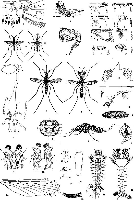
The habitats of mosquitoes are quite diverse. Eggs are laid in various natural as well as artificial bodies of water, sometimes insignificant in size. Mosquitoes of the genera Culex, Uranotaenia, and others glue the eggs they lay side by side, thanks to which the entire clutch takes on the appearance of a bent disk or a little boat (Fig. 20), in which the eggs are arranged vertically (Fig. 18). Mosquitoes of other genera, for example Aedes, lay eggs like Anopheles singly. The eggs of some species of mosquitoes withstand drying well (Aedes argenteus; Fig. 9) and overwinter. The number of eggs laid by Culex pipiens varies from 120 to 400. Aedes argenteus lays 50–120 eggs, and up to 750 in its lifetime. The eggs of some mosquitoes are laid on the ground, in moss in places that are covered in the spring by meltwater (Aedes nemorosus). The larvae of such species hatch very early. It is important that some species of mosquitoes, such as Aedes argenteus, are domestic species, breeding... [Figure 1. Yellow fever mosquito Aedes argenteus (Stegomyia fasciata)—adult larva, posterior end of body: a—siphon; b—gills; c—rudder hairs; d—spinules; d'—the same at high magnification. Figure 2. Yellow fever mosquito Aedes argenteus—female (side view). Figure 3. Hatching of a mosquito from a pupa. Figure 4. Yellow fever mosquito Aedes argenteus—pupa. Figure 5. Morphological differences of the siphon of mosquito larvae Aedinae and Culicinae: 1—Culex pipiens; 2—Culex apicalis; 3—Theobaldia annulata; 4—Theobaldia longiareolata; 5—Theobaldia fumipennis; 6—Orthopodomyia albionensis; 7—Stegomyia fasciata; 8—Taeniorhynchus Richardi. Figure 6. Digestive apparatus of Culex: a—esophageal diverticula; b—proventriculus; c—stomach; d—small intestine; e—rectal bladder with rectal glands; f—Malpighian tubules. Figure 7. Yellow fever mosquito Aedes argenteus (Stegomyia fasciata)—female (slightly magnified). Figure 8. Yellow fever mosquito Aedes argenteus—male (slightly magnified). Figure 9. Yellow fever mosquito Aedes argenteus—egg. Figure 10. Larva of Culex fatigans in the position of breathing in water. Figure 11. Cross-section of the proboscis of a female mosquito: c—labrum; d—mandibles; e—tongue with salivary duct (r); f—maxillae; g and j—labium and muscles; k—tracheae. Figure 12. Salivary glands of the yellow fever mosquito Aedes argenteus. Figure 13. Culex fatigans—pupa. Figures 14 and 15. Chitinous sclerites of the anal cone and penis of Culex fatigans (Fig. 14) and Culex pipiens (Fig. 15). Figure 16. Metathorax: A—with a tuft of hairs; B—bare. Figure 17. A—curved proboscis of mosquitoes (Megarhininae); B—straight proboscis of mosquitoes. Figure 18. Isolated egg of Culex pipiens (magnified). Figure 19. Wing venation of the mosquito Culex; veins: Sc—subcostal; R1, R2, R3, R4+5—radial first, second, etc.; M1, M2—medial first and second; C1, C2—cubital first and second; An—anal; R2 and R3—form the anterior, and M1 and M2—the posterior fork of the wing. Figure 20. "Boat" of eggs of Culex pipiens (side view). Figure 21. Yellow fever mosquito Aedes argenteus—larva (top view). Figure 22. Larva of Culex. The head in front bears dense brushes of long hairs; 1–9—abdominal segments; on the 8th segment on the side is the respiratory siphon; the 9th segment bears a fan of rudder bristles. Figure 23. Culex fatigans: on the left female, on the right male (slightly magnified). (To the illustration of the article Mosquitoes.)]
found in accidental collections of water, often present in households. Such proximity of Aedes argenteus to humans plays a very important role in the spread of yellow fever and dengue fever. A larva emerges from the egg, breaking the eggshell with an "egg tooth" located on its head. The larvae of Culicinae have a long respiratory siphon on the penultimate segment of the abdomen (Fig. 22); due to its presence, the larva hangs obliquely or vertically from the surface of the water body during respiration. The larvae of different genera of mosquitoes differ in the shape and size of the respiratory siphon, its armament with hairs and hooks (Fig. 5), the structural features of the head appendages, the shape of the stigmal plates, and other signs. They pass through four phases of metamorphosis until they turn into a pupa, having undergone three molts. The dietary regimen of Culicinae larvae of different species is diverse. They feed on protozoa, algae, bacteria, small crustaceans, detritus, remains of organic substances, and sometimes on their own kind. The larvae of Culex pipiens and the yellow fever mosquito develop faster in water contaminated with human or animal excrement. The larvae of many of our Aedes, however, inhabit clean waters. Aedes sollicitans lives in brackish waters; the larvae of Culex cantans can live in water with a salt content of 44 g per 1 liter, and Aedes mariae even at 60 g per 1 liter. Highly specialized in their habitat are the inhabitants of tree-hole water bodies—Anopheles plumbeus and Finlaya geniculata, living in water with an unusually high concentration of organic substances (e.g., 8.303 g per 1 liter of water). Being aquatic inhabitants, mosquito larvae breathe atmospheric air, for which they rise to the surface of the water body (Fig. 10). The larvae of Taeniorhynchus Richardi have a modified respiratory siphon, which they insert into the stems of underwater plants to extract air from them. After the third molt of the larvae, pupae appear, breathing by means of two horn-shaped siphons located on the front thick part of the comma-shaped body of the pupa (Figs. 4 and 13). Before the hatching of the imago, the body of the pupa straightens, and the skin on its back bursts. The mosquito gradually crawls out of the slit (Fig. 3), with the skin of the pupa playing the role of a boat; if it is flooded with water, the hatching of the mosquito is disrupted, and it dies. The timing of the various stages of mosquito metamorphosis to calendar dates varies among different species and in different latitudes. The number of their annual generations also varies. Either female mosquitoes, or larvae, or eggs overwinter. Some species overwinter in two forms (for example, eggs and larvae or larvae and imago). The number of generations per year for the middle zone of the USSR (the latitude of England) is two for Aedes caspius, usually one for Aedes nemorosus, two for Theobaldia annulata, and two or more for Culex pipiens. The number of generations for Aed. argenteus is indefinite, since under optimal laboratory conditions, the development of this mosquito "from egg to egg" requires 21 days; practically, however, the yellow fever mosquito can produce 3-4 generations per year in the southern part of the Black Sea coast. The significance of mosquitoes for humans is diverse. The harm caused by mosquitoes is determined by their properties of injecting poisonous saliva into the blood of their host. When huge numbers of mosquitoes are bred, the females, after a "nuptial dance" in the air, attack people and animals in masses. Mosquitoes, together with other blood-sucking dipterans, are known among the people by the collective name "gnus" (midge/blackfly). The "gnus" makes large areas of pastures in Siberia inaccessible to culture and human life. Hogarth (A. M. Hogarth) collected information about cases of human death supposedly from poisoning by mosquito bites in England. In addition to the direct harmful influence, mosquitoes can spread the causative agents of infectious and invasive diseases. During mosquito bites, in some cases, pathogenic bacteria are also transferred, which is more common for Theobaldia, the females of which lay eggs in water bodies contaminated with manure, waste, sewage, etc. For some viruses, mosquitoes are specific vectors. Thus, Aedes argenteus
(Stegomyia fasciata) is a vector of the causative agents of yellow fever and dengue fever (the mosquito Culex fatigans is also accused of spreading the latter). Aedes argenteus (Figures 1, 2, 4, 7, 8, 9, 12, 21) is quite common in some regions of the Black Sea coast of the Caucasus (where it was first discovered in general by E. I. Martsinovsky)—in Batum, Poti, and Sukhumi. In Gudauta, Gagra, Tuapse, and even in Tiflis, this mosquito is from time to time introduced from the above-mentioned places of its habitat. The question of the presence of Culex fatigans (Figs. 10, 13, 23) in our fauna remains unproven. Treponema icterohaemorrhagica, ingested with blood, can maintain its virulence in the stomach of Culex pipiens for about a day. Trypanosoma Evansi, the causative agent of surra in horses, can be purely mechanically transmitted by the mosquito Anopheles fuliginosus, provided that feeding is quickly resumed on a healthy animal after being interrupted on a horse sick with surra. Avian trypanosomes, and possibly Haemoproteidae, are also transmitted by mosquitoes. The causative agent of avian malaria, Proteosoma praecox, is spread by the mosquitoes Culex pipiens, C. fatigans, C. hortensis, Aedes nemorosus, Aedes argenteus, Aedes mariae, and Theobaldia longiareolata. A similar role in relation to various species of human malaria pathogens (Plasmodium vivax, Pl. malariae, and Pl. immaculatum) is played by various species of the genus Anopheles.
Among other protozoa, parasites of mosquitoes, e.g., Culex pipiens, are flagellates—Herpetomonas algeriense, Herpetomonas culicis, Crithidia fasciculata, Leptomonas culicis; sporozoans—Nosema culicis, Thelohania legeri, Stempellia magna, Caulleryella pipientis, Haemoproteus noctuae; Rickettsia, etc., are often parasites of mosquitoes. Among parasitic worms, some filariae can be spread through mosquitoes, in whose bodies the microfilariae ingested with blood undergo some development and reach the proboscis in an invasive form. Such is Wuchereria Bancrofti—the causative agent of human filariasis and also considered the causative agent of elephantiasis (which has recently been questioned); its hosts and vectors can be mosquitoes: Culex pipiens, C. fatigans, Culex annulirostris, Aedes variegatus, Aedes (Finlaya) togoi, Taeniorhynchus africanus, Anopheles rossi, Anopheles costalis, Anopheles algeriensis. Filaria immitis of dogs is transmitted by the mosquito Anopheles bifurcatus, Anopheles maculipennis, Anopheles sinensis, Culex pipiens, Culex fatigans, Aedes punctatus. Aedes argenteus and Anopheles maculipennis transmit another canine filaria—Dirofilaria repens. In addition to this, mosquitoes are hosts to other worms (Agamomermis in various Aedes), trematodes Lecithodendrium ascidia (cysts in Anopheles maculipennis), Distomum globiparum (the same), Cercaria armata (in Culex hortensis), Agamodistomum in Anopheles. In the digestive tract of Culex larvae, the rotifer Philodina parasitica can parasitize. Among ectoparasites, it is necessary to note the larvae of various water mites (Nessea fuscata, Hydrodroma, etc.), as well as the larvae of chigger mites—Trombidiidae. Some Ceratopogoninae parasitize at the expense of adult mosquitoes, sucking blood from their stomach through a puncture of the skin. Mosquitoes can play a peculiar role as distributors of the larvae of the human botfly—Dermatobia hominis, the female of which glues the eggs it lays to the abdomen of a mosquito when the opportunity arises (South America). The benefit of mosquitoes in the economy of nature lies in the fact that their larvae serve as food for fish. Since mosquito larvae can capture bacteria as food, experiments were conducted to clarify the influence of mosquitoes on the number of bacteria in water; mosquito larvae supposedly contribute to the purification of water from bacteria. The enemies of mosquitoes in relation to the larval and pupal stages are waterfowl, fish (see Gambusia), newts, planarians, hydras, predatory insects (larvae of Dytiscus, water beetles, dragonflies, water bugs, predatory mosquito larvae, and others). Adult mosquitoes become prey for birds, frogs, toads, bats, dragonflies, and spiders. Among fungi, Entomophthora sphaerosperma and Empusa culicis are destructive to adult mosquitoes. Control measures against mosquitoes are generally the same as those for Anopheles (see Malaria).
Related articles
Mentioned in
Cite this page
“Mosquitoes.” Soviet Medical Encyclopedia. English translation of Bolshaya Meditsinskaya Entsiklopediya, 1st ed. (Moscow, 1928–1936), ed. N. A. Semashko. https://sovietmedicalencyclopedia.pages.dev/article/mosquitoes/