FILARIAE
Historical document, translated for reference. It reflects medical knowledge of the 1920s–30s and is not medical advice.
Summary
Filariae are nematode worms of the order Filariata that cause various diseases in humans. This article describes the classification, morphology, life cycle, and medical significance of different filarial species, including Wuchereria bancrofti, Loa loa, and Onchocerca volvulus.
Encyclopedia article (1928–1936)
FILARIAE, FILARIIDOSIS. Filariidoses are diseases caused by filariae—nematodes of the order Filariata. Brief characteristics of Filariata: thread-like nematodes of extremely diverse morphological structure. The mouth is simple or provided with lips; often surrounded by special epaulette-like ornamentations. The spicules are of unequal size and different structure. The vulval opening, with rare exceptions, is in the anterior part of the body. Parasites of closed cavities. For development they require an intermediate host. The order includes 3 families: Cystopsidae Skrjabin, 1923, Filariidae Claus, 1885, and Dracunculidae Leiper, 1912. Medical significance is attached to Filariidae and Dracunculidae. Brief characteristics of Filariidae: females are longer than males by no more than 3-4 times. The anal opening is present in both males and females. The vulva is present. Filariidae includes 6 subfamilies (according to Skrjabin and Shikhobalova, 1935): Filariinae, Onchocercinae, Setariinae, Dipiotriaeninae, Aproctinae, Tetracheilonematinae. In humans, representatives of the first three are recorded. From Filariinae in humans, representatives of the genus Wuchereria Silva Araujo, 1877, Dirofilaria Railliet et Henry, 1911, and Loa (Stiles, 1905) are recorded. From the genus Wuchereria, W. bancrofti Cobbold, 1877 [syn. F. sanguinis hominis, F. sanguinis hominis aegyptica, F. bancrofti (fig. 1), Wuchereria filaria, F. sanguinis, F. nocturna and F. philippinensis] parasitize humans. Brief description of Wuchereria bancrofti: mouth without lips, surrounded by 2 rows of concentrically arranged papillae. Length of male 25-40 mm, spicules 0.2 mm and 0.43-0.6 mm. A gubernaculum is present. About 15 pairs of genital papillae. No caudal alae. Female: 50-100 mm in length; vulva 0.4-1.13 mm from the anterior end. Viviparous. Adult parasites live in the lymphatic system (vessels and glands) of various parts of the human body. Intermediate hosts are mosquitoes of the genera: Aedes, Culex, Anopheles and Mansonioides (see Carriers). Microfilariae are provided with a sheath and have a length of 0.127-0.320 mm and a width of 0.007-0.010 mm. The anterior end is provided with a delicate stylet. Microfilariae, by which the diagnosis of the disease is made by finding them in the blood, are characterized by a specific arrangement of nuclei (special large cells with a nucleus), which protrude upon staining. The end of the caudal part is devoid of nuclei, which is a characteristic feature of the microfilariae of W. b., distinguishing them from microfilariae of Loa. In the peripheral blood, microfilariae of W. b. as a rule can be found only at night, which is why they are called Mf. nocturna, in contrast to microfilariae of Loa, which enter the peripheral blood only during the day and bear the name Mf. diurna (see Loa). It is interesting to note that when the night rest regime is changed to day, after a certain period of time, the larvae accordingly change the time of stay in the peripheral blood. FILARIIDOSIS
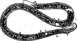
Figure 1. Larva of Wuchereria bancrofti: 1 - excretory cells; 2 - genital cells; 3 - excretory opening; 4 - anal opening; 5 - caudal end; 6 - anterior end.
of the genus Wuchereria in humans are W. bancrofti Cobbold, 1877 [syn. F. sanguinis hominis, F. sanguinis hominis aegyptica, F. bancrofti (fig. 1), Wuchereria filaria, F. sanguinis, F. nocturna and F. philippinensis]. Brief description of Wuchereria bancrofti: mouth without lips, surrounded by 2 rows of concentrically arranged papillae. Length of male 25-40 mm, spicules 0.2 mm and 0.43-0.6 mm. A gubernaculum is present. About 15 pairs of genital papillae. No caudal alae. Female: 50-100 mm in length; vulva 0.4-1.13 mm from the anterior end. Viviparous. Adult parasites live in the lymphatic system (vessels and glands) of various parts of the human body. Intermediate hosts are mosquitoes of the genera: Aedes, Culex, Anopheles and Mansonioides (see Carriers). Microfilariae are provided with a sheath and have a length of 0.127-0.320 mm and a width of 0.007-0.010 mm. The anterior end is provided with a delicate stylet. Microfilariae, by which the diagnosis of the disease is made by finding them in the blood, are characterized by a specific arrangement of nuclei (special large cells with a nucleus), which protrude upon staining. The end of the caudal part is devoid of nuclei, which is a characteristic feature of the microfilariae of W. b., distinguishing them from microfilariae of Loa. In the peripheral blood, microfilariae of W. b. as a rule can be found only at night, which is why they are called Mf. nocturna, in contrast to microfilariae of Loa, which enter the peripheral blood only during the day and bear the name Mf. diurna (see Loa). It is interesting to note that when the night rest regime is changed to day, after a certain period of time, the larvae accordingly change the time of stay in the peripheral blood. FILARIIDOSIS
748 When a mosquito bites a human, together with blood it ingests microfilariae of W. b., which inside it undergo metamorphosis and penetrate into the proboscis, from where, with a new bite, they get onto the human skin; perforating the latter, microfilariae penetrate through the blood into the lymphatic system. W. b. cause in humans the disease—wuchereriosis, which often proceeds very severely. The basis of wuchereriosis lies in changes in the lymphatic system, consisting in inflammation of the vessels and glands and stagnation of lymph. As a result, there is a reverse flow of lymph into the vessels, with subsequent lymphoceles, chylous effusions, etc., and in chronic cases, the development of elephantiasis of various parts of the body, which can reach gigantic sizes (figs. 2 and 3). Geographical distribution of wuchereriosis: the entire tropical and subtropical belt of the globe. In the USSR, wuchereriosis has not been detected. Partsvanidze in Georgia and Pavlovsky in Tajikistan detected cases of elephantiasis clinically resembling wuchereriosis, however without detection of microfilariae. The diagnosis of wuchereriosis is usually made by detecting larvae in the blood or chylous exudate of lymph from ulcers. There is no therapy for wuchereriosis. In individual cases, surgical intervention is applied. Attempts are made to apply chemotherapy. In complications of a septic nature, vaccine therapy is applied. Prevention is not sufficiently developed; measures should be directed against mosquitoes and to protect against their bites. From other representatives of Filariinae parasitic in humans
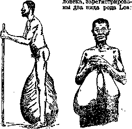
Figure 2.
Figure 3.
Figure 2. Elephantiasis of the scrotum on the basis of wuchereriosis. (According to Meuer.) Figure 3. Elephantiasis of the breast gland on the basis of wuchereriosis. (According to Meuer.) L. loa and L. extraocularis, which are localized in the subcutaneous tissue of various parts of the body, predominantly in the eye area. The first case of Loa extraocularis was described by Skrjabin in 1917 (North Caucasus); to the present time, 4 cases have been registered in the USSR. The diagnosis of loaosis is made upon detection of the parasite under the skin or by the presence of microfilariae in the blood. From representatives of the genus Dirofilaria in humans, 2 species are registered: D. magalhaesi (Blanchard, 1895-1896) and D. repens Rail, et Henry, 1911. Nematodes of the genus Dirofilaria are characterized by a simple mouth, devoid of lips, surrounded by 6 papillae. The vulva is somewhat behind the end of the esophagus. Viviparous. Microfilariae without sheath. Adult parasites inhabit the heart cavity and subcutaneous tissue of various mammals. Microfilariae are found in the blood; no periodicity of their entry into the peripheral blood is noted. D. magalhaesi has been registered in humans only once in Brazil. Dimensions of D. magalhaesi: male 83 mm in length, female 155 mm, microfilaria 0.30-0.35 mm. D. repens, a frequent parasite of dogs. Registered by Skrjabin, Altgauzen and Shulman (1930) in humans under the skin of the lower eyelid. There was a male reaching 58 mm in length with spicules 0.448 mm and 0.176 mm in length. The biology of D. magalhaesi is not precisely deciphered. The intermediate host of D. repens are mosquitoes (genus Stegomyia, etc.). Nematodes of the subfamily Onchocercinae Leiper, 1911, are characterized by ring-like thickenings on the cuticle. The mouth is simple, spicules are unequal, genital papillae are located somewhat asymmetrically. The vulva is in the esophageal region. Adult parasites parasitize in the subcutaneous tissue and walls of vessels. Microfilariae are devoid of a sheath, are found in the blood, lymphatic vessels, or in the subcutaneous tissue near the adult parasite. The carriers are blood-sucking dipterans (Culicoides, etc.). In humans, 2 species of the genus Onchocera Diesing, 1841 are registered: O. volvulus (Leuckart, 1893) and O. caecutiens Brumpt, 1919. O. volvulus in some localities of Africa affects up to 68% of the indigenous population. Length of males about 30 mm, spicules about 0.088 and 0.172 mm in length, the tail is devoid of alae. Length of female about 50 mm. The vulva is slightly behind the end of the esophagus. Microfilariae of different sizes (Faust, 1930): 0.264-0.290 mm and 0.221-0.287 mm. The probable intermediate host is Simulium damnosum. The parasites often cause subcutaneous nodular tumors ranging in size from a pea to a pigeon's egg; in their depth usually lie coiled parasites. The tumors are often very painful, however Faust (Faust, 1930) questions the connection indicated by some authors between the itchy dermatitis craw-craw (see Dermatitis), elephantiasis and skin lichenification with O. volvulus. Localization of O. volvulus in most cases is near the joints. Therapy is not developed. Preventive measures should be reduced to fighting Simulium and taking precautions against their bites. O. caecutiens (Brumpt, 1919) does not differ morphologically from O. volvulus, but has a tendency to affect the face and head, especially the parotid region. About 95% of the indigenous population of Guatemala are affected by fibromatous tumors on the head of onchocercotic origin. The tumors are usually painful; the disease may be accompanied by a rise in temperature. Therapy is not developed. According to observations by practical physicians, excision of nodes gives improvement in cases of complications from the eyes. From nematodes of the subfamily Setariinae in humans, 2 species are registered: Acanthocheilonema perstans (Manson, 1891) and Filaria (sensu lato) ozzardi (Manson, 1897). For Setariinae, the following is characteristic: the anterior end is provided with a peribuccal chitinous ring or epaulette-like formations; microfilariae without sheath. Localization in the body cavities of vertebrates. Ac. perstans (syn. Filaria sanguinis hominis minor, Manson, 1891, Filaria sanguinis hominis perstans, Manson, 1891, F. ozzardi var. truncata, Manson, 1897; Dipetalonema perstans (Manson, 1891) Yorke, Maplestone, 1926. Acanthocheilonema perstans (fig. 4) occurs in
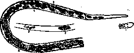
Figure 4. Larva of Acanthocheilonema perstans:
1. Posterior end; 2. Two-tailed end; 3. Genital cells; 4. Anal opening. In South America and various parts of tropical Africa. The male is about 45 mm in length, the female 70-80 mm, they parasitize humans, gorillas, and anthropoid monkeys. Microfilariae are 0.200 mm in length (significantly larger than microfilariae of Wuchereria and Loa). No periodicity of microfilariae entry into the blood has been noted. Intermediate hosts are mosquitoes of different genera. The pathogenesis has not been studied, however some associate A. perstans with varicose inflammation of the lymph glands. F. ozzardi [syn. Mansonella ozzardi (Manson, 1897), Faust, 1929, F. damarquayi, Manson, 1897, Filaria pincea Railliet, 1918, F. tucumana Biglien, Araoz, 1917]. A widely distributed parasite among the indigenous population of some South American countries (Argentina). The length of the female is 65-81 mm. The vulva is 0.71-0.76 mm from the anterior end. The anus is 0.25 mm from the posterior end. On the posterior end, there are 2 projections on both sides. Microfilariae without a sheath. The anterior end is equipped with a stylet. No periodicity of entry into peripheral blood has been noted. Intermediate hosts are Aedes aegypti (Stegomyia fasciata) and possibly Anopheles maculipennis. The pathogenesis has not been studied. Therapy has not been developed. Prevention should be directed toward fighting mosquitoes. Fam. Dracunculidae Leiper, 1912. For nematodes of this family, the significantly larger size of females compared to males is characteristic. The family includes 2 genera: Füllebornius and Philometra. In humans, nematodes of the first genus parasitize. F. medinensis (Linn., 1758) - see Dracunculiasis. - Besides the above filariae described by adult forms, a large number of microfilariae have been registered in humans, i.e., larvae whose adult forms have not yet been found, and therefore their exact position in the zoological system has not been established. Of these, separate mention, as the most widespread, is deserved by microfilariae malayi (Brug., 1927), microfilariae streptocerca (Macfie, Carson, 1922) and some others. In the diagnosis of filariasis by microfilariae, one should take into account, on the one hand, the periodicity of their entry into peripheral blood, and on the other - their morphological features. When analyzing blood, it is recommended to centrifuge it with the addition of Aq. destill. or Ac. citrici. The method of vital staining is often used: a small drop of blood, placed on a slide, is covered with a cover slip and under it a drop of dye is introduced, for example, methylene blue (1:5,000 in physiological solution). In this case, microfilariae behave differently. Thus, living microfilariae of W. bancrofti and streptocerca are not stained at all, microfilariae of Loa loa and Onchocerca volvulus are stained, but not very intensely; microfilariae of perstans have the ability to be stained most vigorously. Therapy for individual microfiliariases has not been developed, and as for prevention, it should in general be directed toward fighting intermediate hosts and protecting against their bites.
N. Tishchenkova. PHYMA (nodus, tubera subcutanea), a tubercle, a bump, a term sometimes used to denote more extensive in size, sharply protruding above the level of the surrounding skin, tumor-like or nodular formations ranging in size from a walnut to a chicken egg and larger. They develop in the deep layers of the proper skin and sometimes in the subcutaneous fatty tissue and appear to be of soft or dense consistency, often with a bumpy surface. Their color sometimes does not differ from the natural color of the skin or is red, reddish-brown, or bluish. They sit on a broad base or hang on a stalk. An example of such formations can be rhinophyma (see). In general, the etiology and course of phymata are different and are reduced to inflammatory or neoplastic processes of the skin. According to Brocq, to this primary lesion should be attributed rheumatic nodules, some gummas in the initial stage of development, etc. The term "phyma" is little used at the present time.
Related articles
Mentioned in
Cite this page
“FILARIAE.” Soviet Medical Encyclopedia. English translation of Bolshaya Meditsinskaya Entsiklopediya, 1st ed. (Moscow, 1928–1936), ed. N. A. Semashko. https://sovietmedicalencyclopedia.pages.dev/article/filariae/