Rhinophyma
Historical document, translated for reference. It reflects medical knowledge of the 1920s–30s and is not medical advice.
Summary
Rhinophyma is a non-malignant hypertrophic growth of the nose that can severely disfigure the face. The article describes its clinical presentation, histological structure, and various surgical treatments developed over time.
Encyclopedia article (1928–1936)
RHINOPHYMA, rhinophyma (from Greek rhis-nose and phyma-growth). Synonyms: elephantiasis nasi, hypertrophy of the nose, wine nose, copper nose, bulbous nose. The term R. was introduced by Hebra, who designated under this name the 3rd stage of acne rosacea of the nose. Trendelenburg distinguishes 3 forms of elephantiasis or hypertrophy of the nose: hyperplastic inflammatory wine nose, fibroma molluscum, and true endemic elephantiasis of the nose in Arabs. Gradually the term R. became established in the literature. Rhinophyma represents a non-malignant, hypertrophic neoplasm that severely disfigures the face. The enormous lobulated tumor described by Hebra (Fig. 1) had changes only in the thickness of the skin, the deeper parts of the nose were unaffected, the tumor hung down on the lip and covered the nostrils. Olier, on the contrary, speaks of a simultaneous increase in cartilage; Joseph in one case during operation found a very enlarged and thickened terminal cartilage. Of 5 cases observed by Rauer, in one case thickening of the terminal and triangular cartilages was found, and in another (markedly expressed case) the cartilage was thinned and had lost its elasticity, in any case this was not a pathological change in the cartilage, but only a consequence of mechanical stretching from the hypertrophy of the skin.
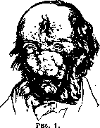
As a rule the tumor has three irregularly shaped round lobes, sitting on a wide base—two on the wings and one on the tip of the nose. In addition, individual lobes are separated by deep grooves into smaller lobules. In markedly expressed cases the middle lobe has the appearance of a bulb, hanging down on the lip or even on the chin and interfering with eating. The tumor has a soft consistency, a fatty surface, a blue-purple, purplish-red or gray unchanging color. When pressed, vermicular plugs of skin fat are squeezed out of it.
Regarding the histological structure of R. Hebra as early as 1845 expressed the opinion that here there is an excessive increase in connective tissue and blood vessels. Research data showed besides the proliferation of connective tissue an increase in the number of sebaceous glands; however Bruns believes that in this case it is not so much about the proliferation of epithelium and proliferation of a large number of sebaceous glands [see separate table (Vol. XXVIII, art. 479-480), Fig. 6], as about a benign and homogeneous-plastic formation of nests, which can be designated as fibroma molluscum or cysto-adeno-fibroma. Olier and other authors believed that the cause of R. lies in chronic alcoholism, however this contradicts the fact that many alcoholics do not have R. and, conversely, R. is present in people who do not drink at all. Alcoholism can only play a predisposing role. R. can also be inherited. The first operation for R. was probably performed by Professor Sennert of Wittenberg. Dieffenbach in 1845
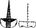
Figure 2.
Figure 3.
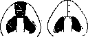
Figure 4.
Figure 5. applied for hypertrophy of the nose a cruciform (Fig. 2, 3, 4, 5) or elliptical excision. Langenbeck (1851) removed the tumor like a shell from the cartilagous framework of the nose. In 1864 Stromeyer cut off the tumor with a knife, in 1904 Bloebaum recommended removing the tumor exclusively with a thermocautery. In 1905 Sick proposed to remove the tumor only to the depth of the sebaceous glands, so that the wound could be covered with skin from the proliferating epithelium of these glands. Braun proposed subcutaneous decortication of the changed tissues through a T-shaped incision with subsequent suturing of the flaps. In 1915 and 1921 Joseph proposed removal of all diseased tissues under local anesthesia and closure of the skin defect with a flap from the cheek or forehead. The operation in the surgical treatment of R. in markedly expressed cases consists in removal of all or most of the pathologically changed skin and reduction of the framework of the nose. In initial forms of rhinophyma, when the nose is slightly enlarged, the skin is thickened and no bumps are found on the surface; through an incision along the anterior edge of the nasal passages, the deep layer of the skin is excised in the form of a plate parallel to the surface of the skin. In case of excess skin, the excess is excised with a spindle-shaped incision. In R. of medium size, when the lower part of the nose is mainly affected, the skin is greatly thickened, divided into lobes and cut by deep folds, sometimes the anterior part of the nose is elongated—the entire thickness of the diseased skin is excised; sometimes the cartilage is reduced, and the defect is closed with a flap from the forehead.
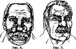
In strongly expressed R. (Fig. 6) under local anesthesia the entire tumor is excised down to the framework of the nose, reduction of the framework of the nose and closure of the entire defect with a flap from the forehead (Fig. 7). The less changed skin not removed at the edges of the tumor is cauterized with a pacelon or diathermic needle.
A. Rauer.
Related articles
Mentioned in
Cite this page
“Rhinophyma.” Soviet Medical Encyclopedia. English translation of Bolshaya Meditsinskaya Entsiklopediya, 1st ed. (Moscow, 1928–1936), ed. N. A. Semashko. https://sovietmedicalencyclopedia.pages.dev/article/rhinophyma/