Elephantiasis
Historical document, translated for reference. It reflects medical knowledge of the 1920s–30s and is not medical advice.
Summary
This article from the 1928–1936 Great Medical Encyclopedia discusses elephantiasis, detailing its pathological tissue growths, congenital and acquired causes, clinical forms, and manifestations across various parts of the body.
Encyclopedia article (1928–1936)
ELEPHANTIASIS (from Latin elephantus — elephant), elephantiasis, a variety of pathological tissue growth in certain organs, acquiring not only a large volume as a result of such growth, but often an irregular, deformed configuration. The process is particularly often observed and was first described in the region of the lower extremities, which thereby take on the appearance of elephant legs. Later, elephantiasis came to mean analogous or only outwardly similar changes in various parts of the body (arms, lips, penis, scrotum, vulva, etc.). The essence of changes in elephantiasis consists of hypertrophic and hyperplastic processes in one or another tissue making up the given organ; of greatest importance is the hypertrophy of connective tissue (Elephantiasis fibrosa), subcutaneous adipose tissue (Elephantiasis lipomatosa), blood vessels (Elephantiasis angiomatosa, or haemangiectatica), lymphatic vessels (Elephantiasis lymphangiectatica), and nerves (Elephantiasis neuromatosa). Consequently, in elephantiasis, there is no uniformly proportional excess growth of all tissues of the given organ, which distinguishes elephantiasis from the so-called partial or local giant growth (see Giants, gigantism). However, gigantism and elephantiasis can also be observed simultaneously, for example, in the region of certain sections of the intestine as a result of disturbances in their embryonic development. A distinction is made between congenital elephantiasis (Elephantiasis congenita) and acquired elephantiasis (Elephantiasis acquisita); the latter is observed significantly more often. In congenital elephantiasis, for example in certain cases of macroglossia, macrocheilia, since excess growth takes place while preserving the basic topographical relationships of the tissues making up the organ, the process itself is essentially close to those forms of developmental malformations (see) which are called monstra per excessum. According to the prevalence of the process, one can speak of diffuse, organ-specific, and localized elephantiasis; however, the first type is very rare; this apparently includes cases of continuous elephantiasis of the skin of newborns, in which case the latter acquire a shapeless appearance (also observed in animals — the Mondkalber of German authors). The main mass of cases of elephantiasis relates to certain organs (completely) or to known areas of them. The causes of the development of acquired elephantiasis can be various factors causing prolonged and progressive disorders of blood and lymph circulation in a certain area. Of great importance are inflammatory processes, for example, chronic leg ulcers, recurrent erysipelas, lupus of the extremities, eczema, then varicose vein expansions (Elephantiasis phlebectatica, or phlebectatic pachydermia) and thrombotic processes in the venous system; finally, the main importance belongs to various processes in the lymphatic vessels and glands causing lymphostasis, e.g., suppuration, tuberculous inflammation, as well as their extirpation, deep scars hindering lymph outflow, and the like. Often the causes of the development of elephantiasis cannot be established (Elephantiasis spontanea), and one has to think of a special predisposition to elephantiasis; perhaps in such cases vasomotor disorders are of importance, creating a prolonged rush of blood, abundant lymph formation, and lymphostasis, to which proliferation of connective tissue joins as a subsequent phenomenon. A predisposition, however, sometimes has to be considered even when the causes seem obvious: all other things being equal, in some subjects these causes are sufficient for the development of elephantiasis, in others they do not produce this effect — thus, one can speak both of congenital elephantiasis in the strict sense of the word and of a congenital predisposition to it. The main form of elephantiasis is Elephantiasis cutis; the skin in this case thickens extremely, becomes dense (Elephantiasis durum), cannot be gathered into folds with the fingers, and is fused with the underlying substrate; with pronounced tissue edema, the skin is, on the contrary, flabby and gelatinous (Elephantiasis mollis). When it develops on the lower extremities (the main localization), significant disfigurement of the leg is noted (see figure), especially the lower leg and foot, which sometimes take on the appearance of a thick pump hose or simply a shapeless block; the transverse folds formed in this way give the leg even greater resemblance to an elephant's leg. In contrast to banal forms, conventionally united by the term Elephantiasis nostras, in Elephantiasis arabum, s. tropica, s. filariosa, bilateral lesion of the extremities is usually observed. The skin surface either remains smooth (Elephantiasis glabra) or becomes uneven, with phenomena of hyperkeratosis resembling ichthyosis, sometimes with wart-like, papillomatous, or nodular growths (Elephantiasis verrucosa, papillomatosa, tuberosa). Sometimes the color of the skin takes on a brown or black shade (Elephantiasis fusca, nigra). Eczemas, cracks, and ulcers (Elephantiasis ulcerosa) are often joined, and the latter secrete a milk-like secret (lymphorrhagia) and show no tendency to heal, which further contributes to the development of elephantiasis. Upon microscopic examination of cutaneous elephantiasis, along with proliferation of connective tissue and expansion of lymphatic vessels, atrophy of elastic tissue and perivascular lymphoid infiltrates are found. The walls of veins are often greatly thickened; sweat and sebaceous glands, and hair undergo atrophy. Along with the proliferation of connective tissue in the skin, analogous phenomena take place in intermuscular spaces; in this case, muscles, adipose tissue, and nerves gradually atrophy and disappear; uneven thickening of the bones (periostitis ossificans) is also noted, which gave reason for incorrect conclusions about the syphilitic
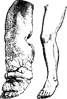
Elephant of the leg (normal leg on the right). (From a preparation of the Museum of the Pathological-Anatomical Institute of the 1st Moscow State University).
nature of the affliction; according to Morris, the Wassermann reaction in such cases is always negative; in rare cases, bone areas (osteomas) are formed in the thickness of the skin itself. When the external genitalia are affected, an analogous picture develops. Mild degrees of elephantiasis of the prepuce, scrotum, and the entire penis [see separate table (p. 671–672), fig. 3] can be observed when the efferent lymphatic vessels become obliterated, for example, during the invasion of inguinal glands by tumors. In pronounced cases, especially in Elephantiasis filariosa, the scrotum sometimes exceeds the size of a human head. The hypertrophied scrotum, penis, labia majora, and mammary glands can turn into powerful tumor-like formations in elephantiasis, hanging deeply downwards, e.g., between the legs, on a thin stalk; sometimes a luxuriant development of adipose tissue occurs in the thickness of these formations. A variety of elephantiasis is also the so-called Hottentot apron (see) of women, developing from the labia minora. Among the causes of gynecological elephantiasis, mention is also made (often, however, without special grounds) of tuberculosis, masturbation, syphilis; the development of Elephantiasis vulvae in connection with chronic (nonspecific) ulcers of it and the rectal area (ulcre elphantiasticum vulvo-anale, s. esthiomene) is also indicated. Elephantiasis of the lips of the mouth (macrocheilia) has a characteristic appearance, especially the upper one; most often this is observed with recurrent facial erysipelas. Macroglossia also belongs to the category of elephantiasis; the latter, like macrocheilia, is also observed in animals, and they can be both congenital and acquired. Elephantiasic growths in leprosy (Elephantiasis graecorum) — see Leproscopy. Elephantiasis with a more or less luxuriant participation of nervous tissue (Elephantiasis neuromatodes) — see Neurofibromatosis. Rare forms of vascular elephantiasis (elephantiasis haemangiectatica) include skin afflictions of the naevus mollusciformis elephantiasticus or naevus vasculosus giganteus type (see Naevus).
I. Davydovsky. Clinical picture and treatment of elephantiasis. The clinical course of the tropical form of elephantiasis (Elephantiasis tropica) is characterized by a sudden rise in temperature, often with chills and symptoms of inflammation of the lymphatic vessels and lymph glands. Upon the subsiding of acute inflammatory phenomena, a certain thickening of the affected organ remains. As a result of repeated exacerbations of the process, a sharp increase in the volume of the affected organ gradually develops, sometimes reaching colossal proportions. In the sporadic form of elephantiasis, the thickening of the limbs, which are affected most often, develops insidiously and imperceptibly. From trivial causes (a small abrasion between the toes, skin excoriation, a corn rubbed by shoes), acute dermatitis flares up, usually treated as erysipelas. The process usually begins at the age of 15–20, rarely after 30, and drags on for years; although the limb does not reach such colossal proportions as in the tropical form of elephantiasis, it nevertheless often deprives the patient of working capacity. To the purely mechanical inconveniences for the patient are added stubborn ulcers on the lower leg and secondary changes in the muscles, bones, and so on. In elephantiasis of the upper limb during recurring cancer of the breast or after extirpation of the axillary lymph glands, the phenomena of lymph stasis and limb edema predominate over the sclerotic phenomena in the skin and subcutaneous tissue. The diagnosis of elephantiasis usually presents no special difficulties. The anamnesis, the course of the disease, the characteristic changes of the skin and subcutaneous tissue, in the absence initially of changes in muscles and bones, make it possible to differentiate elephantiasis from local gigantism. Unilateral involvement speaks against edema on the basis of heart or kidney disease. Treatment. Since the various etiological factors underlying elephantiasis lead ultimately to one and the same pathophysiological picture of the disease—to fibromatosis and lymph stasis—both conservative and operative treatment of the disease are directed at combating lymph stasis. Among conservative methods, rest and the elevated position of the limbs come first. The method yields good results, especially in cases where the phenomena of edema predominate over the phenomena of tissue sclerosis: the volume of the limb decreases, the skin becomes more mobile and begins to gather into a fold, and ulcers (if present) show a tendency to heal. The effect of treatment can be enhanced by the use of tight bandaging with an elastic bandage, provided the skin is intact and there are no inflammatory phenomena. Hot and dry-air baths, and cautious massage with healthy and smooth skin, are also recommended. For skin lesions, eczemas, and ulcers, dressings with an indifferent ointment. With persistent and prolonged implementation of such treatment, it is possible in many secondary or acquired cases to achieve excellent immediate results, even an almost complete return of the affected limb to normal (Eiselsberg). But this result is not stable, and as soon as the patient begins to walk and work, all symptoms return, and after a relatively short time the limb reaches its former dimensions. Further among conservative methods of treatment, mention should be made of injections of various solutions in order to promote the resorption of connective tissue. For this purpose, Castellani proposed injections of fibrolysin; after preliminary preparation of the patient with rest and bandaging, 2 cm3 of fibrolysin is injected into various sites of the skin. After each injection, the limb is tightly bandaged with a flannel bandage. Treatment continues for several months. The immediate results are quite favorable, but long-term cure does not ensue. Instead of fibrolysin, Kültz introduced the use of injections of a 10% iodoform emulsion, the effect of which is attributed to the specific action of iodine on the parasite of the tissue (Filaria sanguinis hominis). Atoxyl has also been proposed for the same purpose (0.45 each week). Schmerz applies treatment with mustard plasters, based on the leukotactic action of mustard oil and the hyperemia achieved thereby. [Dry mustard is mixed with warm water, applied in a thick layer to a piece of canvas, and applied to various areas of the affected limb, first for 5 minutes, and then gradually for an increasingly longer period (up to 2 hours)]. This method gives good results in lesions of the superficial skin layers. There were attempts to treat elephantiasis with iodine (Mikulicz, Simon), a solution of sodium chloride, alcohol (Schwalbe), Ferrum sesquichloratum under the skin and internally (20 drops each), and antistreptococcal serum (Dubruel). However, most of these methods have been abandoned as not justifying themselves in practice. Methods of surgical treatment are divided into 4 groups: 1) amputation of the limb; 2) operations aimed at reducing the volume of the limb by decreasing the blood supply—ligation of the femoral artery (Carnochan), tibial artery (Statham), iliac arteries (Hüter); 3) all types of lymphangioplasty, the purpose of which is to create new pathways for the outflow of lymph (Handley, Lanz, Kondoleon, Sokolovsky); 4) operations to combat the reverse flow of lymph (Oppel). 1. Amputation of the limb can at present be recommended only for those cases in which all attempts at more conservative operations do not lead to the goal, and amputation of the limb is urgently necessary in view of approaching marasmus or disease of the bones and joints. 2. In order to reduce the blood supply to the limb, ligation of the femoral artery in continuity was proposed (Carnochan; 1858). Statham ligated the tibial artery, Hüter the iliac artery. Even if gangrene of the limb does not ensue after these heroic operations and a certain reduction in the volume of the limb is achieved, the long-term results are still poorly satisfactory: according to the statistics of Esmarch and Kulenkampff, out of 77 patients with arterial ligation, long-term success was achieved in only 8. To reduce the risk of the operation, Pieri recommends, instead of ligation, narrowing the vessel lumen by excising an ellipse from its wall. Buchanan achieved success from ligation of the femoral vein. Despite isolated cases of success, the operation of vessel ligation should be abandoned for cases where the question of amputation already arises. In order to obtain lymph drainage, incisions are made in the skin according to Dieffenbach. To reduce the volume of the limb, Winiwarter proposed wedge-shaped excision of pieces of affected skin. This operation gave good results both in the hands of the author and among many other surgeons (including Russians—Kuznetsov, Bogolyubov) not only in cases of sporadic, but also in endemic elephantiasis. 3. At the basis of lymphangioplasty lies the idea of creating a new pathway for the outflow of lymph. Handley proposes passing a bundle of silk threads subcutaneously along the entire affected limb to the limits of healthy tissues: through two incisions on the anterior and posterior surfaces of the upper limb, a double silk thread is passed using a special pointed probe with an eye; from a series of small incisions, the probe is brought out and inserted under the skin again. The end of the loop is brought to the hand, and the free ends of the threads are passed radially under the skin of the scapula. When operating on the lower limb, it is advisable to pass a double loop of threads from the region of the inguinal glands in such a way that on the lower leg the bundle can be split into 4 threads and passed in 4 different quadrants of the lower leg. Handley obtained good results from this operation in edema of the upper limb on the basis of breast cancer or disruption of lymph outflow after extirpation of the axillary glands. In these cases, edema and pain disappear in 2–3 days. Less satisfactory results are given by this operation on the lower limb, as the author himself points out (as well as Draudt, Stieda, and others). Draudt recommends Handley's operation in elephantiasis as a preparation for the subsequent excision of skin that freely gathers into folds; Keisser recommends it as an adjunct to Lanz's operation. The basis of the latter is the idea of creating contact between the network of superficial and deep lymphatic vessels. In order to establish a connection between these groups of lymph vessels, Lanz carves pedicle flaps from the fascia lata of the thigh thickened during elephantiasis, passes them through the thickness of the muscle into the bone marrow canal, where 3 openings are preliminarily drilled in the bone—in the upper, middle, and lower thirds of the thigh. After this, the fascial incision is stitched, and windows are cut out on the sides in the aponeurosis to facilitate communication between the lymphatic vessels of the skin and muscles. The operation yielded a brilliant and stable result in the hands of the author, followed up for three years. The remaining methods of this group (Kondoleon, Rozanov, Oppel, Minkh) are only modifications of Lanz's basic idea. Thus, Kondoleon and Payr propose excising wide pieces of the fascia lata along the entire limb from two incisions on the anterior and posterior surfaces of the thigh. Oppel, when performing Lanz's operation, uses a flap of subcutaneous tissue instead of the fascia lata. Minkh recommends, in addition to excising the fascia lata, also excising intermuscular fascial plates. Rozanov carves triangular flaps from the subcutaneous tissue and fascia of the lower leg, the acute angle of which is passed through the muscles and fixed to the periosteum.
Several similar modifications have also been proposed by Italian authors (Giordano, Melletti). Standing apart is the operation of lymphangioplasty proposed by Sokolovsky at the XVI Congress of Russian Surgeons. The idea consists in the free plastics of lymphatic vessels. The author passes a strip of omentum from the abdominal cavity and fixes it in the subcutaneous tissue of the thigh, while pedicled flaps from the tissue of the region of large vessels, rich in lymphatic vessels, are sutured to the subcutaneous tissue in the region of the great saphenous vein, where the largest trunks of subcutaneous lymphatic vessels are located. (The operation was performed by the author of the method with success twice and once by Vinogradov.) Of the methods of lymphangioplasty described here, the greatest number of observations has accumulated in the literature concerning the operations of Lanz and Kondoleon. Good results from Lanz's operation in the hands of the author himself have been confirmed by observations of other authors, including Russian ones; thus, Fishman cites 4 cases of the operation for elephantiasis of the lower extremity. In 1 case, Lanz's operation on the thigh and lower leg gave an excellent result, followed up for 2 years; in 3 other cases, where Lanz's operation was performed only on the thigh, the results are less brilliant, but in any case entirely satisfactory. Good and stable results were obtained: in one case by Grekov from his own modification of Lanz's operation, by Gusev from a combination of Kondoleon's method (on the lower leg) and Lanz's (on the thigh), by Minkh from Kondoleon's operation. Keyser in fourteen cases of elephantiasis saw good results from a combination of the method of Lanz and Handley. 4. A special position is occupied by the operation proposed by Oppel, which is based on the idea of the dependence of the development of elephantiasis on the reverse flow of lymph (analogous to the reverse flow of blood from the femoral vein to the great saphenous vein in varicose veins of the lower extremities). Oppel explains the success of Winiwarter's operation by the fact that, due to the formation of deep retracted scars, a separation of individual lymphatic trunks from one another occurs and thereby an obstacle to the regurgitation of lymph is created. For a greater effect of the operation, Oppel considers incisions not lengthwise, but across the limb, according to Rindfleisch (spiral incision along the lower leg in 2 to 2½ turns down to the fascia, with excision of the subcutaneous tissue, tamponade, and secondary healing of the wound) to be more appropriate. The operation was performed by the author in seven cases. After the operation, the volume of the limb rapidly decreased. In one of these cases, a good and stable result was obtained, followed up for seven years. In another, after a year, a recurrence and ulceration in the postoperative scars occurred. In the remaining patients, the immediate results are good, but the observation period is relatively short (3 months to 1 year). As for the treatment of elephantiasis of the scrotum, attempts at conservative treatment usually do not achieve their goal here. All types of lymphangioplasty give somewhat better results, but for cases where the scrotum has already reached large sizes, the most radical is amputation of the entire scrotum with preservation of the corpora cavernosa and seminal glands and plastic restoration of the scrotum and skin of the penis. However, with a disorder of lymph circulation in the inguinal regions, recurrences easily occur, which is why it is necessary to supplement the operation with one or another method of lymphangioplasty. For this purpose, Krogius proposed creating a new path for the outflow of lymph from the skin of the scrotum into the lymphatic vessels of the tunica vaginalis of the testis (Winkelmann's operation); with simultaneously existing hydrocele of the testis with impaired absorption by the vaginal tunic itself, Krogius proposes the implantation into the scrotum of a pedicled omental flap, Belitsky proposes the splitting of the spermatic cord along its length in order to use the absorptive capabilities of its initial lymphatic vessel network. For elephantiasis of the penis, wedge-shaped excision of the affected skin of the penis is recommended. In the most severe cases, one has to resort to amputation of the penis. In rare forms of elephantiasis of the vulva, face, and lips, the same principles of lymphangioplasty are applied or partial excision of the affected parts of the skin is performed. The success of the operation depends primarily on the severity of the disease and the degree of sclerotic changes in the skin and subcutaneous tissue. Rosenak advises performing an adrenaline test to develop a surgical plan for elephantiasis: upon injection of 1 cm3 of adrenaline under the skin of the affected limb, normally, anemic stripes begin to slowly spread upwards along the course of the lymphatic vessels. With sclerosis of the vessel walls or their obliteration, the test is negative. In accordance with the results of the test, the choice of operation is outlined. In advanced cases, treatment should begin with preparation by conservative methods, followed by the application of one or another lymphangioplasty and even better, a combination of one or two methods.
F. Karkanova-Müller.

Related articles
Mentioned in
Cite this page
“Elephantiasis.” Soviet Medical Encyclopedia. English translation of Bolshaya Meditsinskaya Entsiklopediya, 1st ed. (Moscow, 1928–1936), ed. N. A. Semashko. https://sovietmedicalencyclopedia.pages.dev/article/elephantiasis/