Adipose Tissue
Historical document, translated for reference. It reflects medical knowledge of the 1920s–30s and is not medical advice.
Summary
This article defines adipose tissue as a specialized connective tissue composed of fat-storing cells, detailing its cellular structure, chemical composition, and distribution throughout the body. It discusses the historical debate regarding its classification, its development from mesenchymal cells, and the characteristics of brown adipose tissue.
Encyclopedia article (1928–1936)
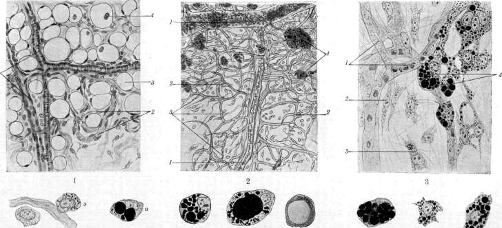
ADIPOSE TISSUE, a name given in histology to a variety of connective tissue in which numerous cells filled with fat droplets are embedded in the basic fibrous substance. Usually, there is one such droplet in fat cells, but in some cases, their number can be significant. Some authors consider the described tissue an independent histological unit (Löwe, Toldt, Ranvier, Kölliker, Hammar), while others accept this term only conditionally and do not separate adipose tissue from fibrous connective tissue (Flemming, Fomin). Accordingly, some authors view the fat cells that make up this tissue as specific elements that are laid down in the early period of embryonic life as a special variety of mesenchymal elements, while others assume that any mesenchymal cell that has fallen into appropriate conditions in relation to blood vessels can become a fat cell. Adipose tissue is widely distributed in the body; it is found in the subcutaneous tissue (panniculus adiposus), in the diaphyses of tubular bones (forming yellow bone marrow), in the cavity of the orbit, in the omentum and mesentery, in the intermuscular tissue, around the kidneys, heart, lymph glands, and neurovascular bundles. A fat cell has large dimensions (25-130 μ) and in humans and mammals usually contains one large fat droplet, which fills the cell body and stretches it into a thin-walled vesicle; the cell nucleus lies to the side, in a small accumulation of fine-grained protoplasm; around the droplet, one can notice a thin rim of the stretched cell body, on which some authors describe their own special membrane (Ranvier, Frey, Policard). The cell nucleus is clearly visible when the fat cell is seen in profile; such a relationship of the nucleus, protoplasm, and fat droplet gives the fat cell the appearance of a signet ring in optical cross-section. Chondriosomes in the form of short rods and grains and the reticular Golgi apparatus (Deineka) have been described in the protoplasm (Chashin, Dubreuil). The fat droplet consists primarily of neutral fats, to which free fatty acids and their calcium salts, as well as the pigment lipochrome, are mixed in small quantities. Depending on which triglyceride predominates in the composition of the fat droplet, it appears either liquid, even at ordinary room temperature (oleins), or proves to be such only at body temperature, and solidifies upon cooling (palmitin, stearin). Each animal has a specific composition of the fat droplet; however, dietary fat can have a significant influence here on the composition and properties of fat in fat cells, as follows from the experiments of Rosenfeld with feeding (previously starved) dogs mutton fat and linseed oil, whereby the fat changed significantly. Fat cells are only rarely encountered singly along the tract of small arteries among the elements of connective tissue (adventitia); usually, they are collected into groups—fat lobules, surrounded by a membrane of fibrous tissue, which in small quantities also penetrates into the interior of the fat lobule; between the cells, one finds a strongly developed network of blood capillaries, originating from a separate small artery that enters the fat lobule and here breaks up into a dense network of capillaries; adipose tissue thus has its own strongly developed network of blood vessels (see separate table, figure 1). Development of adipose tissue. Adipose tissue develops from mesenchyme in the form of islets located in certain places, which gave some authors (Toldt, Löwe, Ranvier, Kölliker) reason to express the opinion that adipose tissue is a specific type of connective tissue and represents something like a gland. In Toldt's opinion, "true adipose tissue" is always laid down in the form of a delimited accumulation of special cells in certain places of the organism ("primary fat organs"), and from here, fat deposits spread to other places in the organism. True, Toldt also admits the transformation of ordinary connective tissue cells into fat cells, but such places, in his opinion, are no longer true adipose tissue and behave quite differently during starvation. This view of Toldt and his supporters met with strong objection from Flemming, who in a series of works showed that the mentioned "primary fat organs" are not genetically connected with all places of fat deposition and that, in general, the formation of fat lobules proceeds simultaneously and independently in very many places of connective tissue. In studying the development of adipose tissue in various representatives of mammals, Fomin managed to show that the formation of fat lobules is closely connected with the formation of a special vascular network, primarily developing in the places of future fat lobules. On thin membranes (omentum, mesentery), it is easy to verify that beforehand, when there are no fat cells at all, a dense network of blood capillaries develops in those places where fat lobules will appear, and only then do individual connective tissue cells appear among this network—Ranvier's milk spots (taches laiteuses Ranvier). Among these cells, there are fibroblasts (mainly), plasma cells, mast cells, and even wandering elements; all of them begin to accumulate fat granules in their bodies and turn into real fat cells, which fold into lobules, in which it is impossible to distinguish cells that originated from fibroblasts from cells of other origin (see separate table, fig. 2). In detail, the transformation of connective tissue cells into fat cells proceeds as follows: initially, larger grains appear in the fine-grained body of the future fat cell, which stain with basic aniline dyes, predominantly safranin (pre-fat granules), then these grains gradually begin to take on the color of special dyes that stain fat, and also give a reaction with osmic acid; at first, the coloring is mixed, and then it becomes a pure fat reaction, while aniline dyes cease to stain such grains (see separate table, fig. 3). Small fat granules gradually merge into larger droplets, separated by protoplasmic partitions; the nucleus remains in the middle of the cell for the first time, but then is gradually pushed to one side, and the fat droplets in the center of the cell body merge into one large droplet. Around such a large droplet, one can see a fairly wide belt of fine-grained protoplasm, in which small fat droplets are constantly formed, merging with the main droplet. As the fat droplet increases, the cell body stretches, and the initially wide belt of protoplasm thins and densifies; however, it is not possible to note any changes in the structure or chemical properties of the cell body that give grounds to recognize the formation of a membrane on the fat cell: the membrane must be considered only a densified, but not altered layer of cell protoplasm (see separate table, fig. a-i). In developing fat cells, one can find karyokinetic figures for a relatively long time, and apparently, the division of the cell body almost always occurs, since binucleate fat cells are encountered extremely rarely. Thus, there is no reason to consider the fat cell a specific element intended for a specific function from the early stages of development: any connective tissue cell that has fallen into appropriate nutritional conditions, into a specific connection with blood capillaries, can turn into a fat cell. The development of fat lobules is not limited to the period of early development of the organism (embryonic and post-embryonic), but can also occur throughout the life of the individual, always being associated with the new formation of a capillary vascular network. Brown adipose tissue. Some authors (Hammar, Auerbach) distinguish brown adipose tissue, or the hibernation gland, as a special variety of adipose tissue. According to Hammar, this tissue occupies a fairly significant space in the organism in the form of cords and accumulations from the small pelvis to the diaphragm along the midline of the body, surrounding blood vessels to the outer edge of the kidneys and adrenal gland; from here, brown adipose tissue passes into the thoracic cavity, where it is also located along the midline of the body in the anterior and posterior mediastinum; in some places, it also lies in intermuscular spaces; on the hind limbs, brown adipose tissue is found in the inguinal fossa, on the forelimbs—between the shoulder blades and in the axillary cavity. This tissue is distinguished by its peculiar dark color and by the fact that its cells, along with a large fat droplet, usually also contain a significant number of small droplets that do not merge with the large one. According to Auerbach's research, brown adipose tissue has nothing to do with hibernation and is found both in animals subject to hibernation and those not subject to it; in addition, Auerbach was able to establish the direct transition of brown adipose tissue into ordinary (white) adipose tissue (for example, in the rat, which is considered by other authors to be typical precisely for this type of adipose tissue). According to Fomin's research, when developing brown adipose tissue, it is impossible to find any features that force it to be singled out as a special type, and the entire process of adipose tissue formation here proceeds according to the same principle. Physiological significance of adipose tissue is threefold. 1.
It serves as a reserve store of fat (one of the main nutritional substances), which is consumed by the organism during starvation. 2. Possessing significant elasticity, it can distribute the pressure falling upon it over a large surface and thus provides protection to the body and organs from mechanical impacts (shocks). 3. Due to poor thermal conductivity, adipose tissue reduces heat loss by the organism, which is why animals in polar countries have thick layers of fat in their skin. When fat metabolism is in equilibrium, fat cells have the appearance indicated above, and it may seem that the fat droplet in the cell lies completely unchanged until a certain moment and appears only as reserve nutritional material. However, a more thorough study of fat cells during changes in the nutritional regimen shows that in the cells, there is a continuous deposition of fat on one hand, and on the other, a continuous expenditure of it and its entry from the cells into the general metabolism of the organism. Thus, with a deficiency of fats in the diet, even under physiological conditions, one can find a picture of atrophy of adipose tissue. Flemming showed that the picture of atrophy of adipose tissue differs depending on whether the starvation is complete, leading to the rapid disappearance of fat from the organism, or incomplete, during which fat cells only slowly and gradually lose their fat. In the first case, the fat droplet rapidly decreases, breaks up into small droplets, which (apparently) dissolve and leave the cell body, while serous fluid accumulates between the fat droplet and the protoplasm; the dimensions of the cell body remain almost unchanged during this process. If the animal does not perish earlier, then all the fat disappears from the cell, and the fat cell appears in the form of a bubble filled with serous fluid, in which the smallest fat grains and remnants of pigment float—serous atrophy of adipose tissue. Leukocytes penetrate into the cells from the surrounding tissue, likewise the nucleus of the fat cell often divides, and as a result, an accumulation of small nuclei appears inside the bubble (Flemming's atrophy with proliferation).
Adipose tissue presents a completely different picture during incomplete starvation. In this case, the fat droplet slowly gives off small fat droplets, which dissolve and leave the cell body; the protoplasm, gradually contracting, does not lag behind the fat droplet, and no accumulation of serous fluid is observed. Gradually losing fat, the cell becomes smaller and smaller and becomes similar to a young fat cell caught by the observer in the process of fat accumulation. In conclusion, the cell loses its fat completely and can again take on the former appearance of an ordinary connective tissue cell, with the only evidence of its former role remaining as a small cluster of yellowish pigment grains. Parallel to the loss of fat and the decrease in the volume of fat cells, the desolation of blood capillaries also occurs; in a far-advanced process, the capillary network undergoes complete regression, and in place of the former adipose lobule, ordinary loose fibrous connective tissue remains, poor in blood vessels—terminal atrophy of adipose tissue (according to Flemming). It goes without saying that the desolation of the capillary network accompanying terminal atrophy significantly reduces the blood channel. Atrophy of adipose tissue in very young animals occurs with great ease: it is enough for sucklings to be deprived of food for a few hours to obtain a significant depletion of fat cells of fat droplets and their replacement with droplets of serous fluid; at the same time, fat droplets very easily break up into small droplets and grains and can simulate the picture of initial fat deposition in cells. The cellular protoplasm becomes coarsely granular, but does not give the typical staining of pre-fatty granularity, and then the picture indeed resembles a gland. Often, atrophy of adipose tissue is accompanied by a change in its color, which becomes darker yellow, which is apparently connected with the thickening and concentration of lipochrome during the decrease in the volume of adipose tissue.
a, b, c, d, e, f, g, h, i
Figure 1. Developed adipose tissue from the stretched intermuscular connective tissue of a calf: 1—blood vessels (arteries and veins); 2—capillary loops between fat cells; 3—fat cell in profile (ring shape); 4—fat cell with a nucleus visible from the plane. (Magnification about 80 times.)
Figure 2. Initial stages of the formation of fat lobules in the omentum of a newborn kitten: 1—larger branches of blood vessels between the forming fat lobules; 2—networks of blood capillaries in the places of future formation of fat lobules; 3—growing capillaries of the forming capillary network; 4—fat lobules of different sizes with the beginning of fat deposition. (Magnification about 25 times.)
Figure 3. Part of a very young fat lobule in the capillary network: 1—blood capillaries; 2—fibroblasts not yet affected by the process of fat deposition; 3—fibroblast with pre-fatty grains and the smallest grains of fat; 4—fibroblasts with a large accumulation of fat droplets, turning into real fat cells. (Magnification about 600 times.)
a—fibroblast with shortened processes and a significant amount of small fat droplets; b and c—further stages of the transformation of a fibroblast into a fat cell; d and e—formation of larger fat droplets and the fusion of newly formed small droplets with the main large droplet; f—young fat cell with a figure of karyokinetic division in the nucleus; g, h, and i—transformation of a rounded cell (histiocyte) into a fat cell. (Magnification about 600 times.) (According to the author's preparations.)
Of the pathological processes in adipose tissue, besides its atrophy and hypertrophy, which lie at the basis of obesity, various inflammatory processes and tumors are encountered. The former proceed in adipose tissue similarly to inflammation of connective tissue; only those cases in which the inflammatory reaction is connected with the destruction of fat cells and the splitting of the released fat differ in their peculiarities; in such cases, there is a proliferation of granulation tissue with giant cells, which is conventionally designated as fatty granuloma, or oleogranuloma. Tumors arising from adipose tissue belong to fatty tumors, or lipomas, and more rarely to liposarcomas and ordinary sarcomas.
Related articles
Mentioned in
Cite this page
“Adipose Tissue.” Soviet Medical Encyclopedia. English translation of Bolshaya Meditsinskaya Entsiklopediya, 1st ed. (Moscow, 1928–1936), ed. N. A. Semashko. https://sovietmedicalencyclopedia.pages.dev/article/adipose-tissue/