Bursitis
Historical document, translated for reference. It reflects medical knowledge of the 1920s–30s and is not medical advice.
Summary
This article from the 1928–1936 Great Medical Encyclopedia defines bursitis as the inflammation of synovial bursae caused by trauma, mechanical irritation, or infection. It details the clinical presentation of acute, chronic, and specific forms like gonorrheal, tuberculous, and syphilitic bursitis, along with their respective treatments.
Encyclopedia article (1928–1936)
BURSITIS (from the Greek bursa—pouch), inflammation of the mucous bursae, both permanent and newly formed, over bony prominences where there is constant pressure or friction. The causes of bursitis are traumatic injuries (bruises), repeated mechanical irritation, various types of infections, and diathesis (gout). The acute form of bursitis is characterized by the formation of a rounded, circumscribed swelling, depending on the accumulation of exudate in the bursa (serous, serous-fibrinous, purulent); its size ranges from a pigeon egg to a fist. In purulent bursitis, pain and tension in the bursa increase; a phlegmonous process develops in the surrounding tissues. Pus from the bursa makes its way outward, and persistent non-healing fistulas form. In serous forms of bursitis, the walls of the bursa are almost unchanged, and the exudate is absorbed; however, cases are observed where, due to the organization of fibrin and the obliteration of a number of vessels of the synovial membrane, persistent changes occur in the wall of the bursa, entailing a special susceptibility of the latter to repeated inflammation from minor traumatic insults; a so-called subacute form of bursitis develops, which easily transitions later into a chronic one. A special form of acute

Figure 1. Changes in the inner lining of the bursa in chronic bursitis (after Lexer).
bursitis is gonorrheal bursitis, which proceeds with a sharply painful swelling of the bursa and with characteristic phlegmonous phenomena in the surrounding tissues. The favorite sites for these bursitis cases are the Achilles and prepatellar bursae. Chronic bursitis develops from an acute one or is the result of repeated mechanical irritation in the area of the affected bursa (occupational bursitis, e.g., in harness makers). It manifests either in the form of "dropsy" with insignificant changes in the mucous bursa, or in the form of "chronic serous synovitis" with significant changes in the bursa. In this case, its walls thicken; the surface, initially smooth, becomes covered with a series of protrusions and cord-like growths along the cavity, giving it a bizarre appearance that defies description (see Figure 1). Among the cords, free "rice" bodies are often found. Sometimes in the walls, at the sites of old hemorrhages, calcifications are encountered. Pathological-anatomical changes in this form of bursitis are explained by some, mainly, by the organization of fibrin precipitating and settling from the exudate; others are inclined to see here a series of degenerative processes in the synovial bursa itself and in the surrounding adipose tissue. The course of the disease is chronic, with temporary improvements, with a tendency to relapses and exacerbations. Most often, the synovial bursae that are easily subjected to trauma become diseased: prepatellar, olecranon
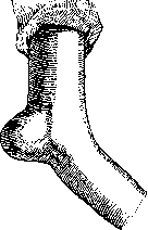
Figure 2. Bursitis of the bursa
of the olecranon process (after Lexer). (see Figure 2), supraclavicular, and trochanteric; all these lesions often turn out to be occupational diseases. Rare forms of bursitis include disease of the hyoid bone bursa (sometimes reaching significant sizes), the ischial tuberosity, and the ilium. Similar phenomena develop in newly formed bursae in cases of hallux valgus and pes varus. Recognition of bursitis is not difficult; they are easily identified by the shape of the swelling, fluctuation, and their location. Treatment of acute bursitis: rest, pressure bandage, compresses. Chronic: puncture and irrigation of the cavity with 2% carbolic acid or Rivanol (1:500). It is more expedient, in view of frequent relapses, to completely remove the bursa under local anesthesia. In purulent bursitis: incision. Tuberculous bursitis is quite rare; the wall of the bursa in these cases is uniformly thickened, and tubercular tubercles are found in it. In the cavity of the bursa is a serous-fibrinous exudate, often with "rice" bodies, which can turn into caseous-purulent and produce tubercular fistulas of the affected bursa. Treatment of bursitis is conservative; puncture with the introduction of 10% iodoform emulsion is appropriate; in other cases, removal of the bursa is indicated.
G. Steblin-Kamensky. Syphilitic bursitis, bursitis syphilitica, is a specific lesion of mucous bursae, natural or accidentally arisen, for example, under a callus; it is observed very rarely. Up to 1909, Churchman (Churchman J. W., American Journal of Medical Sciences, September, 1909) collected only 28 cases from the literature. Later, only isolated observations were published. The mucous bursae most easily subjected to trauma are predominantly affected: under the tendons of the "goose foot" (pes anserinus), the shoulder biceps or femoral biceps, near the head of the fibula, the Achilles tendon, etc. Apparently, accidental trauma also plays a role in the occurrence of specific bursitis. Early syphilitic bursitis, usually arising along with cutaneous syphilides, proceeds according to the type of banal hygroma and is usually distinguished by the absence of subjective sensations. Rarely, having a more acute character, syphilitic bursitis causes significant pain. Specific treatment quickly eliminates all symptoms. Late syphilitic bursitis, mainly in the area of the knee joint, is characterized by the deposition of a gummatous infiltrate around the bursa, which captures the walls of the latter, producing a resistant, often fluctuating tumor. If the disease is left untreated, the infiltrate, capturing the skin, undergoes decay with the formation of a fistulous ulcer leading into the cavity of the bursa. For the most part, syphilitic bursitis develops in isolation from the joint, which remains unaffected; lesions of tendons and their sheaths are often observed along with bursitis. Syphilitic bursitis must be differentiated from professional, traumatic, tuberculous bursitis, or those caused by other chronic infections. Sometimes only a trial treatment establishes the diagnosis.
Related articles
Mentioned in
Cite this page
“Bursitis.” Soviet Medical Encyclopedia. English translation of Bolshaya Meditsinskaya Entsiklopediya, 1st ed. (Moscow, 1928–1936), ed. N. A. Semashko. https://sovietmedicalencyclopedia.pages.dev/article/bursitis/