Gout
Historical document, translated for reference. It reflects medical knowledge of the 1920s–30s and is not medical advice.
Summary
Gout is a complex metabolic disorder characterized by acute attacks of pain and inflammation in joints, particularly the big toe, and deposits of uric acid salts in tissues. The article traces the historical understanding of gout from ancient times to the 19th century, including its terminology, etiology, and epidemiology.
Encyclopedia article (1928–1936)
GOUT, podagra (from Greek pous--foot and agrios-fierce), a complex metabolic disease characterized by acute attacks of pain and inflammatory phenomena, mainly in the joints, and deposits of uric acid salts in mesenchymal tissues (mainly in cartilage, skin, and mucous bursae). Gout has been known since ancient times; the term Gout first appears for the general designation of the disease in Hippocrates, who was well acquainted with the acute gouty attack. Somewhat later, Roman physicians proposed other names for this disease based on the localization of the lesion: omagra^shoulder joint, chiragra-gout of the hand, gonagra-gout of the knee, coxagra-gout of the hip joint, rachiagra-gout of the spine, pechiagra-gout of the meninges, odontagra-gout of the teeth, rhinagra-gout of the nose, ischiagra-gout of the sciatic nerve, tenotagra-gout of the tendon, etc. In Galen, one can find the first good description of the gouty node1, and he is also the first to introduce the term "arthritis" to denote gouty inflammation of the joints, which was later by other authors extended to all kinds of joint inflammation regardless of their etiology. This introduced great confusion in the clinic of joint diseases and in particular gout. The abbess of Bingen Hildegard (12th century A.D.) was the first to use the term gutta (from Latin gutta-drop) to denote gout, thereby expressing the humoral-pathological concept of gout as a disease caused by a poisonous fluid (humor) in the blood, which, being excreted drop by drop into the joints, causes inflammation. (From Latin gutta comes also the French designation for gout-goutte and the English-gout.) In the 16th century, Paracelsus proposed overeating as the cause of gout and created his doctrine of gouty dyscrasia (violation of the proper composition of body fluids), replacing the humor of his predecessors with the concept of a burning mucous fluid-tartarus, which pours into the joint and dries there into crumbly earthy deposits. Sydenham, being a gout sufferer himself, described the clinical picture of a gouty attack and differentiated gout from other joint inflammations. A hundred years later (1787) Wollaston established that the chalk-like deposits in gouty nodes contain uric acid, and another 80 years later (1859) Garrod proved with his famous "thread" experiment that uric acid is present in the blood of gout sufferers. There are no exact data on the prevalence of gout; apparently it is known in all civilized countries. Gout cannot be classified as a frequent disease, and according to various statistics, its frequency ranges from 2 to 4 per 1000 cases. Etiology of Gout. The causes of gout have not been finally clarified, but a number of predisposing factors are well known. Great importance is attached to heredity--already Garrod noted multiple cases of gout in the families of most of his patients, Scudamore confirmed the same in 332 out of 500 cases he studied, Gudzent-in one third of his patients, the American Williamson-only in 12%. Gout often occurs in families whose members also suffer from other metabolic diseases-(obesity, diabetes), migraines, bronchial asthma, exudative diathesis, dermatoses, and the formation of stones in the liver and kidneys, i.e., arthritis in the sense of Bouchard. Gout sufferers are more often hypersthenics than asthenics, men-much more often than women^ (according to Williamson, for 115 cases of gout-1 woman, Gudzent in 76 autopsies did not see a single woman). The age of most frequent onset of gout is from 30 to 50 years; cases of the disease in early childhood and in old age are rare. Among the factors contributing to the development of gout, dietary errors and especially overeating, abuse of alcoholic beverages, as well as a sedentary lifestyle have long been correctly included. Indeed, during the European war and in the first years after it, the clinical manifestations of gout became extremely rare and only now they are beginning to occur more often; the harm of excess meat in the diet is particularly often mentioned. In England, where gout is observed more often than in other countries, they eat especially much meat; Indians-vegetarians do not fear it. B. M. E., XX.V. 771
GOUT
772 leyut P., and those living in the same climatic conditions and eating meat often suffer from Gout. However, it cannot be said that there are no indications to the contrary. For example, the famous expert on gout Cantani asserts that in Italy, vegetarians often suffer from Gout. Abuse of alcoholic beverages, especially port wine, champagne, and beer, is considered of enormous importance. Modern authors (Widal, Leschke) believe that an attack of gout is caused not by alcohol, but by protein impurities from the yeast used in the preparation of alcoholic beverages. An enormous role is attributed to chronic lead poisoning in the development of gout. According to Lichtheim, among 800 workers in lead mines in the northern Harz, 100 cases of gout were observed over 8 years. The role of cold as a factor causing an attack is undeniable; this is evidenced by the frequency of gout attacks in spring and autumn; physical and psychological trauma play a similar role; the significance of random infections, especially those accompanied by high temperature and significant leukocytosis (pneumonia more often than others), is also undeniable. The clinical picture of gout is extremely multifaceted, and if we include here the arthritism of French authors and the atypical form of Goldscheider, then diagnosis becomes extremely difficult. Clinically (according to Herrold and Minkowski), one can distinguish an acute gouty attack, chronic gout, and atypical gout. The acute attack most often begins with the metatarsophalangeal joint of the big toe of the left foot; the first attack can also begin with other joints, and in descending order of frequency, the ankle, talocalcaneal, knee joint, joints of the toes, then joints of the fingers, wrist, sternoclavicular, shoulder, cervical spine, and jaw joints are affected. [This order mainly refers to the first attack; later any joint can become ill—attacks of gout in the larynx have been described, causing inflammatory swelling of the vocal cords and epiglottis with symptoms of threatening suffocation, attacks of gout in the auditory ossicles, tendons (Achilles), joint ligaments, and more rarely, in the skin and muscles.] Gouty equivalents have been described in the eye [attacks of acute pain at night with hyperemia of the eyeball without symptoms of glaucoma (Kruckmann)]. Usually the attack begins suddenly without any prodromal symptoms at night. The patient goes to bed healthy, falls asleep, and suddenly wakes up from the most excruciating drilling pain in one joint. The degree of pain is not always so severe, but even the slightest shaking in the room and even the pressure of the blanket intensify the pain. Chills or even severe chills are often noted, the temperature sometimes rises to 40-41°, the affected joint quickly swells, the skin over it turns red, is tense, shiny, and becomes hot. The redness and swelling spread upward, and the veins also swell up to the knee. The disease of the joint produces the impression of a severe phlegmonous inflammation, and there are known cases of operations due to erroneous diagnosis. By morning the pain usually subsides somewhat, but by evening it resumes again, and this picture continues for 3 to 6 days in a row; finally, the inflammation of the joint gradually subsides, the skin over it peels and slightly itches, and everything passes without serious consequences. The first attack is not always so intense: sometimes there is only slight redness of the joint without an increase in temperature. Sometimes the attack begins with increased sensitivity and redness of the skin in the area of one of the joints, and then swelling and effusion into the joint join in. Sometimes several joints are affected at once or the joints become ill one after another. In such cases, the picture is similar to acute articular rheumatism. Often the attack is not only accompanied by a number of general phenomena, but they can precede it. 1-2 days before it, general malaise, depressed mood, loss of appetite, pressure under the xiphoid process and in the right hypochondrium are noted. The disease can be limited to just one attack: but more often attacks reappear at intervals of 6 months-1 year or more; they can also become more frequent and run in a milder form, and thus the typical acute gout passes into a secondary chronic form (according to Herrold and Minkowski). If attacks repeatedly affect the same joint, a circumscribed persistent tumor appears in its area, which during an attack has a bluish discoloration. Later, the tumor instead of fluctuation gives the impression of granularity and finally forms a dense, translucent through the skin nodule, in the area of which the skin may rupture and then a white, crumbly mass is secreted, which for the most part consists of uric acid salts. Such gouty nodules (tophi) are typical for gout and often develop without an attack or between attacks (and therefore do not depend on it), localizing in the auricle (Fig. 1), less often on the wings of the nose and eyelids; they often appear in the periarticular tissue of joints, on tendons, in mucous bursae (most often in the area of the elbow tubercle) and in muscles. Tophi are pathognomonic for gout, but "one must make sure that they contain uric acid. To distinguish them from nodules due to blockage of the sebaceous glands and atheromas, one should obtain the contents with a needle and
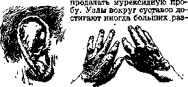
Figure 1.
Figure 2.
deform and disfigure the extremities (arthritis urica) (fig. 2). Despite significant deformities, movement in the joints in G. often—contrary to expectations—remains preserved. Not infrequently, gouty changes are found in the bones. In the epiphyses of the metatarsal, metacarpal, and phalangeal bones, round foci of uric acid deposits up to 1 cm in size are found; on X-rays, these foci appear as amorphous dark spots—cysts—surrounded by a light ring. These cysts are the result of resorption of bone tissue at the site of urate salt deposition, while the salts themselves do not give any shadows. Often the joints themselves are not changed, and the condition is limited to disease of the synovial membrane (synovitis urica) with effusion into the joint, which can be resorbed after an attack. If attacks occur frequently, then deformity of the joint can also occur due to the remaining effusion, due to thickening of the ligaments, or due to destruction of the cartilage. Deposits in periarticular tissue, deposits in cartilage and bones can rupture into the joint, and then the condition leads to ankylosis due to proliferation of connective tissue or due to new formation of bone tissue at the edges of the joint. Often uric acid deposits are found only in one of many affected joints. The deposition of urates in joints in chronic G. can occur painlessly, just like the formation of tophi in the skin. For example, deposits of uric acid were found in joints without any attacks in people suffering from chronic kidney disease, on the other hand, in severe attacks in surgically opened joints, no deposits were found; this is very strong evidence in favor of the fact that during a gouty attack there is dissolution, absorption, and excretion of urates, and not their precipitation in the tissues, as was initially assumed by Garrod and others. After a series of attacks, the joint manifestations often subside and seem to recede to the background, and a number of disorders from internal organs appear. These extra-articular manifestations in goutics are often described under the name of visceral G. In this case, clinical and even patho-anatomical data are so little specific that it is very difficult to prove their gouty nature. Among the visceral manifestations of G., the first place is taken by kidney damage. Ebstein and Umber distinguish 'renal gout' as an independent form on the grounds that they found deposits of urates in necrotic foci of the medullary and less frequently cortical layer of the kidney; these deposits are visible macroscopically as small white dots and streaks, sometimes surrounded by an inflammatory ring; under the microscope, crystals of monosodium and ammonium urate were found in them (not to be confused with uric acid infarction of the kidney in newborns, in which uric acid deposits lie in the lumen of the urinary tubules). According to the description of Ebstein and Umber, the 'renal' form of G. was observed by them among the poor population. If these observations are correct, then this form of G. is still a very rare phenomenon, and diagnosis is complicated by the fact that with kidney insufficiency of non-gouty origin, retention of uric acid in the body and its deposition in any organ is possible. Although deposits of uric acid are very characteristic of G., they cannot in any way be identified with it. Very often in goutics, slight albuminuria and hyaline cylinders are found. Goutzheim in 76 autopsies found no changes in the kidneys in only 6 cases, in the others either non-specific sclerotic changes or shrunken gouty kidney were found. Second in frequency of disease is the cardiovascular system? in particular, arterio- and especially arteriolosclerosis of the kidneys with a typical clinical picture (elevated blood pressure, vascular spasms, intermittent claudication, etc.). Vessel damage in goutics is so frequent (from 35% to 70% according to different statistics) that it is difficult to think of a coincidence, and it is necessary to consider diseases of the blood vessels to be causally related to G. At autopsies, an unchanged heart is rarely found, more often hypertrophy of the left ventricle (result of elevated blood pressure) and approximately in half of the cases arteriosclerotic changes in the myocardium and valves are noted. Legendre described deposits of uric acid salts on the aortic valves, Lancereaux on the mitral valve. Goutics are also very prone to diseases of the respiratory tract. Bronchial asthma is often observed in goutics, however, it is difficult to say to what extent it is correct to consider the latter causally related to a gouty attack. In the literature, cases of 'gouty pneumonia' are described on the grounds that during pneumonia a typical gouty attack occurred. It is more correct, however, to assume that in a goutic who contracted ordinary pneumonia, an attack occurred during pneumonia due to a significant excess of purines during the breakdown of leukocytes. Gastrointestinal disorders are often observed in goutics, which cannot be explained solely by dietary errors. The observations that there is a connection between gouty attacks and the observed gastrointestinal disorders, although not clarified (allergic?), are undoubtedly correct. Deposits of uric acid in the intestinal villi have been described. Umber and Dickworth assert a connection between G. and alveolar pyorrhea and some forms of chronic pharyngitis. Charcot and others pointed to gouty diseases of the liver, recently Schittenhelm and Brugsch attribute great importance to the liver in the genesis of G. Indeed, an enlarged and painful liver is often found in goutics, and if cases of enlargement due to stagnation phenomena, syphilis, etc., are excluded, there still remains a number of cases when no other explanation can be found and one has to recognize the enlargement and sensitivity of the liver as phenomena related to G. (Goldscheider). However, specific patho-anatomical changes in the liver in G. have not been described, deposits of urates in it have never been observed. Pyelitis due to deposits of urates in the mucous membrane of the renal pelvis and gouty cystitis have been described. At first glance, the precipitation of salts in the urine with usually decreased or normal concentration in goutics seems incomprehensible. However, the quantitative excretion of salts in goutics at different times fluctuates within wide limits and it is not this that is decisive. Uric acid, as is known, is generally difficult to dissolve and remains in solution in the urine due to the simultaneously excreted protective colloids by the kidneys. With decreased functional ability of the kidneys, the excretion of colloids also suffers; this obviously explains the frequently observed crystallization of salts in the urine of goutics, this also explains the combination of G. with kidney stones and urinary tract stones (Lichtwitz). To the visceral manifestations of G. are often included the frequently occurring myalgias (lumbar muscle), neuralgias and neuritis, eye diseases (frequent conjunctivitis, episcleritis and iritis), *2Ь 7Д5 77в manifestations of Meniere's disease have been described. To the manifestations of visceral G., authors often include the frequently occurring chronic inflammations of the skin (dry eczema of the eyebrows and psoriasis) found in goutics. Lichtwitz believes that these dermatoses are caused by the irritating influence of excess uric acid in the blood and especially the high content of uric acid in sweat (up to 9.2 mg% according to Adler). In addition to the typical forms of acute and chronic G., occurring with at least one typical attack, there is an atypical form that occurs completely without attacks of joint inflammation; some French clinicians even claim that persons with a gouty heredity are from childhood prone to a number of diseases regardless of whether typical G. will develop in them later or not. In childhood, such subjects suffer from eczema, during the period of maturation they observe persistent catarrhs of the upper respiratory tract, anginas, joint rheumatism, migraines, nosebleeds, conjunctivitis and urticaria; after the period of growth, they observe dermatoses and other phenomena described under the name of arthritism. Goldscheider even asserts that atypical G. occurs much more frequently than typical G., and that only the latter form of G., being the most pronounced, is inherited. For the diagnosis of atypical G. according to Goldscheider, 2 points are sufficient: the presence of tophi (not necessarily in typical locations) and the presence of a fine crunching in the joints, especially in the knees. In the presence of crunching in the joints without tophi, the diagnosis is sufficient with the presence of obesity, cardiovascular and renal symptoms; the presence of obesity and unexplained enlargement of the liver is sufficient to suspect G.
Pathological anatomy. The most characteristic changes are in the joints and periarticular tissue. Upon opening the joint, the surface of the cartilage appears covered with matte-white masses in the form of small separate spots or a continuous covering. The surface of the cartilage is partly smooth, partly uneven and rough. Even macroscopically it is visible that the deposits are located on the surface of the cartilage; in more severe cases, destruction of the cartilage is observed, urates are on its surface and can accumulate inside the joint; when the destruction of the cartilage spreads deeper, the deposits also penetrate into the underlying bone. There are different views on the method of formation and spread of these deposits. Older authors believed that uric acid salts are deposited primarily in the cartilages; according to Munk, uric acid is primarily deposited in the synovial membrane rich in blood vessels, in mucous bursae, periosteal tissue and in the bone marrow, from where it secondarily penetrates into the cartilages. Brogsitter believes that uric acid is deposited primarily in the upper layers of the cartilage, in the cells and in the intercellular substance, but he admits the possibility of primary deposition also in the joint capsules, tendon sheaths and other places. Upon microscopic examination of cartilaginous tophi on sections, it is found that in the upper layers of the cartilage there are no crystal deposits or very few of them, further in depth they spread in the form of radial fountain splashes. 'When studying sections, the impression is obtained that the deposits spread along tissue clefts and lie most densely near the surface. The old view that crystals are primarily deposited in cartilage cells has been refuted by Ebstein. It can be considered established that the deposits begin where the cartilages contain the largest amount of fibers and where blood circulation is very difficult. The structure of the nodules in the periarticular tissue is much simpler: in the center lie needles of sodium urate, around them a ring of connective tissue. In the inner part of this ring, directly around the crystal deposits, are the youngest cells, and among them many multinucleated giant cells that capture and destroy the crystal deposits. Changes in internal organs, not specific for G., are described above. Metabolism (in G.). Since the time of Herrod, it has been believed that the poison causing G. is uric acid, and a huge number of works have been devoted to the study of the question of its excretion and retention in tissues (see Uric acid). Uric acid in normal conditions is excreted mainly by the kidneys and only a small amount is excreted with sputum, sweat, bile and intestinal juice; in pathological conditions a significant amount of uric acid is also excreted by the intestine. It is very difficult to account for the balance of uric acid, since it is not only difficult to account for the amount excreted by the intestine, but even more difficult to determine the amount retained in the tissues. However, the study of purine metabolism has shown that in gouty patients it proceeds in exactly the same way as in healthy people, the difference being only in the excretion of uric acid. It is generally recognized that the content of endogenous uric acid in the blood of a gouty patient is elevated (hyperuricemia) from 5 to 18 mg%. It is especially characteristic that the content of endogenous uric acid in the urine is low while the concentration in the blood is high before an attack. Hyperuricemia in gouty patients does not depend on its increased formation (Brugsch, Schittenhelm, Umber), but on its poor excretion by the kidneys (Thannhauser) due to their functional insufficiency, which at the beginning of the disease fluctuates within wide limits and only later becomes permanent due to the addition of anatomical changes. The excretion of endogenous uric acid in gouty patients proceeds uniformly, at low figures, maximum 0.3 in 24 hours, exogenous uric acid is also excreted more slowly than in healthy people, and only during an attack its excretion sharply increases; immediately before an attack and immediately after it, excretion is reduced. The general metabolism in G. is not disturbed. Protein metabolism processes proceed normally, and only the excretion of products of nitrogenous metabolism is reduced immediately before an attack and increased during it. The theory of pathogenesis must explain the entire complex picture of G.: the phenomena of the attack, the retention of uric acid in the blood and its deposition in tissues. The renal theories of Herrod and Thannhauser do not satisfy these requirements, since at best they only explain the retention of uric acid in the blood and do not explain at all either the violent picture of the attack or the deposition of uric acid salts in tissues. The enzymatic theory of Brugsch and Schittenhelm also explains the entire syndrome little, according to which G. is a disease of metabolism and develops in the absence in the body of an uricolytic enzyme, which in normal conditions converts uric acid into urea. At present this theory has been abandoned, since it has been finally established that in normal conditions humans also lack an uricolytic enzyme and uric acid is the end product of metabolism. Brugsch himself has abandoned the enzymatic theory and now expresses a different view: he believes that G. is the result of a disorder of this complex system of organs participating in purine metabolism from the moment of digestion of nucleoproteins until their conversion into uric acid and excretion. This system begins with the intestine and includes the liver, the small and large circles of blood circulation and the kidneys. As a result of prolonged overload of this system (overeating and alcoholism) G. develops as a disease of purine metabolism in the broad sense of the word. Brugsch also sees the explanation of gouty inflammation in a violation of metabolism. Brugsch's new theory is full of uncertainties, nothing clarifies and therefore does not find recognition. All attempts to explain the pathogenesis of G. by 'special compounds' of uric acid in the blood and 'special structure' of uric acid in gouty patients, due to which the kidneys cannot excrete it, have proved groundless (Minkowski and others). The attempt to explain the deposition of uric acid in the cartilages by a special affinity of the tissues of gouty patients for uric acid - uratohistechia (Minkowski, Umber, Gouzent) - has also not been justified. Thus, we still do not have a theory that could exhaustively explain all the phenomena in G. and connect them with each other. Nevertheless, the latest research by Jones, Lichtwitz, Vidal and Gouzent has significantly expanded and deepened our understanding of the three main moments of gouty disease: the retention of uric acid in the blood, its deposition in tissues, mainly in cartilages, and the picture of the attack. The retention of uric acid in the blood in G. is a consequence of the functional insufficiency of the kidneys and their reduced ability to concentrate and excrete uric acid (Thannhauser, Lichtwitz, Steinitz). - For the deposition of uric acid in tissues, according to Lichtwitz, three conditions are necessary: 1) high concentration of uric acid in the fluids bathing the tissues; 2) the possibility of fluid penetrating into the tissue and 3) the presence of unfavorable conditions in the tissue that promote the crystallization of salts. The first two conditions are present in G., but in the presence of the first two conditions, a normal cartilage is not even encrusted at a higher concentration of uric acid in the blood than occurs in G., and Lichtwitz explains this by the presence of protective colloids in normal cartilage; with prolonged bathing of concentrated solutions of uric acid in cartilage tissue, recessive phenomena appear, as a result of which the protective colloids disappear, and the third condition necessary for the deposition of uric acid is created (Lichtwitz). The picture of an acute attack - sudden onset, chills, fever and violent inflammation - in the light of modern doctrine is considered as a manifestation of allergy (see), i.e., increased sensitivity of the body to allergens, which are either in the external environment or are formed in the patient's own body. It is impossible to specify these allergens exactly, but apparently they are most often components of food products or are formed from them during the act of digestion. In the light of this doctrine, it is understandable that not only meat and alcoholic beverages, but also other products of protein metabolism, bacterial and yeast, as well as those formed in the body during injuries, colds and emotions, can cause an attack; the states of long-term well-being after attacks and a number of so-called 'visceral' phenomena, such as bronchial asthma, unmotivated diarrhea and dyspeptic phenomena, which are observed during attacks and can also be manifestations of allergy, are understandable. In the light of this doctrine, uric acid is not the cause, but only a convenient indicator, a test of true G. The retention of uric acid in the blood and its deposition in tissues are components of the complex complex of G., but from what they depend and the essence of their mutual connection is not yet clarified by this theory. The diagnosis of an acute attack with typical localization rarely presents difficulties. Diagnostic difficulties are possible with accidental inflammation of the joint of the big toe of the foot due to infection; supporting points for recognition are lymphangitis and lymphadenitis in purulent arthritis, the duration of the process and spread to other joints. A gouty attack as a rule is localized and subsides more quickly.
Difficulties may also arise in acute articular rheumatism with initial involvement of one joint of the leg; in gout, the swelling is caused by inflammation of the periarticular tissue, while in rheumatism it is mainly due to inflammatory effusion into the joint, in addition other joints soon become affected. It is rarer to differentiate from acute gonococcal or other infectious (pneumococcal) inflammation; here the diagnosis is clarified by the anamnesis, urological examination, specific reaction or examination by diagnostic puncture. Recognition is significantly more difficult with atypical localization of the attack, especially if there is no history of attacks and if the disease develops directly after trauma; sometimes the effect of using colchicine helps to clarify the diagnosis here—it is specific only for gout. Chronic articular gout (arthritis urica) must be differentiated from chronic infectious arthritis, arthritis deformans, and from endocrine joint diseases. Gout is less common than infectious arthritis; in infections—except for tuberculosis, gonorrhea, and syphilis—the lesions are more often multiple; the temperature curve, examination of organs, specific reactions, puncture of joints with subsequent microscopic, chemical, and bacteriological examination, blood analysis, and X-ray are important; endocrine arthritides are differentiated on the basis of other manifestations of endocrinopathies; arthritis deformans affects large joints and joints of the spine, has a latent and slow course, often a typical X-ray picture (see Arthritis). Special caution is necessary in the diagnosis of atypical gout. Recognizing its existence, one cannot nevertheless diagnose it based on a single joint crack, which occurs in various arthropathies. True, the crack in gout has some characteristic features—it is fine, 'sandy'. In the absence of tophi, it is necessary to examine the blood and urine for uric acid after a purine-free diet, and then determine the excretion of uric acid after a load (nucleic acid or, what is the same, 100.0 thymus): hyperuricemia—the presence of low endogenous uric acid in the urine and its delayed excretion after the load decide the issue. The determination of the uricotoxic coefficient according to Steinitz may also be important: the ratio of the highest concentration of uric acid in the blood to the concentration of uric acid in the urine in a healthy person is about 23, in atypical gout about 16. However, it is completely erroneous to diagnose gout only based on a large deposit of urate in the urine. One should not confuse with gout the deposits of calcium salts in tissues—pseudogout (Kalkgicht of German authors), this includes Duplay's disease—periarthritis humeroscapularis, i.e., deposition of lime in the bursa of the shoulder joint. The prognosis of gout depends on age, the state of the cardiovascular system and kidneys. The mildest cases are those that begin with rare, short attacks in people over 40 years of age without a hereditary predisposition to gout as a result of dietary errors. With proper regimen, complete recovery is possible here. Cases beginning at an age under 30, with a hereditary predisposition, can have a favorable course with proper treatment, but here joint deformities and tophi often remain. In asthenics, severe forms that are resistant to treatment with significant joint deformity are more often encountered than in others. The prognosis is especially serious in persons suffering from chronic lead poisoning and in nephritics. Treatment. In acute attack—bed rest, a heating compress from Bur's solution or from vodka on the affected joint, with severe pain—injection of morphine (0.015-0.02); as a specificum for a gouty attack, autumn crocus preparations—Tincturae Colchici, 15-30 drops 3 times a day or better Colchicin Merck 0.0005 to 0.001 hourly to 0.004 in the first 2 days and to 0.002-0.003 on the 3rd and 4th day. If diarrhea occurs, the autumn crocus preparations are discontinued. At the beginning of the attack, especially with dyspeptic symptoms, it is better to abstain from food for 1-2 days, usually drinking—water, lemonade, fruit drink; with the cessation of dyspepsia, mucilaginous soup, vegetable puree, fruit puree, liquid porridge are prescribed, and then the diet is gradually expanded, adhering to a light purine-free diet. Immediately after the attack, Atophan or Noyatophan is prescribed 3 times a day at 1.0 with Borjomi (1-2 bottles) to facilitate the excretion of uric acid. In chronic gout, main attention should be paid to diet. General moderation in eating is recommended (in the obese about 20 calories per 1 kg of normal weight), with a complete ban on alcohol. It is necessary to find out, if possible, which products are allergens for the patient, and to exclude them. In view of the large role of uric acid in the pathogenesis of gout, everything containing a lot of purines is forbidden—first of all, glandular organs [liver, kidneys, 'sweet meat' (thymus)], as well as extractive substances—meat broth, meat gravies. In severe cases, meat and fish are not allowed earlier than 6-12 months after the last attack, in mild cases it is possible after 3-4 months; meat is given boiled (from the point of view of purine content, white or red is indifferent), not more than 150 g per day, from 2 to 5 times per decade, depending on the severity of the disease. It is better to give meat in the morning hours, since the excretion of uric acid at night is slowed down. The protein ration should not exceed 1 g per 1 kg of normal weight and is given mainly in the form of eggs, cottage cheese, milk; carbohydrates and fats do not contain purines and therefore all kinds of bread, cereals, vegetables and fruits, all kinds of fats are allowed. Peas, beans, lentils, radish and turnip are forbidden. One-sided fruit diets with sensitivity of the digestive apparatus can cause an attack. In the absence of contraindications from the circulatory organs, it is recommended to drink a lot—up to 2-2.5 liters per day, spring water is most beneficial. Weak tea and coffee are allowed. For better excretion of uric acid by the kidneys, Atophan or Noyatophan are prescribed periodically—with frequent attacks first every other day, then after 2,3,4 days, and after the cessation of attacks for 2 days in a row every 2 weeks. The dose of Atophan is 2 g per day (to be taken with Borjomi or soda water); simultaneously—purine-free diet. Severe hepatitis have been described from Atophan (caution is needed). To facilitate the excretion of uric acid, formaldehyde therapy in the form of Urotropin, Citurin has been proposed, Uricedin containing sodium citrate has also been proposed, but the effect of these drugs is doubtful. In stubborn chronic cases, Lichtwitz and others recommend protein therapy: intravenous injections in increasing doses (from 0.2 to 2.0) of Caseosan, Yatren Casein or Novoprotin (from 3 to 5 injections, with intervals of 3-5 days); intramuscular injections of milk from 2 to 10 cm3, intramuscular injections of sulfur suspension in Ol. Olivarum from 1/100 to 1% in increasing doses from 1 to 5 cm3. With a vigorous reaction of the body, treatment gives a better effect. Great assistance for treatment is movement: it improves metabolism, circulation, preserves joint mobility. Sidenham recommended horse riding, walks, gymnastics, any sport are beneficial; depending on the condition of the joints and heart, tennis, golf and even rowing and football are prescribed. Massage is very useful, especially with limited joint mobility. Physiotherapy in the form of dry-air, light, steam, mineral, radon, mud baths, mud cakes, diatherapy gives excellent results due to general and local influence. The effect of these procedures is even stronger at resorts, where the influence of diet, climate and mineral waters is added. Of mineral waters, those containing sulfates act best due to their effect on the liver and intestines. Gies and Gudzent recommended radium in the form of inhaling emanation in chambers, drinking emanation-containing water and injections; the effect proved unreliable, but in stubborn cases it can be tried.
Related articles
Mentioned in
Cite this page
“Gout.” Soviet Medical Encyclopedia. English translation of Bolshaya Meditsinskaya Entsiklopediya, 1st ed. (Moscow, 1928–1936), ed. N. A. Semashko. https://sovietmedicalencyclopedia.pages.dev/article/gout/