Perthes Disease
Historical document, translated for reference. It reflects medical knowledge of the 1920s–30s and is not medical advice.
Summary
Perthes disease is a unique condition affecting the femoral head in children or adolescents, classified as an epiphyseal and apophyseal osteochondropathy. It is characterized by avascular necrosis of the femoral head, leading to fragmentation and flattening, with variable clinical outcomes ranging from complete recovery to limited mobility.
Encyclopedia article (1928–1936)
PERTHES DISEASE (Perthes), a unique affection of the femoral head in childhood or adolescence, belonging to the same category of epiphyseal and apophyseal osteochondropathies as disease of Köhler II, disease of Osgood-Schlatter, etc. According to the surnames of the authors who first pointed out the peculiarities of the clinical and X-ray pictures, the disease is also called disease of Legg-Calvé-Perthes (Legg, Calvé). In 1909, Legg reported on a "mysterious disease" occurring in children on the basis of tuberculosis of the hip, and distinguished from the general mass of tuberculous coxitis an "special form of pseudocoxalgia" with disintegration of the epiphyseal nucleus into separate pieces and with mushroom-like flattening of the head, but without erosion of its cartilaginous covering. Perthes initially considered the disease a variety of juvenile deforming arthritis, and only in 1913, through careful analysis of his clinical material and on the basis of patho-anatomical data obtained during the operation of one case, came to a different conclusion. Despite being more than two years old, structural changes from the capsule and articular cartilage were absent. The cartilaginous covering was depressed, but contrary to the X-ray picture (fig. 1), its appearance and structure remained normal. Consequently, there could be no question of arthritis. Histological examination of the wedge-shaped piece excised from the head revealed numerous subchondrally located cartilaginous islands of spongy bone, thinning of the bone trabeculae, wide cavities with fatty bone marrow. The clearly visible X-ray fragmentation of the epiphyseal nucleus and epiphyseal line into separate blurred light and dark areas found no explanation. Calling the disease osteochondritis deformans juvenilis, Pertes stipulated that the process is not inflammatory, but destructive, caused by a disorder of arterial blood supply. Clinical picture. The disease affects children in the period of growth, predominantly from 5 to 12 years. The incidence varies in different countries, but climatic conditions probably have little importance. Many cases were observed in Scandinavian countries, while in England, judging by literary data, P. d. is rarely encountered. The sick children in general give the impression of healthy and normally developed children. The first signs are usually not subjective complaints of the patients, but functional disorders noticed by parents: children begin to limp slightly, drag the leg, the thigh appears shorter and the greater trochanter protrudes more strongly. Sometimes the condition begins with pains, but it is rarely possible to connect them with a definite trauma. The pains are localized in the knee or in the thigh; the time of appearance, degree and duration of them are very different; there is no special tenderness on pressure. Muscular atrophy - within the former limitation of function. Flexion and extension in the hip joint are completely free, rotation may be difficult. Characteristic is the obstacle encountered when attempts are made to abduct the thigh. Temperature, the white blood cell picture and the erythrocyte sedimentation reaction give no indication of an infectious disease. The course is prolonged, with periodic exacerbations, or uniformly sluggish. The outcome - either complete recovery after several years or some limitation of mobility and slight shortening of the leg. In 10-15% of cases, working capacity remains reduced due to limping and pains. X-ray picture. In the early stages of the disease (fig. 2), up to six months or more from the appearance of the first symptoms, the radiogram shows no morphological changes, but the clear outlines of the epiphyseal nucleus and the absence of osteoporosis allow (which is practically important) to exclude the tuberculous process. In the second stage, with the same clinical signs, radiographically there is determined clouding and flattening of the head (stage of impression fracture), widening of the joint space, sometimes loosening, wavy curvature or bifurcation of the epiphyseal line.
Figure - 2. Schematic representation of changes in the femoral head in different stages of the disease: 1-1st stage; 2 and 3-2nd stage; 4-3rd stage; 5-4th stage; 6-5th stage.
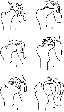
The first structural changes are often found under the epiphyseal line at the upper-lateral edge of the neck in the form of wedge-shaped or irregularly outlined nests of rarefaction. At one time, Waldenstrom and Drehmann considered these neck foci as the primary localization of the process. S. A. Reinberg warns against possible errors when reading X-rays: the shadow of the mushroom-shaped flattened head is projected onto the edge of the neck and can simulate its lesion. However, findings at autopsies and X-rays in the position of abduction and flexion of the hip (Pitzen, Kreund) prove that the neck can also be the initial site of the lesion. The third stage of fragmentation or resorption is characterized by further flattening of the head, its displacement outward and backward onto the neck, which in turn appears shortened and thickened. Most pathognomonic is the fragmentation of the epiphyseal nucleus into segments of various sizes and rich in lime. The epiphyseal line is even more loosened than in the previous stage, is interrupted in places, merging with the nest-like rarefactions of the head structure. The 'resorption' stage lasts the longest, up to 21/2 years or more (S. A. Reinberg), preceding the regeneration of the bony scaffold of the femoral head. Both destructive and restorative processes occur simultaneously in different locations. As the destructive process ends, the fourth reconstructive stage begins. Diffuse sequestra-like shadows are replaced by sclerosed but structural bone, the light intervals fill in, the epiphyseal line reappears, and sometimes the head again takes on a spherical shape. In most cases, however, the known morphological changes remain, and the disease enters the final stage of deformation: the head is mushroom-shaped flattened, cylindrically elongated, protrudes somewhat from the acetabulum, which is tilted upward at the lateral edge (according to Waldenstrom). Along with the deformation of the head itself, curvatures and thickenings of the neck in the sense of coxa vara are also encountered. In some cases, the first X-ray signs of the disease in the form of spotted darkening and loosening of the contour are noticeable on the acetabulum or on the apophysis of the greater trochanter. Secondarily, under the influence of changed static-dynamic conditions, deformations are also observed here. Pathological anatomy is very little studied due to the rarity of autopsy and operative material. In this respect, the best conditions were provided by another osteochondropathy - Kehler's disease {see.} II. For it, Axhausen's theory can be considered confirmed, that the basis of the process is aseptic epiphyseal necrosis. It was natural, based on the analogy of the X-ray picture in P. b. and Kehler's disease II, to assume the identity of the histological changes; however, there are no direct proofs. In view of this, the cases of multiple fresh anemic infarcts described by Axhausen in 1928 in P. b. are of fundamental importance. The infarcts were located in various epiphyses and metaphyses; some had the characteristic wedge-shaped shape and contained Streptococcus brevis anaemolyt. in pure culture. Despite the latter circumstance, no inflammatory changes, for example accumulations of leukocytes, were found anywhere. On the contrary, reactive phenomena in the form of lacunar erosion of necrotic bone trabeculae and proliferation of fibroblasts and endothelium had already begun. True, the case concerned an adult, but the possibility of similar anemic infarcts and aseptically occurring epiphyseal necroses in growing bones cannot be disputed. Thus, it should be recognized that the most important link in the pathogenesis of osteochondropathies has been found. Pathogenesis. It is natural to assume that the process, associated with a local disorder of arterial blood supply, may depend on various causes and must proceed differently, depending on the peculiarities of a given organism. Anamnestic indications of a specific trauma are most often absent, but typical epiphyseal necroses occur only in places subject to special traumatization. Complete or partial necroses of the head due to traumatic epiphyseolysis or fracture of the neck are not disputed by anyone, but are usually considered as moments determining the pathogenesis not of P. disease, but of deforming arthritis, false joints, varus curvatures. Quantitative differences in traumas and the age factor can modify the response reaction and give, as a result, perhaps, a picture of osteochondritis. This viewpoint is persistently defended by the pathologist Lang. Bacterial seeding, not virulent enough for obvious infection, for the development of osteomyelitis, but sufficient for the disturbance of blood circulation in the form of anemic infarct, is, in Axhausen's opinion, the primary cause of P. b. His theory also has numerous supporters and is confirmed by factual material. Cases with a more acute onset, with feverish phenomena, with a positive antistaphyllococcal reaction fall into this category. It is necessary to pay more attention to children's 'coxalgias' among outpatient orthopedic patients. Cases with an unclear anamnesis, functional disorders resembling P. b., are very common, but rarely followed up. The phenomena usually disappear after some time. But it is possible that sometimes against the background of the clinical picture of P. b., coxa vara, deforming arthritis, osteomyelitis of the neck later develop. Even tuberculous coxitis can proceed atypically and for a long time resemble P. b. on X-ray picture. Thus, clinical observations force one to think of the existence of abortive, mixed and transitional forms of P. b. Besides infectious and traumatic moments, congenital and constitutional factors are important. Cases of familial disease and hereditary transmission of P. b. (Kaiser - in four generations, Brill - even in six, and moreover bilateral affection) admit no other interpretation. It is also significant that P. b. occurs 3-4 times more often in boys than in girls, in contrast to congenital dislocation of the hip. Calot's opinion that P. b. is nothing but unrecognized congenital dislocation of the hip is erroneous. However, this does not follow that this developmental anomaly plays no role in the pathogenesis of P. b. S. A. Zilberstein followed 144 patients with congenital dislocation of the hip and found in 57, i.e. in almost 40% of cases, signs of osteochondritis, moreover independently of trauma and during reduction and despite complete restoration of joint function. A number of authors believe that the basis of P. b. is a constitutional anomaly of epiphyseal growth. The peculiarities of blood supply in this place contribute to the manifestation of the general anomaly precisely on the femoral head. Therapeutic and preventive measures should be based on the fact that in typical cases the process develops unnoticed in completely healthy children, proceeds slowly, for years, most often ending spontaneously, without outside intervention, with complete functional recovery. Considering the unclear etiology, but the unquestionable harm of secondary traumatization of the necrotic focus, one should, when clinical signs are present, without waiting for X-ray signs, - take care to unload the affected joint. If every child who sometimes complains of pain in the knee, does not want to stand on one leg and, while standing, cannot actively raise the opposite half of the pelvis (Trendelenburg's symptom), is put to bed, then -abortive cases of P. b. will be more numerous, and -obvious ones fewer. Measurement of temperature and the effect of trial massage will allow to exclude the infectious nature of the disease without resorting to complex diagnostic measures. Unfortunately, parents usually turn to the doctor only in the second or third stage of the disease. Similarly, doctors, having made the correct diagnosis, but guided by favorable statistics, often limit themselves to forbidding 'running and jumping'. Of course, an individual approach and systematic control are necessary. Dispensarization of orthopedic care is called upon to timely identify cases requiring hospital treatment with unloading by traction, or requiring only outpatient application of physio-mechano-therapeutic therapeutic means. Special warning is needed against plaster casts. If they firmly fix and really unload, immobilization is achieved at the cost of muscle atrophy and at the expense of blood circulation. Meanwhile, the basis of the disease is a disorder of blood supply, and improvement of the latter leads to recovery. Operative intervention during the period of the body's reconstructive work is not yet sufficiently justified, although cases of rapid subsidence of P. b. following subtrochanteric osteotomy are noted. In late stages, in the presence of deforming arthritis or coxa vara, operations may be indicated.
Related articles
Cite this page
“Perthes Disease.” Soviet Medical Encyclopedia. English translation of Bolshaya Meditsinskaya Entsiklopediya, 1st ed. (Moscow, 1928–1936), ed. N. A. Semashko. https://sovietmedicalencyclopedia.pages.dev/article/perthes-disease/