Osteochondritis
Historical document, translated for reference. It reflects medical knowledge of the 1920s–30s and is not medical advice.
Summary
Osteochondritis refers to various inflammatory processes affecting both cartilage and bone tissue simultaneously. This article describes different forms of osteochondritis, including syphilitic osteochondritis which is a common manifestation of congenital syphilis in newborns and infants.
Encyclopedia article (1928–1936)
OSTEOCHONDRITIS (from Greek 'osteon-bone and chondros-grain, cartilage), a term designating various processes, mostly inflammatory, which occur in adjacent parts of cartilage and bone and simultaneously involve both these tissues (e.g., the articular cartilage and the adjacent bony part of the epiphysis, the epiphyseal cartilage and the adjacent tissue of the metaphysis, the upper or lower surface of a vertebra and the corresponding intervertebral plate, etc.). In this case, primary changes may arise simultaneously in both these tissues or first in one of them. Among non-inflammatory lesions (at least in their initial phase), certain forms of primary subchondral bone necrosis of unknown etiology, which form the anatomical basis of the diseases of Köhler, Perthes, Schlatter, etc., are included here. Of these diseases, the term osteochondritis is particularly frequently applied: 1) to the lesion of the hip joint first described by Perthes in children (osteochondritis deformans juvenilis coxae) and 2) to the lesion of the spine (osteochondritis deformans juvenilis dorsi). If in any of these diseases, due to subsequent demarcation inflammation, the affected bone-cartilage area completely separates from the underlying bone with the formation of a free joint body, then one speaks of osteochondritis dissecans (König). A significantly larger number of bone-cartilage lesions arise from the very beginning on an inflammatory basis. Thus, purulent osteochondritis can develop in severe purulent lesions of the joints (as a result of the spread of the purulent process to the articular cartilage and the underlying bone), as well as in purulent epiphyseal and metaphyseal osteomyelitis (in people with unfinished bone growth). In the latter case, the process sometimes ends with the complete separation of the epiphysis from the diaphysis. Among non-purulent inflammatory processes, tuberculosis and especially syphilis are of the greatest importance. The first gives changes characteristic of osteochondritis mainly in joint lesions, namely in the bone form of joint tuberculosis and in the so-called dry tuberculous caries of joints; as for syphilitic osteochondritis, it is one of the most typical manifestations of congenital syphilis in newborns and children in the first months of life. The importance of syphilitic osteochondritis is very great not only due to its frequency (according to some statistics, it occurs in 97% of all newborn syphilitics and syphilitic fetuses, starting from the 7th month of embryonic life) but also because it can be determined even on a macerated corpse when other syphilitic changes are no longer distinguishable. Syphilitic osteochondritis can arise in any bone developing from cartilage, at the border of its epiphyseal and metaphyseal part, i.e., in the area of the ossification strip, but the most typical and frequent sites of its localization are the corresponding areas of long tubular bones, especially the ossification strip at the lower epiphysis of the femur and the upper epiphysis of the tibia, and mostly the bones are affected symmetrically on both sides. In moderately severe cases, the macroscopic changes are expressed by the fact that between the epiphyseal cartilage and the spongy bone mass, in place of the normally thin, white, sharply defined and even line corresponding to the zone of preliminary calcification of the cartilage, a more or less wide (up to 2 mm or more in width) strip of yellowish color with uneven zigzag and indistinctly distinguishable edges (so-called Wegner's strip) appears. As microscopy shows, this is a significant but uneven thickening of the zone of preliminary calcification of the cartilage, as well as a violation of the regularity in the arrangement of the cartilage layers and a decrease in osteoblastic activity of the bone marrow. In places, the calcification of the cartilage matrix reaches the columnar zone*; on the other hand, in the parts of the cartilage immediately adjacent to the bone-forming zone, there are areas without lime. Bone marrow vessels penetrate the cartilage to varying depths, and the resulting bone trabeculae turn out to be very rare, thin, and contain numerous remnants of unused cartilage tissue, mostly impregnated with lime. The main characteristic feature of the entire picture, sharply distinguishing these changes from others, including rachitic ones, is that the total amount of lime in the area of the ossification strip in osteochondritis is always clearly increased. In more severe cases, between the forming bone trabeculae and the cartilage, either separate foci or a continuous, unevenly wide strip of specific granulation tissue of grayish-yellow or even greenish-yellow color appear, often completely breaking the connection between the epiphysis and diaphysis, which as a result of this, with the slightest violence, and often spontaneously, separate from each other. In the latter case (i.e., with spontaneous intra-vital separation), a picture of the so-called pseudoparalysis of Parrot (Parrot, 1869) is obtained. This is most often observed at the distal end of the radius.
M. Skvortsov. Parrot's disease gives the highest percentage of diseases in children under 3 months of age, after which this percentage gradually decreases and at the beginning of the second year of life this disease occurs only as an exception. According to data from the syphilis department of the State Scientific Institute for the Protection of Mother and Infancy of the People's Commissariat of Health (Moscow) out of 750 children with congenital syphilis, Parrot's disease was noted in 8.5%, of which 70% were in children under 3 months, 25% from 3 to 6 months, and only in 5% was this disease observed in children from 6 months to 1 year. Approximately the same data are provided by Hochsinger based on his material, as well as Parrot himself, Finkelstein and other authors. The favorite site of joint lesions in Parrot's disease are the upper extremities. Material from the syphilis department of the State Scientific Institute for the Protection of Mother and Infancy of the People's Commissariat of Health shows that the upper extremities were affected in 50%, the lower in 12.5%, and in 37.5% simultaneous involvement of upper and lower extremities was noted, most frequently affecting the shoulder and elbow joints, followed by the knee, wrist, and hip joints. According to Hochsinger's data, involvement of the upper extremities was noted in 72%, of the lower in 18%, and simultaneous involvement of upper and lower extremities in 10%. Parrot's disease has a quite definite clinical picture. In severe cases, the affected limb of the child lies limply in the crib, an attempt to make passive movement is accompanied by crying or screaming, even slight movement, and sometimes even touching causes pain. If the limb is lifted and left in an elevated position, the limb falls back onto the bed, and the child reacts with crying. Often the affected limb is thrown backward, which is especially noticeable if the child is placed in a sitting position (Fig. 1). If the patient is lifted, the upper extremities usually hang like rags. However, the limb is not paralyzed; the child makes protective movements with the fingers in pain; sometimes movements in the forearm muscles can be noted, less often flexion movements in the elbow joint of the affected limb. Limp state is observed mainly in the upper extremities, while the lower ones are drawn to the abdomen and show signs of contractures (Fig. 2). This position of the limbs depends-on the place of attachment of the muscular apparatus; while the muscles of the upper extremities in most cases are attached to the epiphyses, the lower extremities have their attachment mainly to the diaphysis of the femur and the pelvic bone. In addition to the above-mentioned symptoms (disorders of the motor sphere, paininess), swelling of the areas of the affected joints, i.e., those places where the integrity of the bone tissue is disrupted or separation occurs, is often added; besides that,
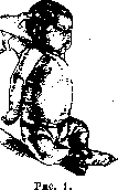
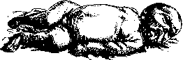
Figure 2.
especially in fresh cases, upon examination of the affected limb, one can note the following signs. All the listed symptoms gave Hohzinger reason to view this disease as a fracture, albeit not always complete. This disease does not appear immediately, as is the case with spinal paralysis, but gradually: usually the mother notes that at the beginning of the illness the child moves the limb on its own, but when an attempt is made to make a passive movement, the child begins to cry; only gradually do active movements cease, swelling of the joints appears, and finally not only movement but even touching causes pain. In most cases of Parrot's disease, in addition to disorders of the motor apparatus, there are other specific manifestations of congenital syphilis: firm liver and spleen, runny nose, skin manifestations, less often hydrocephalus, etc. It should also be noted that in Parrot's disease there is a complete absence of disorders of the sensory sphere, the muscles give normal electrical excitability and react to a prick. Parrot's disease also has a clearly expressed X-ray picture. X-rays made of the affected limb make it possible, in the absence of both clinical symptoms and serological data, to diagnose congenital syphilis. In the photographs in typical cases, a violation of the integrity of the bone tissue in the metaphysis is usually noted, and it is clearly visible that the separation occurs in that part where the bony trabeculae are few or completely absent. The band where separation occurs is dark on the X-ray, light on the print, compared to the rest of the bone. The separated part often shows significant displacement, resembling a complete fracture. Separation in the metaphysis is not always complete, and then one can observe, on the one hand, partial separation, on the other, the undamaged integrity of the bone, which, as the X-ray shows, due to the expanded dark band (light on the print) where the bony trabeculae are absent, can separate at any moment. In more severe cases, the separated parts are layered on the posterior surface of the metaphysis, giving a thickened irregular configuration, often with subsequent bone tissue growth on the sides. This phenomenon, as shown by the material from the syphilis department of the same institute, is noted on the femur, especially at its distal end. Often the separated and displaced parts are connected to the diaphysis by thin white strips, representing nothing other than delicate periosteal new formation. In addition to these minor changes in the periosteum, ossifying periostitis are observed in Parrot's disease, which give massive compact shadows on X-rays surrounding the diaphysis in the form of a shell. X-rays in Parrot's disease in newborn children are particularly interesting: the separated parts form wavy bands in the form of lace [see separate plate (Vol. XIV; pp. 159-160), Fig. 2]. X-rays in Parrot's disease often indicate multiple joint involvement of both upper and lower limbs. The diagnosis of Parrot's disease is made on the basis of clinical data, mainly on the basis of disorders of the motor sphere, on the basis of the flaccid, paralysis-like condition of the upper limbs and contracture phenomena in the lower limbs, as well as on the basis of swelling and pain in the area of the affected joints. Parrot's disease must be differentiated mainly from Barlow's disease (see), in which there is also separation of bone in the metaphysis area, causing both disorders of the motor sphere and severe pain. However, Barlow's disease is observed less frequently at the end of the first year and more often during the second year of life (usually in artificially fed children and under conditions of avitaminosis), i.e., at a time when Parrot's disease is extremely rare. In addition, Parrot's disease is often accompanied by other symptoms of congenital syphilis, which significantly facilitates the diagnosis. Sometimes a flaccid condition of the limbs, especially the upper limbs, is observed in birth injuries, especially if operative intervention was used (forceps, turns). In such cases, there are bruises at the site of manipulation or the surrounding circumstances themselves indicate trauma as the cause of the disease. Peripheral paralyses (besides the traumatic one mentioned) occur as an independent condition in early childhood very rarely; they can accompany general diseases, such as meningitis, but then they are recognized by the picture of the main condition. Parrot's disease can be confused with Heine-Medin disease. The distinguishing features are the acute febrile onset, absence of pain and bone changes. Cerebral paralyses are usually of a spastic nature. The prognosis in Parrot's disease is usually good, unless there are simultaneously manifestations of visceral syphilis, i.e., an enlarged and firm liver and spleen or severe kidney damage. Specific treatment leads to rapid recovery and by the end of the course, complete restoration of motor function of the limb. - Treatment - specific antisyphilitic. Usually combined courses are prescribed, consisting of neosalvarsan and mercury or neosalvarsan and bismuth (see Syphilis). After the first course, the manifestations of Parrot's disease disappear. Courses must be repeated every 2 months to avoid relapses of the disease. The patient should receive at least 4 such courses with 2-month intervals. After 7 years or after a year, a 5th, and less often a 6th course is carried out. - Prevention of this disease coincides with the prevention of congenital syphilis. For the prevention of Parrot's disease, it is also important to treat children without signs of the disease, born from untreated syphilitic parents, especially if the sick mother did not undergo courses during pregnancy; as shown by the experience of the syphilis department of the State Scientific Institute for the Protection of Mother and Infant of the People's Commissariat of Health, such children can during the first year of life give symptoms of congenital syphilis, including manifestations of Parrot's disease.
Related articles
Mentioned in
Cite this page
“Osteochondritis.” Soviet Medical Encyclopedia. English translation of Bolshaya Meditsinskaya Entsiklopediya, 1st ed. (Moscow, 1928–1936), ed. N. A. Semashko. https://sovietmedicalencyclopedia.pages.dev/article/osteochondritis/