Osteomyelitis
Historical document, translated for reference. It reflects medical knowledge of the 1920s–30s and is not medical advice.
Summary
Osteomyelitis is an inflammation of the bone marrow that typically extends to the bone cortex and periosteum. This 1930s article details the classification, causes, pathophysiology, and anatomical changes associated with both acute and chronic forms of the disease.
Encyclopedia article (1928–1936)
OSTEOMYELITIS, osteomyelitis, inflammation of the bone marrow. As a rule, however, the inflammation does not limit itself to the bone marrow alone, but thanks to the close connection of the vessels, it extends to the cortical substance of the bone (Haversian canals) and the periosteum, especially when the process begins in the bone marrow. Thus, the concept of O. includes both osteitis and periostitis. If the process begins with the tissues adjacent to the bone, with the periosteum, it does not always lead to true O., the inflammation may be limited to the periosteum (periostitis), involving only the more superficial layers of the bone (osteitis) and leaving the deeper layers and bone marrow tissue untouched. Usually, O. are suppurative. According to their development and course, suppurative O. are divided into acute and chronic; acute ones are further divided into so-called acute (osteomyelitis purul. acuta, diffusa) septic forms with very violent, rapid and severe course, and acute and subacute ones. In chronic O., they distinguish primary chronic suppurative O. and chronic consecutive forms following acute ones; a special group includes O. caused by specific pathogens - tuberculous, syphilitic, actinomycotic, etc. (see Bone). - Diseases with suppurative O. are observed, starting from the earliest childhood (O. of newborns), during the period of bone development and growth, up to 20-21 years of age, most frequently from 13 to 17 years (75% according to Krasnobaev). According to some statistics (Haag), men get sick 3.38 times more often than women. Certain regions are known where severe acute suppurative O. occurs in the form of epidemics (Switzerland and the adjacent southern mountainous Germany, northern coast of Germany). Diseases are more frequently observed in cities, during the rainy season. The severity and rapidity of development and course of O. depend 1) on the nature of the infection, in particular its virulence, and 2) on the degree of resistance of the body. All pyogenic microorganisms can cause suppurative O.; most commonly involved are staphylococcus (golden, white, lemon), less frequently streptococcus, mixed infections with various cocci (especially severe course), Ebert's typhoid bacillus, N-paratyphoid, intestinal, diplococcus, and many other microorganisms, both aerobic and anaerobic. Sources of infection in O. can be all kinds of local infectious processes: furuncles, paronychia, phlegmons, suppurative inflammation of joints, carious teeth, inflammation of the paranasal sinuses, middle ear, erysipelas, infected wounds, superficial and deep, angina, etc., as well as various infectious diseases - measles, smallpox, typhoid, diphtheria, scarlet fever, septicopyemia, etc. In newborns and infants, the infection is very often associated with postpartum illness of the mother, illness of the navel. The infection penetrates the bone marrow either 1) directly through trauma, for example, in complicated fracture, operation, gunshot wound, or 2) spreads to the bone and bone marrow from an adjacent purulent focus (phlegmon, suppurative periostitis, etc.); in these cases, the spread of infection is facilitated, in addition to lymphatic pathways, by purulent thrombi and inflammation of the veins around the focus, communicating with the veins of the bone and bone marrow (osteophlebitis); 3) the infection is carried into the bone marrow from some distant purulent focus by the blood flow either in the form of emboli, softened thrombi or due to the presence of bacteremia (hematogenous O.).-It is not always possible to establish the presence of a primary purulent focus (spontaneous, genuine O.). Here one has to assume the carrying of infection from the tonsillar lacunae or from the mucous membrane of the respiratory tract, intestines, or finally the flare-up of a latent infection existing somewhere, which had not manifested itself until a convenient moment. Dissemination of infection from a purulent focus or the manifestation of a latent infection are favored by all causes that weaken the body's resistance, reduce the bactericidal properties of tissues - overexertion, cooling of one or another extremity and general, malnutrition, debilitating diseases, and especially trauma, which in 50-70% of cases according to various statistics (Barrie, Kocher, etc.) precedes O. It is very probable that traumatic O. without damage to the coverings reveals a pre-existing bacteremia. On the other hand, the development of the disease is facilitated by the circumstance that in various infectious diseases, bacteria are often present in the bone marrow, which do not cause a clinical picture of O. until a certain time, but when favorable conditions occur, they show considerable viability, and suppurative inflammation of the bone marrow develops. Of all kinds of bones, suppurative O. most frequently affects the metaphyseal part of long tubular bones. According to Lexer, the metaphyses and epiphyses of tubular bones are richly supplied with vessels built on the type of terminal ones. During the period of bone growth, this area is abundantly supplied with blood, the flow of which slows down in the expansions of capillaries, as it were provided with bulges, and in the expansions of the blood vessel of the spongy part of the bone, where bacteria carried by the blood easily settle. Experiments with the introduction of bacterial cultures (golden staphylococcus) into the bloodstream repeatedly indicated the development of purulent foci similar to osteomyelitic ones, mainly in the metaphyseal parts of bones, especially the femur and tibia. The relatively large size of the arteries supplying the bone, their numerousness in young age, their departure at an acute angle from the main trunk, their shortness - all this creates favorable conditions for the entry of an embolus. In young age, red bone marrow predominates in the bones, which actively reacts to infection. In old age, there are fewer vessels in the bones, they are tortuous, the bone marrow is mainly yellow and reacts sluggishly (research of the school of Moskalenko). Experiments have also confirmed the role of trauma in the development of O. of one or another bone. Certain types of infection have certain peculiarities of course. For example, staphylococcal more often spreads to the bone marrow of the diaphysis, streptococcal is usually localized near the epiphyseal line, more often causing separation of the epiphysis, inflammation of the joint. Streptococcal O. easily occur in children and are severe in adults, accompanied by strong general infection. Pneumococcal infection of the bone does not cause hyperemia of the skin for a long time. Typhoid bacilli mainly affect the diaphysis of the bone and predominantly in the company of other microbes - golden staphylococcus, streptococcus, intestinal bacillus. The severity of typhoid fever does not affect the frequency and strength of post-typhoid bone lesions, which develop more often during recovery, less often - in the first days of the disease or after months, years after it. Most frequently, suppurative O. affects the lower end of the femur, the upper end of the tibia, its lower end, and much less frequently the humerus, radius, fibula, ulna, clavicle, etc. In early childhood, the most frequent is the lesion of the epiphysis, running in the form of suppurative inflammation of the joint. The order of frequency of lesions of flat bones: pelvis, lower jaw, scapula, skull, etc. According to the statistics of Trendel, the femur and tibia account for about 80% of diseases with osteomyelitis. Usually one bone is affected, less frequently several. Pathological anatomy. In the bone marrow, in the mildest cases, the infection causes inflammatory edema, hyperemia, and this may be limited to this, without reaching suppuration, necrosis, etc. Low-virulence microorganisms either die or may remain in the bone marrow tissue in the form of latent infection. A sufficiently virulent infection causes in the bone marrow inflammatory edema, stagnant hyperemia, small hemorrhages, thrombosis of vessels, inflammatory infiltration. The bone marrow at this time is penetrated by separate purulent foci, which gradually increase, merge with each other, forming either a limited abscess or diffuse infiltration of a larger part or the entire bone marrow and spreading along the vessels into the Haversian canals. In some cases (more severe, septic), the lesion of the bone marrow has the character of an infiltrating phlegmon without a tendency to rapid softening, in others - the infiltrate resolves quite quickly into suppuration. Sometimes gangrenous inflammation occurs with rapid necrosis of the bone, gangrenous decay of tissues, foul-smelling pus, presence of gas in the pus (anaerobes). Pus with an admixture of fat droplets from the destroyed bone marrow, located in the bone marrow cavity sometimes under considerable pressure, rushes into the Haversian canals, already captured by inflammation and somewhat expanded due to bone resorption. At this time, the periosteum of the diseased bone appears hyperemic, the bone itself is pinkish-gray, with a matte or slightly rough surface, with sharply expressed openings of the Haversian canals, visible as small red dots densely covering the inflamed area of the bone. At first, bloody exudate appears in small drops from the Haversian canals, later, with the development of suppuration in the bone, drops of pus are secreted through them, the bone becomes increasingly porous, loose. The pus penetrates from the channels under the inflamed periosteum, stripping it off to a greater or lesser extent from the bone. The periosteum is sharply hyperemic, thickened, in it are visible ecchymoses, hemorrhages; due to thrombi and ruptures of vessels supplying the bone, necrotic areas are visible. The detachment of the periosteum finally disrupts the nutrition of the affected part of the bone.
Inflammation from the periosteum spreads through the vessels or through a rupture in the periosteum to the surrounding soft tissues, resulting in intermuscular phlegmon, which gradually thins the skin and ruptures outward. The infection can develop so rapidly that it almost simultaneously affects the bone marrow, Haversian canals, periosteum, and surrounding soft tissues. In these cases, bluish, grayish-pink spots on the bone are a sign of deep bone damage and the presence of pus within it. When the bone marrow is affected near the epiphyseal line, the process can extend toward the epiphysis to the growth cartilage, which in some cases provides a sufficient barrier to the spread of infection. Sometimes, however, the suppuration destroys the growth cartilage, causes the epiphysis to be shed off, or penetrates the joint directly through the epiphysis or through lymphatic vessels, or first affects the adjacent joint capsule with subsequent purulent inflammation of the joint. If the inflammatory process is limited to the epiphysis without rupturing into the joint, a reactive serous or serofibrinous inflammation still occurs in the joint. The integrity of the diseased bone is impaired depending on the extent of the process, its severity, and the degree of disruption of bone nutrition (thrombosis of bone vessels, periosteum, and detachment of the periosteum). In mild cases, the inflammation is limited to the bone marrow tissue and subsides without extending beyond it, while in more severe cases, the nutrition of both the bone marrow tissue and the cortical layer is severely disrupted. Deprived of nutrition, a portion of the bone dies and becomes an infectious foreign body to the surrounding healthy tissues. At the boundary between dead and healthy bone, in the intact Haversian canals, and in the bone marrow cavity, granulations appear, lacunar resorption of bone substance begins, a demarcation belt is formed, and the process becomes chronic. The development of granulations with the deposition of new bone also occurs from the side of the periosteum. If the periosteum and bone marrow are severely destroyed, the bone does not regenerate (e.g., finger phalanges in panaritium). The dead bone gradually loses its connection with the healthy bone, is shed off, becomes mobile, and forms what is called a sequestrum. Sequestra are distinguished as: 1) central, occupying the bone marrow cavity in the form of an amorphous, isolated island of bone tissue, or sequestra of the inner layers of compact bone in the form of a tube, groove, adjacent to the bone marrow cavity; 2) cortical (superficial, peripheral, external), located on the surface of the bone, involving the cortical layer to varying depths and extent; 3) cylindrical, involving the entire thickness of a long bone over a greater or lesser extent. If the bone is affected throughout its entire length and thickness, a total sequestrum results. Multiple sequestra may also be observed; in several places of the same bone. A detached sequestrum, if there are no conditions for its rapid exit outward, remains for a long time among the granulations and adjacent infected bone tissue; the sequestrum is a focus of infection, maintaining inflammation, pus formation, and thereby causing fistulas. Thanks to the reaction of the periosteum, which produces new bone, and the preserved bone marrow (endosteum), a kind of sheath, called the sequestrum box or capsule, is formed around the sequestra. The adjacent intact bone, under the influence of chronic inflammation, becomes sclerosed and thickened. The newly formed bone from the periosteum is initially loose, brittle, and thin, then it thickens, becomes very dense and strong, and ultimately fully compensates for the strength lost by the diseased bone due to inflammation and necrosis. In cortical sequestrum, the sequestrum capsule covers the larger or smaller part of the bone periphery above the sequestrum. In cylindrical sequestra, the sequestrum box is especially voluminous as it surrounds the bone completely. The sequestrum capsule is lined with granulations and usually has one or more openings through which pus is discharged outward. These bone fistulas (cloacae) form where the periosteum has died, have varying sizes, and end on the skin as fistulas surrounded by granulations. With more extensive destruction by suppuration of the soft tissues, superficial sequestra can sometimes be clearly seen, and through bone fistulae, central ones as well. Sequestra may even protrude outward from the wound. A sequestrum left to itself can maintain inflammation and fistulas continuously for many years. If the infection subsides and goes into a latent state, the inflammation subsides and the fistulas close for an indefinitely long time (so-called incorporation of the sequestrum) until a new flare-up of infection under the influence of one cause or another or reinfection. Small sequestra, with good development of surrounding granulations, can be resorbed, albeit extremely slowly. An abundance of pus hinders the resorption of the sequestrum, as pus has no effect on dead bone. After the resorption of the sequestrum, as well as after its surgical removal, the cavity in the bone is filled with granulations, fibrous tissue, in which lime is deposited, and finally bone tissue is formed. In mild cases of osteomyelitis, the condition may not progress to sequestration, with the affected area of bone partly remaining intact and partly gradually being replaced by new bone. In other cases, a sequestrum does form, but the weak infection quickly dies off, and the sequestrum becomes encapsulated as a foreign body. Such is the patho-anatomical picture of chronic consecutive osteomyelitis that developed after an acute episode. Primary chronic osteomyelitis, caused by weakly virulent pus-forming microorganisms (so-called osteomyelitis of reduced virulence), manifests as inflammatory (caries, osteitis) osteoporosis; the bone marrow tissue is OSTEOMYELITIS
is filled with richly vascularized granulation tissue, spreading into the Haversian canals (ostitis granulosa, fungosa). Lacunar resorption of bone occurs. The Haversian channels turn into cavities, new channels perforating the bone may form. The process proceeds without (or almost without) suppuration. Among the granulations, detached sequestra are found. The bone surrounding the affected area thickens due to periosteal deposits and becomes denser. In other cases, the chronic form of O. manifests as a slow thickening and hardening of the bone (ostitis ossificans, hyperplastica), involving all the spongy substance of the bone (eburnation). In this dense mass and on it, foci of granulations with small sequestra are found in places. Such a hypertrophic process, involving the entire bone without suppuration, is sometimes caused by typhoid infection. Primary chronic O. is rarely encountered in the form of the so-called Brodie's bone abscess (Brodie), usually caused by golden staphylococcus. The abscess spreads in the metaphysis of long bones (most often the tibia) near the epiphysis, in most cases adjacent to the joint. Around the abscess, the bone is swollen and sclerosed, the adjacent periosteum is slightly thickened. The cavity in the bone is lined with a dense, thick, pyogenic, very sensitive membrane. In the cavity, liquid or clotted pus is found under pressure. The proximity to the joint leads to fairly frequent recurrent exudative synovitis; the abscess rarely ruptures into the joint and outward. Clinic of O. Typical acute hematogenous purulent O. usually begins with a prodromal period lasting 1-3 days: general malaise, aching in the limbs, muscle and headaches. This is followed by a sharp chill with persistent elevation of temperature to 39° and higher; chills may recur. The tongue is coated, dry, weakness, headache, sometimes vomiting, loss of consciousness, the pulse lags behind the temperature, severe tearing pains in the area of the affected bone(s), local elevation of temperature, impaired function, forced position of the affected part, a rapidly developing very painful swelling, edema of the soft tissues in the affected area. After several days, softening, fluctuation becomes noticeable, the skin takes on a congested red color, is edematous, veins are dilated, regional lymph nodes are enlarged. Left to itself, the abscess finds a way outward, empties, the general phenomena gradually weaken. The mortality in this form of O. is relatively small (10-12%), the organism more often copes with the infection. In cases of death, the cause is septicopyemia, either primary, causing the process in the bone, or secondary from the bone focus. In the most severe cases (acute septic O., osteomyelitis septica), which often occur without any prodromes, in the nearest hours after injury, cooling, wounding, and proceeding with temperature up to 40-41°, strongest chills, rapid loss of consciousness, with a picture of septicemia (enlargement of liver, spleen), local phenomena sometimes do not have time to develop sufficiently, as death occurs in the very first days of the disease (in 30-33% of cases) from complications from the lungs, pleura, pericardium, kidneys, liver (jaundice), brain. If the organism copes with the infection, the further course is the same as in the acute form, but with a more pronounced and rapid manifestation of individual symptoms. Epiphyseal O. of early childhood begins with restless behavior of the child due to pain when turning, bathing, examination, with elevation of temperature to 38-39°. In the nearest days, swelling in the area of the diseased epiphysis and adjacent joint can be noticed, the limb seems paralyzed, the child moves it poorly, holds it in a protective position. Several joints are often affected. The course is often benign with complete recovery. In more severe cases, deformation of the joints or death with symptoms of septicopyemia develops. Subacute hematogenous O. can develop with satisfactory general well-being, moderate pain in the affected part, low temperature, absence of chills. Local phenomena develop slowly, are not pronounced. Complications and fatal outcomes are rare and are associated with exacerbation of the process. 2-3 weeks after the onset of acute purulent O., with proper reaction and resistance of the organism, in the absence of complications, acute phenomena gradually subside, temperature decreases, general condition improves, inflammatory phenomena decrease, a fistula remains, discharging pus, due to the presence of a detaching bone sequestrum. The separation of the sequestrum, depending on its size, sometimes drags on for several weeks and even months. During this time, the temperature either gives evening elevations or is normal, if there are no pockets and retention of purulent discharge. The inflammatory swelling of the tissues subsides, and the thickening of the affected bone becomes more noticeable due to the development of the sequestrum capsule. Chronic consecutive O. proceeds, if there is no retention of pus, usually without elevation of temperature, in some cases without pain, in others with pains at night or in connection with weather changes. The soft tissues at the affected site are dense, somewhat edematous, near fistulas there is a small inflammatory swelling. The amount of pus discharged is variable. If the fistulas heal, they form scars adherent to the bone, drawn in. With a new exacerbation of the process, temperature rises, severe pains appear, swelling, in short, the picture of the beginning of acute O. repeats, and fistulas form again either at old sites or new ones. Such exacerbations occur if an unremoved sequestrum is present sometimes even after several decades, but they are also possible after performed operations. Primary chronic O. proceeds almost as subacute, with low temperature or without it. All processes develop slowly, sluggishly. Pains in the bones, thickening of the bone, some denseness of the adjacent soft tissues are noted. Small abscesses near the surface of small sequestra are accompanied by a sluggish inflammatory reaction of the tissues, slight pain, they gradually thin the skin, after removal of the sequestrum they sometimes heal quickly, but nearby new sequestra and abscesses may form. Figure 1. X-ray of a cyst of the neck and head of the femur with sharply defined edges and a cavity filled with a homogeneous mass. Figure 2. X-ray of a cyst of the middle part of the humerus. Sharp atrophy of bone substance with thinning of the cortical substance, in places almost to complete disappearance. Figure 3. Local fibrous osteodystrophy of the tibia. Foci of osteodystrophy are scattered along the entire length of the tibia. Figure 4. X-ray of both tibias in acute purulent osteomyelitis. Formation of sequestra, porosity of the bones and significant thickening of the periosteum. Figure 5. X-ray of the tibia in acute purulent osteomyelitis on the second day of the disease. No changes are visible. Figure 6. X-ray of the tibia in acute purulent osteomyelitis on the 10th day of the disease. Porosity of the metaphysis and part of the diaphysis of the tibia; slight thickening of the periosteum. Figure 7. X-ray of a healthy (on the right in the figure) and diseased (left) tibia in acute purulent osteomyelitis at the beginning of the disease. Porosity of the bone in the lower third and small periosteal deposits. Figure 8. X-ray of the bones of the lower leg. Restoration of the fibula after resection of a segment due to acute purulent osteomyelitis. Figure 9. X-ray of the femur in chronic osteomyelitis. Large cylindrical sequestrum in the lower third of the femur. The middle third of the femur is significantly deformed and sclerosed. The articular end of the bone is affected. Figure 10. X-ray of the femur in chronic ossifying osteomyelitis. Thickening and sclerosis of the bone. Small foci are visible in the bone and at the periphery. (For illustration of the article Osteomyelitis, Fibrous osteitis.)
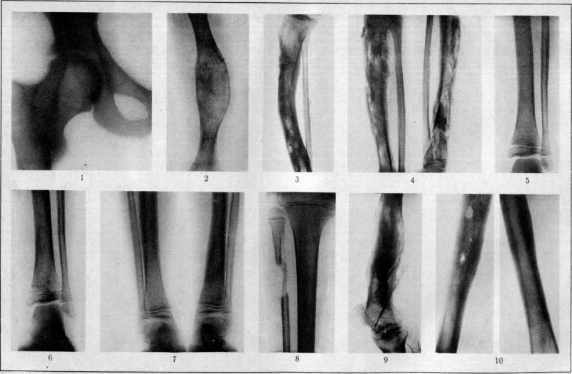
Osteomyelitis. Suppurative osteitis. 10(5 In Brodie's abscesses, which are more often associated with past trauma, it is very difficult to determine the beginning of the abscess in the bone, since at the beginning of the disease there are no symptoms; later, with the development of the abscess, pains appear, stronger at night, thickening of the bone in the area of the abscess. Without significant1 exacerbations, the thickening of the bone increases, periodic swellings of the adjacent joint appear, pains intensify, mainly in connection with movement, with injuries. From the complications of acute purulent O., the possibility of general infection or its metastases to individual organs, and joint diseases have already been mentioned. Various deformations, curvatures of the affected bone due to its porosity and weakness, fractures either after curvatures or following necrosis of the bone, before a sufficiently strong new bone has had time to form, are also observed. In some cases, these fractures lead to the development of a false joint, in others they end with the consolidation of the newly developing bone. Destruction of the epiphyseal cartilage in an earlier period of life, in addition to the separation of the epiphysis, usually leads to deformation of the bone, shortening of the limb, since the epiphyseal cartilage of the healthy end of the bone cannot always sufficiently compensate for the growth of the bone. Deformations are especially sharp when the epiphyseal cartilage of one of the bones of the forearm or tibia is affected at an earlier period of life, since the resulting delay in growth of one bone leads to gross curvatures of the other, significantly impairing function. On the other hand, the prolonged presence of an inflammatory focus or sequestrum near the epiphyseal line can irritate the epiphyseal cartilage and cause accelerated growth of the bone, lengthening of the limb. From the complications of chronic O., it is necessary to note the possibility of general infection from the existing chronic focus, slowly developing metastatic abscesses (particularly in the brain), exhaustion of the body from prolonged abundant suppuration with subsequent amyloid degeneration of organs (kidneys, spleen, adrenal glands, intestines, etc.). 1 The diagnosis of acute purulent O. can present significant difficulties at the very beginning of the disease. At this time, it is easy to confuse O. with other infectious diseases, for example, typhoid fever (most often), pneumonia, meningitis, etc. The most careful examination of the entire body of the patient, attentive comparison of symmetrical places on both sides, is necessary. With careful palpation, one can find a specific, painful even in an unconscious state of the patient, area of the affected bone. It is also necessary to remember that pains in the bone, but of a more diffuse character and less severe, occur in some infectious diseases (smallpox, typhoid, etc.). Important indications of the nature of the disease are given by blood examination: increase in leukocytosis (neutrophils), shift of the formula to the left according to Schilling, presence of bacteria in the blood (seeding!). In the first days after the onset of the disease, the diagnosis is facilitated by the appearance of local symptoms: strict localization of pains, inflammatory phenomena, local increase in temperature, limitation of movements, etc. In this period, O. can be confused with deep phlegmon. Bone involvement can be established by trial puncture^-the needle in depth encounters the exposed bone. After the formation of fistulas, the diagnosis is easily established by careful examination with a probe.-Recognition of sequential chronic O. usually does not present difficulties on the basis of the anamnesis, nature of the pus, sequestration. Primary chronic O. is not always easy to recognize and without an X-ray- it can be easily confused with other bone diseases: tuberculosis, syphilis, bone cyst, bone neoplasm, etc. X-ray examination gives much valuable data also in acute O., partly helping to establish the diagnosis of the disease, and mainly giving the possibility to see the size, nature of the bone lesion and to follow the course of sequestration and bone regeneration. In the first days of the disease, changes in the structure of the bone cannot be seen on the X-ray (Fig. 5). After 5-10 days in children, and in adults after 10-20 days, the thickened shadow of the periosteum becomes noticeable on the X-ray,- the pattern of the bone in the affected area is as if blurred, indistinct, the clear picture of the bone structure is lost (bone trabeculae are poorly visible) (Fig. 6 and 7); gradually the shadow of the bone becomes more transparent, variegated, the shadow of the periosteum thickens, densifies, sometimes its separation from the bone is visible. The boundary between the diseased and healthy bone becomes sharper due to the resorption of bone on the demarcation line and the densification of the shadow of the adjacent healthy bone; the shadow of the sequestrum capsule also begins to densify (Fig. 4). With a lesion in the area of the epiphyseal line, irregularity, notching of the bone adjacent to this line is visible. The shadow of the sequestrum, rich in lime salts, is dense, usually sharply stands out (Fig. 9) against the surrounding shadow of the sequestrum box and the loosened bone. Later, defects in the shadow of the sequestrum box corresponding to the position of bone fistulas (cloacae) are well visible. Chronic sclerosing O. gives a massive, very dense shadow of the bone with irregularities (osteophytes) along the edges, small foci of bone rarefaction in the thickness and on the surface (Fig. 10). In these foci, shadows of small sequestra may be noticeable. Chronic fungous O. gives a shadow of porous, eroded bone, thickening of the periosteum. In Brodie's abscesses, a sharply contoured dark shadow of a cavity adjacent to the epiphysis, occupying the swollen part of the bone, surrounded by a wider or narrower light border of densified bone, is visible. O. of flat and small bones of the spine, skull due to weak regeneration is not always easily distinguished radiologically from tuberculous lesions of them. In the treatment of acute purulent O., general anti-inflammatory measures (rest, elevated position of the affected part, heat) are not sufficient. Surgical treatment is necessary to create good conditions for the outflow of pus, reduce the tension of soft tissues, weaken the entry of infection from the inflamed bone marrow into the bloodstream, and reduce the increasing pressure there by timely trepanation of the bone. Attempts to establish statistically the best method of surgical treatment of acute purulent O. have not yielded tangible results, since they are based on too different material in different authors. It is necessary to be guided by the general condition of the patient, the severity and rapidity of development of general phenomena, the local process, age, patho-anatomical changes. In children, bones are softer, more porous, richer in blood vessels, the cortical layer is thinner, the bone is destroyed faster by the purulent process, the bone marrow cavity sooner has the opportunity to empty itself of pus and thereby the pressure in it is weakened and the danger of general infection is reduced. Therefore, in children, especially of younger age, one can limit oneself to a simple incision of the soft tissues to the bone under freezing with ethyl chloride, and then either wait for sequestration and remove the already separated sequestrum, or after the cessation of septic phenomena, remove the necrotic area of bone, carefully preserving the healthy bone (early necrotomy). Similarly, it is better to limit oneself to an incision or even to aspirate pus with a syringe in epiphyseal O. in newborns and infants. In adolescents, young people, and especially in adults, where bones are dense and the cortical layer is unyielding, one incision of the soft tissues is in most cases insufficient, early trepanation of the bone over the affected area is necessary to empty the focus of the bone's spongy substance, because otherwise the pus spreads along the bone marrow tissue, affects it throughout, and as a result the bone itself suffers more. In adults, one can limit oneself to one incision only with low-virulence infection, small size of the bone lesion. It is absolutely necessary to make a sufficiently wide incision to the bone in an adult to reduce septic phenomena, for some reason or other, if trepanation cannot be performed. With pneumococcal infection, wide incisions increase the access of air, and thereby improve the conditions for growth and virulence of the aerobic pneumococcus; therefore, for pneumococcal O., small incisions or even puncture with a syringe with the use of antiseptic substances are recommended. It is especially important to trepanate the bone early in cases with a large development of severe general phenomena, when no pus is found in the soft tissues and under the periosteum at the time of incision, as well as with a close location of the bone lesion focus to the joint (danger of rupture into the joint). The operation in acute purulent O. should be performed as quickly as possible. It is better to operate under general anesthesia with ether or ethyl chloride, avoiding chloroform, especially in severe septic cases. The use of a tourniquet to deplete blood is generally undesirable, since even temporary depletion of blood lowers the resistance of tissues to infection. One has to resort to a tourniquet in cases where it is especially necessary to avoid blood loss-in anemic, exhausted, weak patients, in small children. For the incision, they follow the direction most convenient for the outflow and drainage of the wound, spare muscles, nerves, large vessels, very carefully preserve the periosteum and its connection with the bone, separating it from the bone only over the necessary extent for trepanation.
During an operation in the area of the growth plate, its integrity is preserved to avoid disruption of bone growth, care is taken not to disrupt the connection between the epiphysis and diaphysis, and the integrity of the epiphysis and joint capsule is maintained. The detached epiphysis is removed in cases of purulent joint involvement. If during the detachment of the periosteum no clear indications of the area of greatest bone involvement are found (see above), trial holes can be made with a drill or small chisel to locate the abscess, and once found, the bone is widely opened in this location with a chisel (or a burr, circular saw). It is preferable to open the abscess of the spongy substance throughout its entire length. The pus is carefully removed with a sharp spoon or gauze, preserving healthy tissue. At this point, one can stop, leaving the removal of the affected bone until sequestration and a secondary operation, or the affected, softened part of the bone can be thoroughly curetted with a sharp spoon down to the boundaries of healthy bone, the walls and edges of the cavity are smoothed, the opening is made wide, and the cavity is tamponed with gauze (iodoform or aseptic after greasing the cavity with tincture of iodine). A splint bandage is applied. If there are no special indications—high temperature, severe general condition, signs of spread of the process—then the tampons are left unchanged for as long as possible (up to 20 days or longer), when the bone cavity is already well covered with granulations. Further treatment can be carried out without tampons according to general rules. In cases of bone involvement throughout its entire thickness, it is sometimes more advantageous to resect the affected area, and when the entire diaphysis is involved, it should be removed. This method is mainly applicable in childhood and adolescence, when the regenerative capacity of bone is great [see separate table (art. 103-104), fig. 8]. In adults, the process of bone regeneration proceeds slowly, defects and deformities of bones may occur, leading to impaired function. Resections give better results in cases of involvement of flat bones (scapula, pelvis). In the treatment of acute purulent osteomyelitis, careful attention must be paid to the general condition of patients, to infection in the blood, and to combat these manifestations of the disease (vaccines, protein therapy, autogenous blood therapy, etc.). If, despite all measures, the general condition remains severe and threatening, the affected part of the limb represents a diffuse phlegmon with signs of thrombophlebitis, severe circulatory disturbance, a large joint is involved, then amputation or disarticulation of the limb has to be performed to save life. This measure also has to be resorted to in cases of prolonged chronic osteomyelitis, when the extent of the lesion, the nature of the infection, profuse suppuration, or a noticeable decline in the patient's strength are accompanied by severe exhaustion threatening amyloid degeneration of organs. Usually in chronic osteomyelitis, the matter is reduced to the removal of the already detached sequestrum, and it is necessary to remove also the adjacent part of the bone, affected by chronic inflammation, porous, impregnated with granulations and pus, because otherwise the operation will not achieve its purpose. Access to a superficial sequestrum is in most cases simple, while to a central one it is sometimes very difficult, especially in old cases with a very thick sequestrum capsule. After an incision in the soft tissues over the necessary area, the periosteum is carefully stripped. It is important not to expose the bone excessively, whose nutrition is already disrupted by inflammation and which can easily be infected from the surrounding tissues and give rise to a new necrotic area in the future. Then, either existing bone fistulas are enlarged, or the bone is opened sufficiently widely in the most favorable place (X-ray) for removal of the sequestrum, always trying to affect healthy bone as little as possible. After removal of the sequestrum (necrectomy), the cavity is thoroughly cleansed of any small sequestral fragments, granulations are removed, diseased non-sequestrated areas are removed, roughnesses and irregularities of the cavity are smoothed, and it is given the shape of a shallow groove, most convenient for filling with adjacent soft tissues, otherwise healing is delayed, exacerbations with formation of new sequestra, etc., may occur. The wound is closed tightly or open treatment is carried out, in which it is better to change the dressing less frequently if there are no special indications. For more certain and rapid healing of bone cavities, a number of methods are used , of which in osteomyelitis the most certain is the transplantation of a flap of adjacent muscle on a sufficiently wide pedicle. The muscle takes root well despite continued suppuration in the wound for some time. Where there is no muscle nearby, wrapping of a skin flap into the cavity for its epithelialization is used; tight closure with a blood clot (Schede) rarely gives good results. Bone-plastic closure is less suitable, because in an infected wound, even a bone transplanted on a pedicle does not always take root. When a significant part of the thickness of the diseased bone is removed, careful handling during and after the operation (splint) is necessary to avoid fracture. In chronic sclerosing osteomyelitis with many small foci and sequestra, with significant thickening of the bone, it is impossible to clean all foci without causing new ones to appear. In extreme cases, resection of the affected bone can be performed with subsequent traction during its regeneration, but it is better to achieve subsidence of the process by conservative treatment, enhancing the body's protective mechanisms by autovaccination and autogenous blood therapy, antivirum according to Besredka, physical therapeutic measures (baths, sun, etc.). These same measures are useful to apply as preparatory to an operation for chronic osteomyelitis and to accelerate healing after the operation. The use of autovaccination helps in cases of chronic osteomyelitis of low virulence, and material for seeding can be obtained by drilling the bone with a thin drill through a small skin incision. In Brodie's abscess, treatment consists of opening the bone cavity, removing the pyogenic membrane, and closing tightly. It is necessary to mention the first described by Ollier so-called albuminous osteomyelitis (albuminous periostitis, albuminous osteitis, osteomyelitis serosa, s. albuminosa, serous periostitis), which runs in a mild acute and subacute form and mainly involves the periosteum and bone. The detached periosteum is sometimes surrounded by a considerable amount of blood-tinged, serous, tenacious, sticky, protein fluid, similar to synovial fluid. The bone is thickened, formation of sequestra and other changes characteristic of osteomyelitis and periostitis is possible. Albuminous osteomyelitis is caused by low-virulence staphylococci and streptococci, apparently unable to cause purulent inflammation. Dor found in albuminous periostitis Bacillus cereus citreus, and with experimental injections into animals of cultures of this bacillus, he obtained a picture of albuminous periostitis. Garre believes that initially in this form of osteomyelitis, pus is formed, which with subsidence of the process turns into a mucous fluid, possibly due to mucous degeneration of leukocytes. The course, symptoms, X-ray picture, treatment—do not differ from those in ordinary osteomyelitis. Albuminous osteomyelitis should be classified as osteomyelitis of low virulence.
Related articles
Mentioned in
Cite this page
“Osteomyelitis.” Soviet Medical Encyclopedia. English translation of Bolshaya Meditsinskaya Entsiklopediya, 1st ed. (Moscow, 1928–1936), ed. N. A. Semashko. https://sovietmedicalencyclopedia.pages.dev/article/osteomyelitis/