Maxillary Sinusitis
Historical document, translated for reference. It reflects medical knowledge of the 1920s–30s and is not medical advice.
Summary
Maxillary sinusitis is an inflammation of the mucous membrane of the maxillary sinus cavity, characterized by purulent discharge from the corresponding nostril, nasal obstruction, and cheek pain. The article describes acute and chronic forms, their causes, symptoms, diagnosis, and treatment approaches from a 1930s Soviet medical perspective.
Encyclopedia article (1928–1936)
MAXILLARY SINUSITIS, highmoritis (synonyms: maxillitis, sinusitis, antritis maxillaris), inflammation of the mucous membrane of the maxillary sinus cavity; first described by Runge in 1755; the most characteristic feature of maxillary sinusitis is the discharge of pus from the corresponding half of the nose, nasal obstruction, and pain in the cheek area. The causative agents of maxillary sinusitis enter the maxillary sinus cavity from the nose, especially during nose-blowing and sneezing, or microbes previously present in the maxillary sinus cavity become pathogenic due to weakening of the body. The type and variety of microbes are not particularly significant. Maxillary sinusitis is divided into acute and chronic. Acute maxillary sinusitis is most often observed after acute rhinitis, influenza, less frequently with pneumonia, typhoid, scarlet fever; even less frequently, maxillary sinusitis is a consequence of osteomyelitis of the upper jaw and trauma. Regarding the mechanism of infection penetration, there are two theories: 1) the theory of nasal origin of maxillary sinusitis, i.e., the transition of inflammation from the nasal mucous membrane, and 2) the theory of hematogenous origin. Both methods of infection penetration should be considered possible. - The pathology and anatomy of acute maxillary sinusitis have been little studied. By analogy with diseases of other paranasal sinuses, catarrhal and purulent forms are distinguished. The first is characterized by edematous impregnation of the mucous membrane and its swelling almost to the obliteration of the cavity lumen, along with serous-mucous discharge; the second is characterized by small-cell infiltration of the mucous membrane and the discharge of pus into the cavity. - Clinical picture. The disease often proceeds without any symptoms, or patients complain of pain of varying intensity, localized not only within the boundaries of the maxillary sinus cavity but also involving the forehead, less frequently the temple and the entire half of the face. Pain during chewing and a feeling of elongation of the teeth, nasal obstruction are noted. The pain depends on the retention of discharge and compression of nerves by collateral edema. In the initial stage of maxillary sinusitis, general symptoms may be sharply expressed: elevated temperature, chills, and poor general condition. Among the objective symptoms, the presence of pus in the middle nasal passage and swelling of the mucous membrane on the turbinate of the affected side are characteristic. Less frequently, swelling of the cheek and lower eyelid is noted. - Diagnosis is made based on the patient's complaints, history data, and objective examination of the nasal cavity. The presence of pus in the middle nasal passage (under the middle turbinate), which reappears after wiping and when tilting the head to the opposite side (Frenkel's position), indicates its origin from the maxillary sinus cavity. To facilitate the finding of pus, it is recommended to cocaineize the middle nasal passage or move the turbinate to the midline with a Killian mirror (middle rhinoscopy). Suction of pus is also used. Additionally, for diagnosis, transillumination (diaphanoscopy) and X-ray are used. During transillumination, the cheek and lower eyelid on the affected side are darkened, the pupil does not glow, and there is no sensation of light in the patient's eye. On the X-ray, the affected side blocks the rays and appears lighter on the negative. - The prognosis for acute maxillary sinusitis is favorable, as most cases resolve without any treatment. In the initial stage of maxillary sinusitis, general treatment is used: bed rest and diaphoretics; locally, a heating compress or warmth on the cheek. Nasal drops with 1-2% cocaine are used. Smearing the middle nasal passage with 10% cocaine-adrenaline is also used to cause its dilation by narrowing the vessels of its mucous membrane and thus promote the outflow of secretions. If the process drags on, puncture of the maxillary sinus cavity and washing are recommended, as described below. In case of dental origin maxillary sinusitis, immediate removal of the affected tooth is indicated. - Statistics. In autopsies of those who died from influenza, Frankels and Geipel found that in 70% the maxillary sinus cavity was affected. Clinical observation shows that with influenza and acute rhinitis, acute maxillary sinusitis is diagnosed less frequently, as maxillary sinusitis often occurs without symptoms. Autopsies usually involve the most severe cases of influenza. C h r o n i c maxillary sinusitis usually develops from acute, and its causes are the same as for acute maxillary sinusitis. Chronic forms of maxillary sinusitis more often come to specialized consultations. The reasons why in some cases acute maxillary sinusitis resolves, while in others it becomes chronic, are partly local (congenital narrowness of the outlet of the maxillary sinus cavity, bends of the septum, enlargement of turbinates, polyps), and partly general - weakening of the body's resistance. The question of dental origin maxillary sinusitis deserves special attention. At present, most authors believe that diseased teeth cause maxillary sinusitis significantly less frequently (5-8%) than was previously claimed (up to 50%, according to Nihsmann's data). Pathologically and clinically, four forms of chronic maxillary sinusitis are distinguished: 1) Catarrhal form. The mucous membrane is pale, edematous, often with bulgings and the formation of polyps that fill not only the maxillary sinus cavity but also grow into the nose. Infiltration is slight. In the further course, development of connective tissue and sometimes sclerosis of the mucous membrane occurs. The discharge is mucous and mucopurulent, odorless, and temporarily disappears after washings, only to reappear after their cessation. With hyperplastic changes in the mucous membrane, limited swelling of the mucous membrane of the lateral wall of the nose in the area of the anterior fontanelle is sometimes observed. With gradually increasing light during transillumination, a delay in transillumination of the affected side is noted. 2) Purulent form. Characterized by thickening of the mucous membrane due to infiltration and its red-gray color. Under the microscope, there is significant small-cell infiltration, and in the further course of the disease, transition to scar tissue. Sometimes cysts form from compression of the excretory ducts of the glands by scar tissue. The discharge is purulent. Pus in the middle nasal passage, darkening during transillumination and X-ray. The disease is usually unilateral. There are no polyps. These changes are characteristic of simple purulent maxillary sinusitis (maxillitis purulenta chronica simplex). In other cases, significant thickening of the mucous membrane with the formation of folds and polypoid bulgings is noted - maxillitis purulenta chronica hypertrophica (A. F. Ivanov). In the postoperative period with maxillitis pur. chron. simplex, pus disappears quickly; in the hypertrophic form, due to significant changes in the mucous membrane, pus discharge usually drags on. In the presence of a fistula in the maxillary sinus cavity after Cooper's operation through the alveolar process and prolonged wearing of a pin, a form of limited purulent maxillary sinusitis (maxillitis purulenta circumscripta) is observed. Granulations develop around this fistula, which maintain the suppuration. In such cases, sometimes removal of the pin is sufficient, but sometimes radical surgery is necessary. 3) Atrophic form (A. F. Ivanov). The mucous membrane of the nose and maxillary sinus cavity is atrophic. There is purulent discharge with an odor in the cavity. It is rare. 4) Mixed form. It develops from catarrhal and purulent forms due to secondary infection or weakening of the body. In addition to edema and polyps, small-cell infiltration of varying degrees is observed. The discharge is mucopurulent. C l i n i c a l picture. Subjective complaints may be absent or consist of a feeling of tension or pressure in the maxillary sinus cavity or the corresponding half of the forehead and head. Nasal obstruction, odor from the nose, and loss of smell are noted. Objectively, the formation of polyps and the discharge of purulent or mucopurulent secretions, often with an odor. The amount of discharge varies; in the morning it is usually greater. Pus flows into the nasopharynx and is the cause of the development of chronic pharyngitis and laryngitis and stomach diseases. - Diagnosis is made based on the same symptoms as in acute maxillary sinusitis. In the absence of pus, a final decision presents great difficulties, and sometimes the question is resolved only by trial treatment with washings. - In addition to purulent inflammation of the maxillary sinus cavity, so-called pyosinus can also be observed, i.e., the accumulation in the maxillary sinus cavity of pus that forms in some other cavity [frontal, ethmoidal (anterior cells)] and only flows into the maxillary sinus cavity. The diagnosis of pyosinus is made based on the results of washing. If after washing the maxillary sinus cavity, pus reappears in the middle nasal passage after a short time, this indicates that it comes from another cavity. The disease of the maxillary sinus cavity can be combined with inflammations of other nasal cavities - frontal, ethmoidal, sphenoidal; in the process, all paranasal sinuses of one or both sides of the nose can also be involved - pansinusitis (pansinuitis). In differential diagnosis, it should be kept in mind that the excretory ducts of three paranasal sinuses open into the middle nasal passage - maxillary, frontal, and anterior ethmoidal cells, and into the upper nasal passage - the sphenoid sinus and posterior ethmoidal cells. The presence of pus under the middle turbinate is characteristic of diseases of the first group of sinuses, above the middle turbinate - of the second group. Further differentiation consists in finding the openings of the maxillary and then frontal sinuses (which is achieved in 50% of cases with trial puncture) and washing the maxillary sinus cavity.
In addition, diagnosis is aided by transillumination and X-ray images. - Complications from maxillary sinusitis affecting the cranial cavity are observed very rarely. Individual cases of meningitis and brain abscess have been described. More commonly observed are a boil of the lower eyelid (especially in children) and orbital cellulitis. - The choice of treatment method depends on the severity and duration of the disease and the patho-anatomical changes in the mucous membrane, with either irrigations or surgery being used (see figures 1, 2, 3, 4). Irrigations are performed with disinfectant solutions through the middle or inferior nasal meatus. If access through the middle nasal meatus is difficult, the middle turbinate can be displaced according to Killian or its anterior end can be resected. Success in treatment can only be expected in the initial stages of inflammation when the mucous membrane is not yet too altered. Surgical treatment methods. Intranasal operations consist of creating an opening in the inferior or middle nasal meatus. Under local anesthesia (cocaine), the anterior end of the inferior turbinate is either displaced toward the midline or resected, and correspondingly curved trocar is used to create an opening into the maxillary cavity through the lateral wall of the nose, which is then enlarged by a conchotome. Although the operation does not present great difficulties, it must be performed at considerable depth, and if bleeding occurs, the maxillary cavity cannot be examined, i.e., the essential condition for performing any operation is lost.
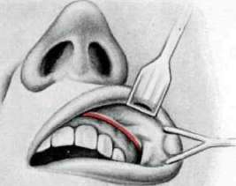
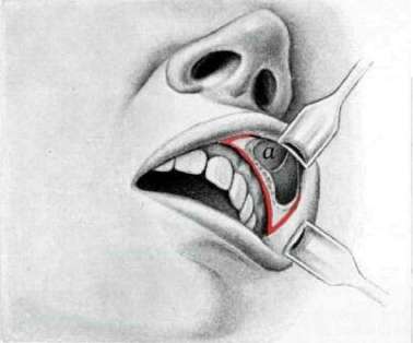
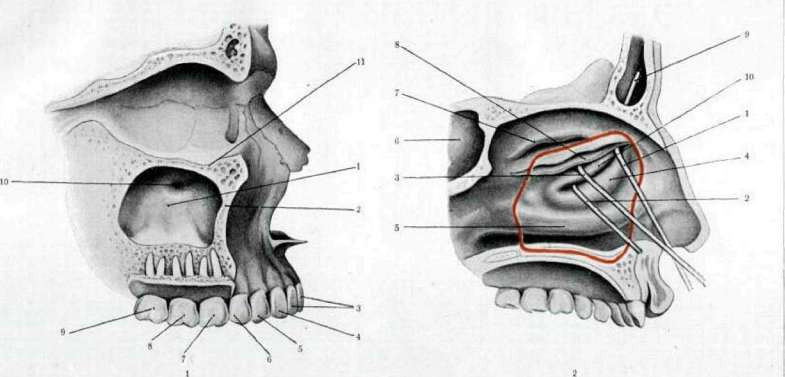
The results of the operation are questionable, as the opening often grows over. The operation is used in the early stages of maxillary sinusitis, where irrigations often suffice. The radical operation (antrotomy) (see figures 5 and 6) consists of wide opening of the maxillary cavity through the canine fossa (fossa canina) and creating an opening into the nose in the inferior nasal meatus. The operation is usually performed under local anesthesia, using two methods: Caldwell-Luc (1893-97) and Denker (1902-1905). The difference between these methods is that in the Denker operation, the medial wall of the maxillary cavity is removed along the inferior meatus as far as the apertura piriformis. The maxillary cavity is carefully examined, and severely altered areas of the mucous membrane (polyps, granulations) are removed. The mucous membrane of the lateral wall of the nose is either removed or a flap is fashioned from it, with the base downward, which is laid on the floor of the maxillary cavity. The incision in the cheek mucosa should be sutured. Radical operations for chronic M. in the vast majority of cases result in complete recovery, if there is no simultaneous disease of other cavities. - Statistics. Inflammations of the accessory sinuses belong to the most frequent human diseases. In autopsies following severe infectious diseases, the accessory sinuses were found to be affected in 30-50% (according to Niihsmann); in cases of death from accidental causes, diseases of the maxillary cavity are rarely encountered. Most frequently the maxillary cavity is affected, less frequently the ethmoid cells, then the sphenoid, and least frequently the frontal. Combined diseases of several sinuses and pansinusitis occur.
Related articles
Mentioned in
Cite this page
“Maxillary Sinusitis.” Soviet Medical Encyclopedia. English translation of Bolshaya Meditsinskaya Entsiklopediya, 1st ed. (Moscow, 1928–1936), ed. N. A. Semashko. https://sovietmedicalencyclopedia.pages.dev/article/maxillary-sinusitis/