Maxillary Sinus
Historical document, translated for reference. It reflects medical knowledge of the 1920s–30s and is not medical advice.
Summary
The maxillary sinus is the largest paranasal sinus, located in the upper jaw. This article describes its anatomy, development, physiology, and various pathological conditions including inflammation, tumors, and traumatic injuries.
Encyclopedia article (1928–1936)
MAXILLARY SINUS (antrum Highmori, sinus maxillaris), first described in 1651 by Highmor, is the largest paranasal sinus, located in the body of the maxilla. Its walls are distinguished as follows: superior, inferior, anterior, lateral, posterior, and medial (see Figure 1). The superior wall (roof), rather thin, also forms the inferior wall of the orbit; within it passes the infraorbital nerve canal (n. infraorbitalis), which often forms a bulge toward the maxillary sinus. The inferior wall (floor) is formed by the alveolar process of the maxilla and the hard palate. The alveolar process, thicker anteriorly, thins posteriorly, so that the roots of the posterior teeth (the second premolar and three molars) may sometimes protrude into the maxillary sinus or be separated from it by only a thin layer of bone. The anterior wall (fossa canina) is oriented in the frontal plane in 1/8 of cases, at a 45° angle to the frontal plane in 2/8 of cases, and thus directly continues into the lateral wall, which is located in the zygomatic process of the maxilla. The anterior wall, when located in the frontal plane, is usually thin. When obliquely oriented, its inner part contains a significant layer of compact bone. The medial wall corresponds to the middle and inferior nasal meati; its lower part is thicker, while the upper part is very thin and in places completely absent, forming so-called fontanelles where the mucous membranes of the maxillary sinus and nose are directly adjacent to each other. Two fontanelles are distinguished—anterior and posterior. They are located in the middle nasal meatus and are separated from each other by the uncinate process (processus uncinatus). The opening of the maxillary sinus (ostium maxillare), of a slit-like shape, is located in its uppermost part and opens into the nose in the middle nasal meatus, beneath the middle nasal concha (see Figures 2 and 3). In addition to the permanent opening, an accessory opening (ostium maxillare accessorium) is noted in 10% of cases, located below the permanent opening, above the middle of the inferior concha, in the middle nasal meatus.
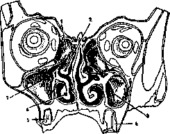
Figure 3. Maxillary sinus: 1-cellula ethmoidalis; 2-middle nasal concha; 5 and 7-sinus maxillaris (probe in the opening); 4-first molar; 5-second premolar; 6-inferior nasal concha (after Braus).
The capacity of the maxillary sinus varies from 5 to 29 cubic cm. On average, in men it is 18.5, in women 12 cubic cm (according to Schurch).-The maxillary sinus begins to develop at the end of the second month of intrauterine life as a downward protrusion from the middle nasal meatus, forming a shallow depression with an excretory duct at its upper-anterior end. Further growth proceeds forward and laterally, and by the end of the first year of life, the maxillary sinus acquires a round shape. Until 7 years of age, growth is very slow, but from this age rapid growth begins, forming the nasal and facial surfaces, and by 12-14 years of age, the maxillary sinus assumes its normal shape. The maxillary sinus is lined with mucous membrane covered by stratified ciliated epithelium. The mucous membrane is very thin and lies directly on the periosteum. Its structure in general resembles that of the nasal mucous membrane. The difference is the absence of a cavernous layer and a small number of glands.-The physiological significance of the maxillary sinus, like other nasal cavities, has not been clarified. The main theories are: the cavities serve 1) to warm and humidify the inhaled air, 2) to aid olfaction, 3) to lighten the weight of the facial part of the skull, 4) represent rudimentary organs having no physiological significance in humans, etc. Pathology. In addition to inflammations (see Maxillary Sinusitis), the development of mucoceles due to obstruction of the cavity's opening should be noted. Tuberculosis and syphilis occur rarely. Among new growths, cysts of the alveolar process and jaw that invade the maxillary sinus should be noted. They bulge and thin its walls, causing a 'parchment crackle' upon palpation. From the nasal side, nasal and nasopharyngeal fibromas and osteomas may grow in. Among malignant new growths, carcinoma and more rarely sarcoma are encountered. Traumatic injuries to the maxillary sinus in war, according to statistics by Bruckner and Weingartner, occur in one-third of all injuries to the accessory sinuses (300-100 per 1000). In peacetime, injuries to the maxillary sinus apparently occur more frequently than to other accessory sinuses, but exact figures are not available. Injuries to the maxillary sinus result from blows, contusions, falls, and penetrating wounds. Accidental injuries during nasal operations and tooth extractions are also observed. Penetrating injuries from the facial side are accompanied by damage to the soft tissues of the face, bones of the facial skeleton, and the mucous membrane of the maxillary sinus. Combined injuries of the maxillary sinus with adjacent—the ethmoid and frontal sinuses or even with the cranial cavity—are often encountered. After injury, swelling of soft tissues and edema of the mucous membrane are observed, especially when foreign bodies enter the maxillary sinus. Symptoms in injuries limited to the maxillary sinus are minor—pain, swelling, bleeding from the wound and nose, emphysema, anesthesia of the cheek when the n. infraorbitalis is damaged. In injuries to adjacent cavities and especially the brain, the symptoms are more severe. The course of the injury depends on whether the wound heals by first intention or suppuration develops, depending on the penetration of infection from outside or from the nasal cavity. In this respect, it is important whether the injured maxillary sinus was previously healthy or diseased. Diagnosis presents no great difficulties; inspection of the wound and nose, diaphanoscopy, X-ray, etc., are used. Among complications, osteomyelitis of facial bones and pyemia should be mentioned. Injuries to the maxillary sinus are considered as open fractures and are treated according to the general principles of surgery. Irrigation of the maxillary sinus after injury is irrational. If surgical opening of the maxillary sinus is necessary to remove bone fragments, foreign bodies, stop bleeding, etc., the approach through the facial wall is recommended.
Related articles
Mentioned in
Cite this page
“Maxillary Sinus.” Soviet Medical Encyclopedia. English translation of Bolshaya Meditsinskaya Entsiklopediya, 1st ed. (Moscow, 1928–1936), ed. N. A. Semashko. https://sovietmedicalencyclopedia.pages.dev/article/maxillary-sinus/