Olfaction
Historical document, translated for reference. It reflects medical knowledge of the 1920s–30s and is not medical advice.
Summary
An overview of the physiological and chemical process of olfaction in humans and animals, detailing the anatomical structure of the peripheral and central olfactory pathways, olfactory centers, and neural connections as understood in 1930s medicine.
Encyclopedia article (1928–1936)
OLFACTION, a physico-chemical process by which man and animals receive the perception of odor. Olfaction plays a particularly important role in animals, and in some of them the sense of smell (syn. scent) is very highly developed; the ability to distinguish the faintest odors is for many of them a powerful weapon in the struggle for existence, and olfaction also has a very great significance in the sexual life of animals. Parallel with this development of olfaction, the olfactory part of the brain is particularly well developed (see Rhinencephalon). The significance of olfaction in human life is no longer so great, and in connection with this, many parts of the olfactory brain are in a rudimentary state. The olfactory conduction pathways consist of two parts: peripheral
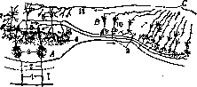
Figure 1. Olfactory pathways: A - bulbus olfactorius; B - tractus olfactorius; C - trigonum olfactorium; 1 - cellulae olfactoriae in the nasal cavity; 2 - nervus olfactorius; 3 - glomeruli olfactorii; 4 - stratum moleculare; 5 - mitral cells; 6 - granule cells of the olfactory bulb; 7 - tufted cells; 8 - collaterals of the axial cylinders of mitral cells in the olfactory bulb; 9 - the same in the olfactory tract; 10 - pyramidal cells in the olfactory tract; 11 - fibers terminating in the olfactory bulb.
and central. The peripheral part includes the olfactory nerve, while the central part includes the olfactory bulb (where the peripheral and central pathways meet), a series of cortical and subcortical centers, and the pathways connecting them. Regarding the peripheral olfactory pathway, see Olfactory nerve. From the mitral cells of the bulb begins the central olfactory pathway (Fig. 1), which runs in the superficial or molecular layer of the olfactory tract (often incorrectly called the "olfactory nerve"), then into the olfactory trigone, sending collaterals to the underlying cell layers and being continually reinforced by fibers emerging from there. From the olfactory trigone, a large portion of the olfactory fibers passes into the lateral olfactory stria (stria olfactoria externa), the main site of termination of which is the anterior section of the hippocampal gyrus (regio olfactiva); a smaller portion of the olfactory fibers passes through the intermediate stria (or middle olfactory stria, poorly developed in humans and inconstant) into the anterior perforated substance; the medial olfactory stria (stria olfactoria interna), or upper olfactory root (not recognized by all authors), passes to the frontal lobe and apparently serves as an association system between the olfactory and frontal lobes. Thus, the second olfactory neuron, having begun in the olfactory bulb, terminates in the hippocampal gyrus, in the cortical layer of the olfactory tract and lateral olfactory striae (partly also in the anterior perforated substance), which are therefore secondary olfactory centers. The anterior white commissure connects them with the centers of the opposite side. Its anterior section (pars olfactoria) connects the bulb, olfactory tract, and perforated substance of both sides, while the olfactory regions of both hippocampal gyri are connected by the posterior section (pars temporalis, s. hemisphaerica) of the commissure (Figure 2). Projection systems connect the cortical olfactory centers with the subcortical ones; these include 1) the main olfactory bundle of Wallenberg (radiatio olfactiva basalis, s. profunda), running from the olfactory bulb and from the cortex of the olfactory tract through the deep layers of the anterior perforated substance (where the number of fibers in it increases) to the mamillary bodies; according to some authors, this bundle can be traced from there to the substantia reticularis of the midbrain (tractus olfacto-mesencephalicus); 2) fibrae olfacto-habenulares, separating from the previous bundle and running as part of the medullary stria to the habenular trigone and terminating in the habenular nucleus; 3) the stria cornea, s. terminalis, s. taenia semicircularis, the corneal or terminal stria, which runs from the uncus of the hippocampal gyrus and from the amygdaloid nucleus, describing an arc along the upper wall of the inferior horn of the lateral ventricle, lies between the caudate nucleus and the optic thalamus, and then passes with its anterior end into the anterior perforated substance; some authors admit the existence of fibers in this system running in the opposite direction. In addition to projection systems, there are also association fibers connecting the olfactory centers with other parts of the cortex, mainly with the cortex of Ammon's horn (cornu Ammonis, s. hippocampus). These fibers include the tractus olfacto-ammonicus, the cingulum, and the longitudinal striae. The tractus olfacto-ammonicus, the olfactory bundle of Ammon's horn (the oldest bundle in the vertebrate brain), begins in the anterior perforated substance and partly in the anterior section of the hippocampal gyrus; via the subcallosal gyrus of Zuckerkandl, it is directed toward the septum pellucidum, receiving reinforcement from fibers of both these formations, then at the posterior edge of the septum it joins the fornix and, running as part of its fibers in the reverse direction, reaches Ammon's horn. The cingulum, s. fornix periphericus (cingulate bundle), runs from the olfactory lobe in the white matter of the fornicate gyrus and terminates in Ammon's horn; according to most authors, the cingulum consists of short fibers interrupted in the cortex of the lower divisions
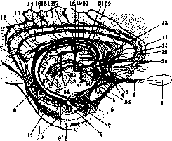
Figure 2. Olfactory pathways: 1 - bulbus olfactorius; 2 - tractus olfactorius; 3 - trigonum olfactorium; 4 - stria olfactoria intermedia, s. profunda; 5 - stria olfactoria externa; 6 - uncus gyri hippocampi; 7 - nucleus amygdalae; 8 - gyr. hippocampi; 9 - alveus; 9' - fimbria; 10 - fascia dentata; 11 - cingulum; 12 - fornix and commissura interfornicata (lyra Davidis); 13 - corpora quadrigemina; 14 - fibrae perforantes; 15 - gyrus fornicatus; 16 - thalamus opticus; 16' - nucl. anter. thalami optici; 17 - tract. mamillo-thalamicus; 18 - striae longitudinales indusei grisei; 19 - fornix; 20 - tract. olfacto-ammonicus; 21 - stria cornea; 22 - striae medullares; 23 - induseum griseum; 24 - habenula; 25 - commissura alba anterior; 26 - striae olfactoriae internae; 27 - tract. septo-thalamicus; 28 - pedunc. septi pellucidi; 29 - fibr. olfactoriae to tuber cinereum; 30 - main olfactory bundle to corpora mamillaria; 31 - corpus mamillare; 32 - tract. mamillo-tegmentalis; 33 - fasc. retroflexus Meynert'a; 34 - gangl. interpedunculare; 35 - pedunculus cerebri; 36 - pedunculus corp. mamillaris; 37 - fasc. longitud. dorsal. Schütz'a.
lov gyri fornicati, where new fibers originate, passing partly into the hippocampus and partly into the septum pellucidum. Striae longitudinales indusii grisei begin in the olfactory lobe (in Broca's carrefour olfactif), partly in the gyrus subcallosus, run backward along the upper surface of the corpus callosum, pass at the splenium of the corpus callosum into the fasciola cinerea, then into the fascia dentata and into the hippocampus. Part of the fibers entering the striae longitudinales originates from the cells of the indusium griseum; some of them are directed backward to the hippocampus, others forward to the olfactory lobe; some fibers divide into anterior and posterior branches. The striae longitudinales communicate by means of fibrae perforantes corporis callosi with the tractus olfacto-ammonicus running as part of the fornix beneath the corpus callosum; on the lower surface of the corpus callosum, these fibers form a bundle, the so-called fornix longus—the centripetal pathway from the substantia perforata anterior to the hippocampus. The existence of a direct connection between the olfactory centers (gyrus hippocampi) and the hippocampus, located very close to each other, is assumed. Finally, the fornix also belongs to the conducting pathways of the olfactory sphere; it consists of projection-commissural fibers connecting the hippocampi with each other and with the corpora mamillaria; the axons of the pyramidal cells of the hippocampus pass into the alveus, then into the fimbria, the direct continuation of which is the fornix (see); it lies beneath the corpus callosum, then curves downward and backward and ends in the corpus mamillare of its own side, and partly also of the opposite side (tractus cortico-mamillaris). A certain part of the fibers of the fornix passes into the striae medullares thalami and as part of them goes to the ganglion habenulae under the name tractus cortico-habenularis. Finally, a part of the fibers of the fornix runs in a transverse direction as part of the commissura hippocampi, s. interfornicatae (s. psalterium, s. lyra Davidis) and ends in the hippocampus and in the corpus mamillare of the opposite side. In view of the extensive connections of the hippocampus with the olfactory centers, it must unconditionally be classed among the olfactory sphere, but the question of its functional significance in this sphere still remains unresolved; whether it is a higher associative center (since it is connected only with secondary subcortical and cortical centers) or an ordinary cortical center of olfactory sensations. Negative experimental results and the lack of positive clinical data do not yet allow this question to be resolved. The question of the significance of the gyrus fornicati is in the same position. Subcortical olfactory centers, namely the corpus mamillare, have a connection with many formations of the diencephalon and brainstem. From the medial nucleus of the corpus mamillare arises the fasciculus mamillaris princeps (principal bundle), which is directed upward and divides into two branches: 1) tractus mamillo-thalamicus, or fasciculus thalamo-mamillaris (Vicq d'Azyr's bundle), ending in the nucleus anterior thalami optici, and 2) tractus mamillo-tegmentalis, or fasciculus tegmento-mamillaris (Gudden's tegmento-mamillary bundle), going into the tegmentum of the cerebral peduncle partly to the ganglion profundum tegmenti and partly to the gray matter of the cerebral aqueduct; fibers descending as far as the substantia reticularis of the pons have been described. In the lateral nucleus (nucleus accessorius) of the corpus mamillare originates the pedunculus corporis mamillaris, which also goes into the tegmentum of the cerebral peduncle to the ganglion dorsale tegmenti and to the gray matter of the cerebral aqueduct; in these formations arises the dorsal longitudinal bundle (Schütz's fasciculus longitudinalis dorsalis), which extends through the gray matter of the brainstem to the nucleus of the hypoglossal nerve and to other cranial nerve nuclei. E. Kononova. Under normal conditions, odorous substances together with inhaled air enter the nasal cavity, and thanks to eddy currents, the inhaled air stream carries part of the odorous substances to the regio olfactoria, where, upon coming into contact with the olfactory cells, they cause irritation that is processed into a corresponding nerve impulse. Exhaled air also produces olfactory sensations. This occurs especially during eating, when after swallowing, the soft palate opens the entrance to the nasopharynx and air filled with the smell of food enters from the fauces. These sensations, joining with pure gustatory sensations from the oral cavity, provide a criterion for evaluating food and drink and stimulate appetite. Usually, gustatory and olfactory sensations are very closely mixed, and it seems to us that the latter emanate from the oral cavity. The normal stimuli for the olfactory surface are the smallest particles of odorous substances dispersed in the air, and the degree of sensitivity, even in humans, is very high. Thus, the smell of musk is sensed at a concentration of one 20-millionth part of a milligram in one liter of air. Zwaardemaker gives the following table of threshold values found by him for various odors: acetone 0.4×10⁻⁸; camphor 1.6×10⁻¹⁰; nitrobenzene 4.1×10⁻¹¹; ionone 1.0×10⁻¹⁰; mercaptan 4.4×10⁻¹⁴; ethyl bisulfite 3.0×10⁻¹³; valeric acid 2.1×10⁻¹²; pyridine 4.0×10⁻¹¹; skatole 4.0×10⁻¹². The values are expressed in g/cm3. The absolute amount of odorous substance necessary for the emergence of an olfactory sensation is also small, since the capacity of the nasal cavity is only 10-15 cm3. Zwaardemaker's olfactometer is used to measure the acuity of the sense of smell. The intensity of olfactory sensations, other conditions being equal, depends on the size of the contact surface of the substance with the olfactory region, on the frequency of delivery of odorous substances to the olfactory cells, and on the concentration of the odorous air mixture. The question of whether there is a differentiation of terminal nerve apparatuses for individual olfactory sensations is apparently resolved in the negative sense, although certain facts speak for a certain specialization of the peripheral olfactory apparatuses. Among such facts is, for example, the phenomenon of fatigue of the nerve apparatus for a given odor during prolonged smelling of it and the appearance of partial anosmias and hyposmias, i.e., the complete or partial elimination of the ability to perceive individual odors.
S. Zeitlin. Pathology. Olfactory disorders are observed in various diseases of both central and peripheral origin. The fila olfactoria, bulbus, and tractus olfactorius of both sides, owing to their close proximity, are usually affected simultaneously, whereas in diseases of the centers of olfaction, the impairment is ordinarily less severe due to the very rich connections that exist not only between the centers of one side, but also with the opposite side. Olfactory disorders can be of various types: anosmia, hyperosmia, parosmia (paraesthesia olfactoria), and finally olfactory hallucinations. Anosmia may be incomplete (hyposmia), sometimes affecting only certain smells; it is more pronounced when combined with a taste disorder. Aside from local causes and diseases of the first cranial nerve, anosmia is observed in diseases of the fifth cranial nerve, or more correctly the parasympathetic system (the fibers of which, entering into the composition of the fifth nerve, serve for the trophic, vasomotor, and secretory innervation of the nasal cavity mucosa), as well as in certain diseases of the central nervous system (see below). Anosmia may also be: 1) congenital, as a result of underdevelopment of the olfactory nerves or olfactory centers; 2) senile, as a result of atrophy of the first cranial nerve; and 3) associated with menopause. In hyperosmia, the weakest smells are perceived as strong, and generally all smells are very unpleasant and cause reflex phenomena—headaches, nausea, and vomiting. Parosmia (dysosmia), a perversion of olfactory sensations, may be accompanied by anosmia and hyperosmia; in mild degrees of parosmia, the patient confuses smells, while in severe degrees, pleasant smells cause unpleasant sensations and vice versa. Finally, olfactory hallucinations also occur: patients perceive various smells (usually unpleasant ones) without any apparent cause. Very frequently, olfactory disturbances, especially anosmia, proceed unnoticed and are discovered only under accidental circumstances. Among diseases of the central nervous system, olfactory disorders are observed in meningitis and brain tumors in the frontal lobe or at the base, wherein hyposmia and olfactory hallucinations existing initially are subsequently replaced by anosmia. Anosmia has also been described in cerebellar tumors accompanied by markedly increased pressure. In progressive paralysis and tabes dorsalis, perversions of olfactory sensations and hyperosmia crises have been described at the beginning of the disease, and blunting of olfaction in later stages; complete anosmia is rare. In epileptics, an olfactory aura in the form of olfactory hallucinations is very frequently observed; quite often, hyperosmia is present in the intervals between seizures. In neurasthenia, hyperosmia is encountered; in hysteria, the most diverse olfactory disorders occur, often combined with impaired taste and hearing. In idiots and imbeciles, olfaction is blunted. Olfactory disorders are also observed in many mental illnesses—olfactory hallucinations in severe forms of schizophrenia, hyperosmia in manic-depressive psychosis in the excitation stage, and so on.
Related articles
Mentioned in
Cite this page
“Olfaction.” Soviet Medical Encyclopedia. English translation of Bolshaya Meditsinskaya Entsiklopediya, 1st ed. (Moscow, 1928–1936), ed. N. A. Semashko. https://sovietmedicalencyclopedia.pages.dev/article/olfaction/