Ringer's Solution
Historical document, translated for reference. It reflects medical knowledge of the 1920s–30s and is not medical advice.
Summary
Ringer's solution is a physiological solution developed by English physiologist S. Ringer for experiments with isolated tissues and organs. It maintains isotonicity with blood plasma and closely resembles its mineral composition, allowing tissues to retain their vital properties for longer periods than ordinary saline solution.
Encyclopedia article (1928–1936)
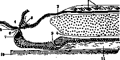
Ringer's Solution was introduced into physiological practice by the English physiologist S. Ringer for experiments with isolated tissues and organs. Its advantage over ordinary physiological saline solution lies in the fact that, in addition to being isotonic with blood plasma, it is also similar in its mineral composition. Due to these properties, tissues placed in Ringer's solution retain their vital properties for significantly longer periods. Ringer provides two recipes: 1) for experiments with frog tissues: NaCl-7.5; CaCl2-0.125; KCl-0.075; NaHCO3-0.125; Aq. destill.-1,000.0 and 2) for mammals: NaCl-9.0; CaCl2-0.24; KCl-0.42; NaHCO3-0.3; Aq. destill.-1,000.0. (See also Physiological solutions.) RHINENCEPHALON, the olfactory brain, is phylogenetically the oldest part of the central nervous system and is usually contrasted with the younger part - the cerebral mantle. In comparative anatomy, the first part is therefore designated as archipallium or archicortex (old cortex), the second as neopallium (new cortex). R. in turn consists of two parts: the olfactory lobe and the marginal convolution. The first of these, i.e., the olfactory lobe (lobus olfactorius), is an outgrowth of the forebrain and embryologically divides into two areas - anterior and posterior, separated in the embryo by a deep groove (incisura prima His). The anterior part includes the olfactory bulb and olfactory tract. The posterior part consists of the anterior perforated substance or lamina, in the anterior part of which in animals there is an elevation in the form of the so-called olfactory tubercle. The second part of R., the so-called marginal convolution (lobus limbicus), is located on the inner surface of the hemispheres and consists of two convolutions: the fornicate, or cingulate (gyrus fornicatus, s. cinguli) and the pear-shaped, or hippocampal (gyrus parahippocampalis, s. hippocampi), connected to each other by means of a isthmus. In addition to the marginal convolution belong: a) the dentate gyrus (gyrus, s. fascia dentata), b) the gray layer of the corpus callosum (induseum griseum corporis callosi), which passes into the fimbria, and c) the septum pellucidum-see also Olfaction (fig. 1). In different animals, R. as a whole has varying degrees of development, depending on which all animals are divided into macroosmatic and microosmatic. Most small animals (edentates, marsupials, rodents) belong to the first group; the second group includes cetaceans and pinnipeds (walruses, seals), in which remnants of the olfactory lobe are found only microscopically. An intermediate or third group is formed by ruminants and carnivores, as well as some species of lower primates (Prosimii, Lemuridae). In primates and humans, R. is greatly reduced. It should be borne in mind that in microosmatic animals, along with the reduction of the olfactory lobe, there is atrophy of the lobi pyriformis (lobi hippocampi).-Turning to the description of R. in various animal species, it should be noted that in invertebrates (e.g., in amphioxus) one can see only a single olfactory pit and the olfactory nerve (so-called complete monorhinia; fig. 2). In cyclostomes (lamprey) both halves of the forebrain are more or less clearly differentiated into anterior and posterior lobes. The olfactory nerve originates from the anterior lobe, usually designated as the olfactory lobe, although in its internal structure it most closely corresponds to the olfactory bulb. From the olfactory nerve, secondary fibers extend, going to the posterior lobe of the forebrain, which should therefore be considered as the secondary olfactory lobe (this lobe is the rudiment of the hemispheres of higher vertebrates). 841
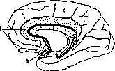
Fig. 1. 1-Lobus olfactorius anterior and gyrus fornicatus; 2 - lobus olfactorius posterior and gyrus dentatus. (According to Williger.)
In another cyclostome animal-myxines-the hemispheres of the endbrain are very poorly developed and consist almost exclusively of olfactory lobes, so closely fused with the diencephalon that it is impossible to draw a boundary between them. In fish, the primary olfactory lobes are usually in the form of an elongation of the anterior parts of the endbrain, from which they are more or sharply delimited (fig. 3). In some sharks and rays, the olfactory lobes begin from below or to the side of the endbrain, while in dipnoi the olfactory lobes adjoin the hemispheres themselves from the front (fig. 4). In cases where the primary olfactory
Fig. 2. Amphioxus: 1-large dorsal cells; 2-central canal (pore); 3-olfactory nerve; 4-olfactory pit; 5-connective tissue; 6-recessus neuroporicus; 7-pigment spot; 8-central canal (ventral); 9-so-called funnel; 10-notochord; 11-white mass of fibers. (According to Bütschli.)
lobe is very long (as observed in Chondropterygii), a division into 1) bulb, 2) long tract, and 3) secondary olfactory lobe adjacent to the endbrain is noticeable. The powerful bulbs of the mentioned fish species lie in the nasal cavity mucosa (as in myxine). In cartilaginous fish, the anterior lobe of the ventral part of the hemispheres (so-called lobus postolfactorius of Burkhardt) probably corresponds to the so-called lobus parolfactorius of higher vertebrates, and the inferior lobe to the pear-shaped lobe (lobus hippocampi); in the latter, a cortical structure can already be distinguished in this species of fish. In addition, in fish, the presence of the anterior commissure of the brain (commissura cerebri anterior) can also be established, containing fibers: 1) from the olfactory lobe and bulb, and 2) from the basal ganglia. In dipnoi, the so-called anterior commissure of the mantle (commissura pallii anterior) is also added to this commissure, containing connections between the olfactory formations of the mantle. In amphibians, the olfactory lobes are not separated from the hemispheres of the endbrain and pass into them without a sharp boundary (fig. 5);
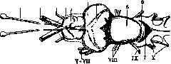
Fig. 3. Fish brain: I-olfactory nerve; II-XII cranial nerves; 1-cerebral mantle; 2-striated body; 3 and 4-visual lobe; 5-cerebellum; 6-unpaired lobe; 7-rhomboid fossa. (According to Bütschli.)
only in gymnophions and tailless amphibians they are more sharply defined due to the presence of a transverse groove. The olfactory lobes of amphibians (as well as in some species of fish) contain an extension of the lateral ventricles of the brain; in addition, in tailless amphibians, the olfactory nerve has two roots. Both commissural systems noted above in fish are also clearly expressed in amphibians, with commissura pallii anterior (previously designated as the corpus callosum) sending its fibers to the inner wall of the hemispheres, and commissura anterior to the ventral and lateral.-In reptiles, the primary olfactory lobes are a continuation of the anterior end of
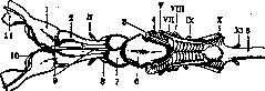
Fig. 4. Shark brain: 1 and 2-olfactory lobe; 3 and 4-rhombencephalon; 5-medulla oblongata; 6-cerebellum; 7-visual lobe; 8-inferior lobe; 9-epiphysis; 10-terminal nerves; 11-olfactory bulb; II-XII cranial nerves. (According to Bütschli.)
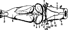
the hemispheres; the lateral ventricles usually reach the olfactory lobes themselves (fig. 6 and 7). Many reptiles (e.g., crocodiles) show a division of the olfactory lobe into: 1) bulb, 2) tract, and 3) secondary olfactory lobe. In addition, in reptiles, a longitudinal groove is noted, cutting through the medial wall of the mantle - the so-called fissura arcuata (fissura hippocampi, fovea limbica interna), causing a small fold to appear in the wall of the ventricle. This thickening, which occurs
Fig. 5. Frog brain: 1-medulla oblongata; 2-cerebellum; 3-anterior olfactory root; 4-olfactory lobe; 5-choroid plexus; 6-epiphysis; 7-visual lobe; 8-ventricle; IX-XI cranial nerves; II-spinal nerve. (According to Bütschli.)
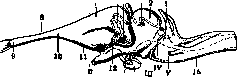
in the medial wall of the mantle - the so-called fissura arcuata (fissura hippocampi, fovea limbica interna), causing a small fold to appear in the wall of the ventricle. This thickening, which occurs
Fig. 6. Lizard brain: 1-cerebellum; 2-visual lobe; 3-posterior commissure; 4-epiphysis; 5-temporal lobe; 6-velum transversum; 7-cerebral hemisphere; 8-olfactory tract; 9-olfactory bulb; 10-olfactory lobe; 11-lobus parolfactorius; 12-anterior commissure; 13-funnel; 14-hypophysis; 15-medulla oblongata; II-V cranial nerves. (According to Bütschli.)
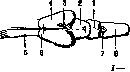
Fig. 7. Snake brain: 1-cerebellum; 2-visual lobe; 3-epiphysis; 4-cerebral hemisphere; 5-olfactory tract; 6-olfactory lobe; 7-rhomboid fossa; *-medulla oblongata. (According to Bütschli.)
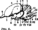
Bird brain: 1-cerebellum; 2-epiphysis; 3-visual lobe; 4-temporal lobe; 5-valecula; 6-olfactory bulb; 7-olfactory lobe; 8-hypophysis; 9-medulla oblongata; 10-flocculus; II-XII cranial nerves. (According to Bütschli.)
first observed in tailless amphibians, serves as the rudiment of the hippocampus and the fimbria of mammals. This structure in all reptiles sends fibers to the anterior commissure of the pallium, to which also two bundles of fibers from the subcallosal area (homologs of the legs of the fornix of higher vertebrates) extend. Centrally from the mentioned commissure is located the anterior commissure. The olfactory lobes of birds are very small in size compared to the volume of the hemispheres (fig. 8). If these lobes are strongly reduced (e.g. in parrots), they may not be visible at all from the dorsal surface; additionally, sometimes they may fuse with each other. Further, behind the olfactory lobes in birds, two small elevations are observed (so-called lobus parolfactorii), first noted in reptiles, but expressed in the latter extremely weakly. Commissura pallii anterior in birds is strongly reduced. In mammals, the basal part of the hemispheres, containing in front the olfactory lobes and behind the powerful arrays of the temporal lobes, is separated from the rest of the pallium by means of a horizontal groove (fissura rhinalis externa, s. fovea limbica). This groove is clearly expressed in dipnoan fishes and tailless amphibians. Depending on the powerful development of the pallium, the olfactory lobes, which in many mammals extend beyond the anterior edge of the hemisphere, in humans and monkeys are completely covered by the massive frontal lobes of the hemispheres; this same feature is also seen in cases of strong atrophy of R. in whales and pinnipeds. In macrosmatic animals, the olfactory lobes are very large; the distal end of them thickens into a massive bulb, to which numerous olfactory filaments approach, penetrating the holes of the ethmoid bone; only in the platypus is a whole complex of such filaments observed in the form of a separate bundle. With strong development of R.: Hedgehog brain: A-from above-3-cerebellum; 4- fovea limbica; 5-cerebral hemisphere; b-olfactory bulb; 7-lobus parolfactorius; 8-pituitary gland; 9-flocculus; 10- velum medullare post.; 11-medulla oblongata; II and V-cranial nerves; B-from below: 1-flocculus; 2-cerebellum; 3-temporal lobe; 4-lobus parolfactorius; 5-olfactory bulb; 6-pituitary gland; 7-pons; 8-medulla oblongata; II and V-cranial nerves. (According to Büchli.)
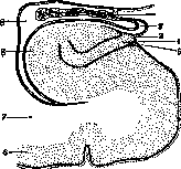
each olfactory lobe usually continues as the cavity of the lateral ventricle. Directly behind and somewhat inward from each olfactory tract in all mammals is located a more or less significant bulge-the so-called lobus parolfactorius (tuberculum olf., area olfact.), occupying in insectivores and rodents 1/2 of the entire base of the brain (fig. 9). It is expressed much less sharply in carnivores, reaching medium size in ruminants. According to the latest research, this structure has a close connection with the nucleus of the trigeminal nerve and is most developed in those animals in which particularly strong innervation of the muzzle is observed (for example in the tapir), in birds-with a very large beak. In humans, lobus parolfactorius is represented as a small structure located in the middle of the anterior perforated space. The commissures described above in mammals are in the following state: commissura anterior, s. ventralis, located in reptiles in lamina terminalis, changes little; already in lower mammals it is represented quite distinctly and in it two parts can be distinguished: Figure 10. Hippocampus and dentate gyrus: 1-choroid plexus; 2-fimbria; 3-sulcus fimbrio-dentatus; 4-gyrus dentatus; 5-fissura hippocampi; 6 and 7-gyrus hippocampi; 8-alveus; 9-inferior horn of the ventricle. (According to Williger.)
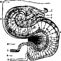
Figure 11. Hippocampus and dentate gyrus: 1-fimbria; 2-gyrus dentatus; 3-fissura hippocampi; 4-subiculum; 5-molecular layer; 6-layer of pyramidal cells; 7-layer of polymorphic cells; 8-white matter; 9-alveus; 10-inferior horn of the ventricle. (According to Williger.)
in mammals, commissura pallii anterior undergoes further development, expressed in the connection of the hippocampus. We have seen that in amphibians and reptiles, the so-called hippocampus is represented as a ganglionic thickening of the medial wall of the hemisphere, which in turn is depressed into the cavity of the lateral ventricle, forming the so-called hippocampus. On a cross-section through the latter in lower mammals (platypus), the same relationships can be seen as are characteristic of scaled reptiles, with the difference that fissura hippocampi in them is more deeply indented and thus the depression into the ventricular cavity is expressed more sharply (fig. 10). The dorsal lip of this depression, which due to its spiral curvature is directed into the ventricular cavity, has a special structure (alveus). The ventral lip of fissurae hippocampi is designated as fascia dentata and also shows another feature of its cortical structure (fig. 11).
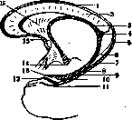
In non-placental mammals, the hippocampus is located in the anterior part of the brain (fig. 11); in higher mammals, this structure is displaced far backward into the area of the temporal lobe, representing an important point of R.: fiber bundles from lobus olfactorius and parolfactorius go here (see Olfaction). The fibers going from the hippocampus inward and forward form the fringe (fimbria); these fibers reach commissura pallii anterior; here they, partially crossing over, pass to the opposite hemisphere. Thus, the mentioned commissure represents the connection of the hippocampi in lower mammals. In the latter, starting from monotremes, in addition to this, fibers extending from area praecommissuralis, located before commissura anterior, as well as fibers from the subcallosal area, also extend to commissura pallii anterior. In higher mammals, in connection with the appearance of the corpus callosum, the size of commissurae ant. becomes smaller; at the same time, the ventral part of the original commissurae pallii ant. transforms into that structure which is designated as the fornix (fornix). In lower placental mammals, the corpus callosum and fornix remain very short and are located dorso-caudally; as the brain hemispheres increase backward, the fornix and corpus callosum also stretch and acquire a horizontal position. These structures reach their largest size, obviously, in primates and humans. The fornix represents the continuation of both fringes forward, forming in the middle the crossing of their fibers (commissura fornix). As the corpus callosum of placental mammals grows backward, it narrows the part of the primary hippocampus located above it to the degree of a narrow plate (striae longitudinales Lancisii); the lower part of the hippocampus, located within the temporal lobe, however, retains its original massive volume. Between the corpus callosum and the fornix a thin membrane is visible, forming on both sides the transparent septum, between the Figure 12. 1-induseum griseum; 2-corpus callosum; 3-fornix; 4-fornix transversus; 5-fasciola cinerea; 6-gyrus fasciolaris; 7-fissura hippocampi; 8-fimbria; 9-gyrus dentatus; 10-sulcus fimbrio-dentatus; 11-fissura hippocampi; 12-tenia chiasmatis; 13-gyrus intralimbicus; 14-corpus mamillare; 15-gyrus subcallosus; 16-septum pellucidum. (According to Williger.)
walls of which there is usually a cavity, having nothing in common with the cavities of the brain ventricles (fig. 12 and 13).
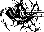
In addition, the so-called amygdala, observed in all mammals, also belongs to R. This nucleus, phylogenetically the oldest, is already noted in birds (epistriatum 2 Edinger) and on the basis of cytoarchitectonic studies contains in humans 3 main nuclei (Gil-Figure 13. 1-tenia chiasmatis; 2-gyrus intralimbicus; 3-fornix; 4-corpus callosum; 5-fornix transversus; 6-fasciola cinerea; 7-gyrus fasciolaris; 8-fissura hippocampi; 9-fimbria; 10-gyrus dentatus; 11-sulcus fimbrio-dentatus; 12-fissura hippocampi; 13-tenia chiasmatis; 14-gyrus intralimbicus; 15-corpus mamillare; 16-septum pellucidum. (According to Williger.)
As for the peculiarities of R., the most detailed comparative anatomical data in this area belong to Brodmann (see Cortex architecture). This author, dividing the cortex into homo- and heterogeneric, attributed to the latter all structures of R. Thus, as cortex primitivus (cortex not forming layers) he considered bulbus olfactorius, pedunculus olf., tuberc. olf., subst. perf. ant. and nucl. amygdalae; cortex rudimentarius (cortex containing individual layers) includes: hippocampus, fascia dentata, subiculum, induseum griseum, septum pellucidum, area praeterminalis (25); to cortex striatus he attributed: area praesubicularis (27), area retrosubicularis (48), area entorhinalis (28, 34), area perirhinalis (35) and area praepyriformis (51). The cortex of the limbic gyrus Brodmann attributed to the homogeneric, distinguishing here subregiones postcingularis (23, 31), praecingularis (24, 32, 33) and retrosplenialis (26, 29, 30). In humans, this author could identify fields: 27, 28, 34, 35, 31, 32, 33, 24, 25, 26, 29 and 30 (fig. 14); in Cercopithecus, besides the fields of the limbic gyrus, fields 27 and 28 are described; in Hapale
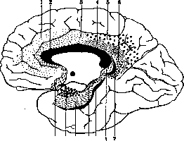
Figure 14. Fields of the olfactory area of the human brain according to Brodmann: 1- field 32; 2-field 33; 3- field 21; 4- field 23; 5- field 31; 6-field 30; 7- field 29; 8-field 26; 9- field 35; 10-field 27; 11-field 28; 12-field 34; 13-field 25, fields 27, 35, 28 and 48 (area retrosubicularis). In lower monkeys, he marked fields: 27, 28 and 51 (area praepyriformis); he also described these fields in bats, in which he also indicated the existence of fields 48 (area retrosubicularis) and 49 (area praesubicularis). Brodmann gave a detailed description of the heterogenetic cortex in the hedgehog, distinguishing in it field 51 (a,b,c,d), where field 51 d corresponds to the olfactory tubercle; the latter consists of three nuclei: anterior, middle, and posterior; further fields 28 (a and b), 27, 35, 48 and 49 are noted. O. Voit, on the basis of myeloarchitectonic studies, combines most of the formations of R. under the term allocortex, denoting by this term those areas of the cortex in which radial fibers reach the 1st layer. Thus, allocortex will include: 1) bulbus, tractus and tuberculum olf., 2) gyrus olf. medialis (fields 13 and 14 of Voit), 3) gyrus olf. lateralis, 4) substantia perforata ant. and gyrus. subcallosus (area diagonalis), 5) septum pellucidum, 6) regio hippocampica (cornu Ammonis, fascia dentata, uncus), 7) induseum cor-poris callosi, 8) gyrus semilunaris, 9) gyrus ambiens with three subfields (a1, aa and PNA), 10) gyrus hippocampi with ten subfields (Я6-чЯ15) and isthmus gyri limbici with five subfields (Я1-Я5). Rose, on the basis of ontogenetic and phylogenetic data, gives such a division of the cortex R. (1926): I. Cortex semiparietinus (cortex of 3 layers)--regio praepyriformis, tuberculum olf., regio periamygdalaris, septum pellucidum, area diagonalis. II. Cortex toto parietinus: a) schizopro-toptychos-a) parumstratificatus-regio praesubicularis, area parasubicularis, area perirhinalis, /8) multiformis-regio entorhinalis; b) holo-protoptychos - a) bistratificatus-cornu Ammonis (h1-№), subiculum, taenia tecta, fascia dentata, area retrobulbaris; /J) quinquestrati-ficatus-mesocortex. Touching on individual details, it should be noted that the olfactory tubercle is absent in birds, in reptiles it is found only in crocodiles and turtles; regio peri-amygdalaris (cortex covering the amygdala) has no homolog in birds and reptiles; in mammals it is expressed and divided into several subfields (in monotremes 3, in chimpanzees 5); the septum pellucidum is observed in all birds, reptiles and mammals; the horn of Ammon is best seen in predatory and swimming birds, while in parrots it is absent at all; reptiles possess a well-expressed horn of Ammon, as well as all mammals with its division into 3-5 subfields *; the dentate fascia is not observed at all in birds, among reptiles it is not expressed in all species, in mammals it can be found, starting from monotremes and ending with man; regio entorhinalis (field 28) or hippocampal cortex has a 4-layered structure and is already clearly expressed in birds (especially singing and swimming) and reptiles. In mammals, this field belongs to the most constant, breaking down in monotremes, marsupials, insectivores and rodents into 3 fields, in the dog into 8, in lower monkeys into 11 and in man into 23. As for the ontogenesis of R., its rudiments appear very early in the embryo: thus, in a chick on the 7th day of incubation, and in man on the 5th week (His) on the anterior part of each frontal lobe a small bulge directed forward is formed. It gradually takes the form of a bulb, the expanded part of which corresponds to the bulb, and the stalk-to the olfactory tract. This swelling contains a cavity inside, connected with the cavity of the lateral ventricle. As the embryo develops, this cavity gradually decreases and finally disappears altogether, while in many animals it remains for life. The division of the olfactory lobe into anterior and posterior becomes more noticeable as incisura prima (His) becomes deeper. The anterior olfactory lobe has the appearance of a small pyramid, the elongated and narrow base of which extends from the Sylvian fossa to the inner wall of the hemisphere. This lobe is bounded behind by incisura prima (His), outside by a groove forming a continuation forward of the Sylvian fossa, and in front by a shallow fissura sero-tina (His). The described anterior lobe gradually thickens at its apex and gives rise to the olfactory bulb, and then becomes detached from the base due to the development of a hollow stalk (pedunculus olfactorius), which appears the longer and thinner the older the embryo. As the olfactory stalk elongates, the olfactory bulb moves more and more forward, and by the 3rd month of intrauterine life it is located under the frontal lobe. The narrow base of the anterior olfactory lobe gives rise to the olfactory triangle, which in turn forms on the outside the outer olfactory root, and on the inside the inner one. On the brain of a 4-5 month old embryo, one can see how the outer olfactory root, turning almost at a right angle, goes outward to the Sylvian fossa (anterior stalk), and then goes along the medial edge of the mentioned fossa backward (posterior stalk); the outer root ends in small thickenings, of which the middle is designated as gyrus semilunaris Retzius'a, and the lateral-gyrus ambiens Retzius'a; the groove separating both thickenings is called sulcus semiannularis (Fig. 15). Due to the further development of the frontal and temporal lobes, the angle formed by the anterior and posterior stalks becomes more and more acute. In later stages, both stalks approach each other more closely, as a result of which the former continuity in the course of the stalks is disrupted, and at the same time the boundary between them and the insula is smoothed out. The inner olfactory root in its further development has a close relationship to the cingulate gyrus (see Olfaction). The posterior olfactory lobe is located between incisura prima His and the groove forming a continuation of the Sylvian fossa. This lobe gives rise to the anterior perforated plate and the diagonal ligament of Broca, then it continues on the inner surface of the hemisphere, where it merges with area trapezoides His. The embryonic development of the septum pellucidum in the embryo refers to the middle of the 3rd month, and of the arch-to the beginning of the 4th; the dentate fascia on the 5th month is grooved by numerous teeth, which in this period of embryonic life already make it resemble this formation in the adult; to the same time (5th month) belongs the appearance on the medial surface of each hemisphere of sulci calloso-margina-lis, limiting the gyrus marginalis from above. Cytoarchitectonically, the characteristic structure of Rinencephalon (allocortex) appears very early; in an embryo of 2-3 months it is already clearly differentiated from the rest of the cortex. Physiology-see Olfaction.
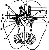
Figure 15. Head of a 5-6 month old human fetus: 2-bulbus olfactorius; 2-tractus olfactorius; 3-diagonal ligament of Broca; 4-insula; 5-trigonum prae-commissurale; 6-gyrus semilunaris; 7-substantia perforata ante-rior; 8-sulcus inferior rhinencephali; 9-gyrus ambiens; 10-angulus hippocampi; 11-gyrus olfactorius lat.; 12-gyrus olfactorius orbitalis; 13-sulcus parolfactorius post. (according to Williger), the outer root ends in small thickenings, from which the middle is designated as gyrus semilunaris Retzius'a, and the lateral-gyrus ambiens Retzius'a; the groove separating both thickenings is called sulcus semiannularis (Fig. 15).
Physiology-see Olfaction.
Related articles
Mentioned in
Cite this page
“Ringer's Solution.” Soviet Medical Encyclopedia. English translation of Bolshaya Meditsinskaya Entsiklopediya, 1st ed. (Moscow, 1928–1936), ed. N. A. Semashko. https://sovietmedicalencyclopedia.pages.dev/article/ringer-s-solution/