Kyphosis (or roundback, see)
Historical document, translated for reference. It reflects medical knowledge of the 1920s–30s and is not medical advice.
Summary
Kyphosis, or roundback, refers to an excessive outward curvature of the spine. This article describes the anatomical structures and functions of tendons in relation to muscle movement and support.
Encyclopedia article (1928–1936)
KYPHOSIS, or roundback, see Spine. TENDON (Latin: tendo), a component of striated muscle, closely connected with it. Tendons may be present at both ends of muscle bodies, run along one or the other lateral edge of the muscle, and also be found within the muscle mass, covering it on one or the other, sometimes very extensive, portions, splitting the muscle into layers, dividing it along its length, or being located between muscle bundles (figs. 1 and 2). Thus, a series of forms of T. can be established. The initial T. is one that goes from the bone to the origin of the muscle (origo). Initial T. may have the appearance of very short tendon fibers or a broad plate, or a narrow ribbon, cord, spindle, or cone. The initial T. may be double or multiple if the muscle originates from two or many bones or their parts. The terminal T. goes from the end of the muscle to its place of attachment (insertio). It can also be of various sizes and shapes: from short tendon fibers and flat plates to bundles of spindle-shaped, ribbon-like, conical, cylindrical, fan-shaped and other forms; it can reach considerable length. The terminal tendon can also be double or multiple if it splits and approaches two or several bones or areas of bone. A special type of terminal T. is the fibrous expansion (lacertus fibrosus), by which the biceps brachii muscle attaches to the bones of the forearm. The terminal T., like the initial T., can cover the body of the muscle over a very extensive portion, enclosing it on all sides like a sheath, and gradually transitioning without any
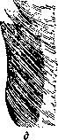
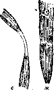
Fig. 1. Forms of muscles (from Toldt): a-spindle-shaped; b-periopodous; c-bi-periopodous; d-bicipital; e-broad; f-biventral; g-long with parallel fibers.
boundary into the outer connective tissue sheath (perimysium externum).-T. can cover the surface of the muscle over its entire extent or over a significant part of it; this is the aponeurosis of the muscle (tendo latus, s. aponeurosis). Muscle fibers can attach to it or originate from it. Such is, for example, the aponeurosis of the temporal muscle - the temporal fascia (fascia temporalis), which consists of two layers: superficial and deep (lamina superficialis and profunda). T. can run along one of the lateral edges of the muscle, and muscle fibers can attach to it; in this case, the muscle will be unipennate (m. unipennatus). If T. runs along the midline of the muscle and its fibers attach from both sides, the muscle will be bipennate (m. bipennatus). Tendon fibers can run along the length of the muscle in several longitudinal plates, dividing the muscle into separate regular or irregular bundles (for example, the bundles of the subscapularis muscle). Tendon fibers can run within the muscle mass in plates parallel to its surface, splitting it into layers. Such is, for example, m. masseter, consisting of outer and inner layers. Between intermediate T. (tendo intermedius), a muscle can be divided into two bellies; this is a biventral muscle (m. biventer). Such intermediate T. can divide the body of the muscle along its length into separate segments. These are so-called tendon bridges or inscriptions (inscriptiones tendineae) of the rectus abdominis muscle and the sometimes occurring bridges of the sternohyoid muscle. In these cases, they represent traces of the segmental development of muscles from myomers and remnants of myocommata, separating myomers from each other.-A special form of T. is the sheath of the rectus abdominis muscle (vagina m. recti abdominis), which consists of two layers - outer and inner (lamina externa et interna vaginae m. recti abdominis) - and is composed of tendon fibers of the external and internal oblique and transverse abdominal muscles. A special form of T. is also the central tendon of the diaphragm (centrum tendineum diaphragmatis).-Under the name of coronary T. (tendo coronarius) is meant the fibrous ring of the heart (annulus fibrosus cordis), to which the muscle bundles of the heart attach. Further, in the heart there are also chordae tendineae (see Heart, anatomy). The tendon fibers of the valve of the inferior vena cava are also called T. (tendo valvulae venae cavae). This term is also used to denote the ligaments of the eyelids (lig. palpebrale medium), calling it T. (tendo palpebralis, s. palpebrarum). Some T. end in the skin, in the subcutaneous tissue, in tendon structures of other muscles, in bursae or their additional tendon or fibrous structures. Sesamoid bones, fibrous and mucous sheaths, mucous bursae and mesenteries are associated with T. (see figs. 3 and 4). T. often pass through bone-fibrous canals, formed by depressions on the bones and fibrous masses associated with them - these are fibrous sheaths of T. (vaginae fibrosae tendinum). The significance of these structures is that they strengthen T. in their position and prevent them
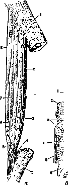
4, u Fig. 2. Diagrams of striated muscles: a-between two bones, approximately like the brachial muscle (m. brachialis); muscle fibers are shown too enlarged: b-muscle fiber at higher magnification (from Shimanovich-Krause). a: 1-periosteum; 2-muscle fiber, starting and ending tendinously; 3-outer perimysium; 4-subtendinous bursa; 5-tendon fiber; 6'-muscle fiber, one end originating from the muscle; 7-muscle fiber, both ends connected to muscles. b: 1-muscle fibers (fibrils); 2-Conheim fields; 3-nuclei of muscle fibers; 4-nuclei of connective tissue cells; 5-perimysium of individual muscle fibers; 6-sarcolemma.
from shifting. In addition, T. can lie in synovial, or mucous sheaths (vaginae tendinum synoviales, s. mucosae), in which two layers are distinguished - inner - visceral, adjacent to T. (lamina visceralis), and outer - parietal (lamina parietalis). The visceral layer fuses with T. and at its ends passes into the parietal. The parietal layer consists of two layers: outer - dense fibrous, and inner - of flat cells having the character of endothelium. Between the parietal and visceral layers there is a small amount of fluid resembling synovia. Between both layers, tendon fibers can pass, which have the character of mesenteries of tendons (mesotenon, s. vinculum tendinis). In some places between T., on one side, and bones or skin,-on the other, mucous bursae of T. can be located (fig. 4) (bursae mucosae subtendineae), which play the same role as in muscles.-According to their histological structure, T. consist of bundles of primary tendon fibrils, glued together by interfibrillar collagenous substance and having tendon cells; the nucleus of such a cell is located peripherally (figs. 5 and 6). Primary bundles, through interbundle collagenous substance, merge into secondary bundles, which are surrounded by connective tissue layers (peritendineum, s. peritendineum internum). From such secondary bundles are formed tertiary bundles, also surrounded by their own connective tissue sheath. Finally, on the outside, T. is in turn covered by a sheath (peritendineum, s. peritendineum externum). The connection between tendon and muscle fibers is extremely strong, due to the direct transition of tendon fibers peritendineum int. into perimysium int. As for the direct transition of muscle fibrils into tendon fibers, as some authors accept, it is not recognized by all, however. The physiological function of T.- transmission of movements originating from muscles. Movement - sliding of T.-in comparison with other tissue, e.g. bone, skin,-is more extensive; it depends for T., devoid of a tendon sheath, on its being surrounded by loose connective tissue, for T., surrounded by a sheath, on the presence of synovia in the latter. The movement of these T. occurs under great pressure, especially under ligaments. The blood supply to T. occurs through blood vessels of the muscles;
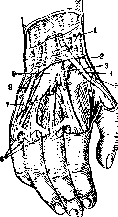
Figure 3. Synovial sheaths of the dorsum of the hand (from Vorobiev): 1-dorsal transverse ligament of the wrist; 2-synovial sheath of the abductor pollicis longus and extensor pollicis brevis; 3 and 4-synovial sheaths of the extensor carpi radialis; 5-synovial sheath of the extensor pollicis longus; 6-tendon bridges; 7-tendon sheath of the extensor of the fifth finger; 8-tendon sheath of the extensor carpi ulnaris; 9-tendon sheath of the common extensor of the fingers and extensor of the index finger. It should be noted that the blood supply to the tendons is very limited; tendons are poor in blood vessels. These anatomical-physiological features must be taken into account in the treatment of diseases of the tendons and their sheaths. The function of the tendons can be impaired due to 1) injuries to the tendons, 2) inflammatory processes, 3) tumors of the tendons or as a result of complications and improper treatment of these processes. Injuries to the tendons can be closed (subcutaneous) and open. The first group includes bruises, dislocations, and contusions. Bruises of the tendons occur with severe trauma, when the tendon is pressed against the bone and subjected to crushing; with mild bruises, the tendon mostly escapes due to its elasticity and the presence of the surrounding subcutaneous tissue. Often with trauma, there is a hemorrhage into the tendon sheath and its surroundings. Some authors (Kilttner) observed isolated hematomas of the tendons. A characteristic sign of the latter is swelling along the course of the tendon, which appears immediately after the injury. When the tendon moves, the swelling repeats the movement of the latter. Puncture confirms the diagnosis. Treatment consists of complete rest of the injured limb (splint), heating compress, tight bandaging followed by massage. - Severe violence in trauma can lead to dislocation of the tendon. The dislocated tendon is not in its proper place, its function is impaired; usually with dislocation of the tendon, there is a large bruise due to the rupture of the ligaments that hold it in its usual place. An example of tendon dislocation can be dislocation of the tendon of the peronaeus longus muscle. A predisposing factor for the dislocation of the latter may be to a certain extent subcutaneous ruptures of the tendons.
Figure 4. Bursae in the area of the anterior surface of the knee (from Vorobiev): 1-anterior superior iliac spine; 2-sartorius muscle, cut off; 3-iliopsoas muscle; 4-inguinal ligament; 5-iliac fossa; 6-pectineus muscle; 7-adductor longus muscle; 8-gracilis muscle; 9-lamina vaso-adductoria; 10-m. vastus medialis; 11-its tendon bundles; 12-fascia lata; 13-subcutaneous bursa in section; 14-sartorius muscle, cut off; 15-tendon of the patella; 16-fascia lata; 17-patella; 18-fascia lata; 19-fascia lata; 20-m. vastus lateralis; 21-rectus femoris muscle; 22-fascia lata (cut off); 23-musculus tensor fasciae latae.
degrees of underdevelopment of the superficial flat groove in which the tendon lies. Dislocations of the tendons on the extensors of the fingers of the hand often occur. Sudden muscle contraction is sometimes the cause of dislocation. Treatment of tendon dislocation does not present great difficulties and consists in fixing the tendon in its normal place by applying a fixing bandage or surgery. Subcutaneous ruptures of the tendons occur due to severe trauma with overstretching of the tendon or due to sudden contraction of the muscle while simultaneously contracting its antagonist. Usually the tendon ruptures either at its attachment to the bone, in which case part of the periosteum or bone often comes off with the tendon, or at its transition into the muscle and then part of the torn muscle fibers comes off with the tendon. Rupture of the tendon is accompanied by acute severe pain, at which patients, and sometimes those around them, hear a crackle, after which the tendon is disabled from work. An example of spontaneous rupture of the tendon can be rupture of the tendon of the quadriceps muscle when trying to stay on one's feet during a fall backward, rupture of the Achilles tendon when jumping, rupture of the extensor of Figure 5. Longitudinal the thumb of the hand. In section through the calf muscle for ETIOLOGY of tendon rupture MO-play a role as prof. muscle fibers in changes, for example in workers, grinders; muscle fibers; 3-cells of the tendon; 4-perimysium; ™ Degenerative and necrotic changes in the tendons, which leads to weakening and loosening of the latter. Rupture of the tendons occurs more often in middle age, when their elasticity begins to decrease. Treatment of spontaneous ruptures of the tendons can be conservative and surgical, the latter is preferable, although a number of cases with good results have been described with conservative treatment. Nevertheless, the possibility of repeated rupture is noted. Open injury to the tendons is mainly the result of injury by sharp objects (glass, knives-household injuries) or the result of rupture when caught in a machine. It should be noted the great strength and elasticity of the tendons, thanks to which the tendons often remain intact despite the action of great force that destroys bones, joints and muscles. When caught in a machine, cases of complete avulsion of the thumb together with the long extensor and flexor tendons are noted, which are torn off together with the muscular belly at their place of attachment. With open injuries, there can be either isolated injury to the tendons, or injury along with other organs, nerves and vessels. When injured by sharp objects, the edges of the incision usually have a smooth surface, when injured by moving parts of the machine, the tendons are torn and appear frayed. With simple transection of the tendon, its ends diverge, with the proximal end usually going deeper centrally, in the direction of the shortened muscle. The peripheral end, if it separates, does so very little. Injury to the tendon can be incomplete and then its function can be partially preserved. Often with minor skin damage, there are significant disorders of the tendon-holding apparatus, so it is necessary to especially carefully examine the function of the tendons with the slightest suspicion of the possibility of its injury and to sufficiently expand the wound when the tendon is injured, giving the possibility of detailed examination and free operation in the injured area. With insufficient expansion of the wound, especially on the forearm and hand, one can miss and not notice the separated ends of the injured tendons and not understand the anatomical relationships, in which one must always be well oriented when suturing the tendons. On the upper limbs, injury to the tendons occurs much more frequently compared to the lower limbs, which should be attributed to the greater participation of the upper limbs in labor processes and the more superficial location of the tendons of the forearm and hand. - Diagnosis of tendon injury does not present difficulties. With careful examination in the case of a small skin wound and with sufficient expansion of it, the severed ends of the tendons are visible, examination of the function confirms the diagnosis. Treatment of tendon injuries should consist in restoring the continuity of the tendon by applying a primary suture (see Tendon suture). Most authors (Salomon, Bior, Hueck) on the basis of experimental data note the extremely insignificant regenerative ability of the tendons inside the tendon sheaths, and Bior believes that the presence of synovial fluid promotes regeneration. According to others (Wehner), synovial fluid has no significance for tendon regeneration. Most authors believe that regeneration of the tendons occurs by the formation of connective tissue scar, although the function of the tendons is fully restored. Inflammatory processes of the tendons of a specific nature. Acute inflammatory processes of the tendon sheaths-see Tendovaginitis. Gonorrheal lesions of the tendons are rarely isolated, they are more often of a secondary nature with simultaneous joint involvement. The favorite place of lesion is the common extensors of the fingers of the hand; cases of lesion of the extensors of the thumb and extensors of the little finger have been described. Metastatic lesion is proved, on the one hand, by the presence of the primary focus, on the other-by the finding of gonococci in the exudate of the tendon sheaths. Clinically, the disease is characterized by the appearance in the area of the affected tendon of a painful swelling, gradually without a sharp border passing into healthy tissue with elevated body temperature. The swelling is very painful on pressure; there is impairment of the function of the organ due to severe pain on movement. Gonorrheal inflammation of the tendon sheaths is more often encountered in the form of a serous form. There are cases when serous lesion passes into purulent, then opens into the subcutaneous tissue, forming an abscess or phlegmon. Diagnosis of gonorrheal lesion of the tendon sheath does not present significant difficulties, especially in the presence of
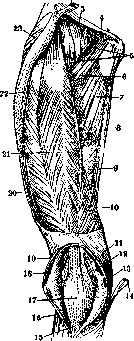
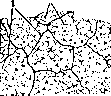
Figure 6. Part of a cross-section through the tendon of the human popliteal muscle (from Shimanovich - Krause): 1 - connective tissue septa; 2 - tendon cells; 3 - primary bundles. 10a of the primary focus; puncture and examination of the exudate confirm the diagnosis. Treatment in the early stages consists of complete rest of the limb, application of heat; in cases that progress to suppuration, surgical intervention is required. Tuberculous involvement of the tendon sheath was first described by Carre in 1891; although before him this disease was known under the name of rice bodies of the tendon sheath and hygromas, the tuberculous origin of such diseases was established only in 1876 by Geftman (NoeShpap). At present, tuberculous involvement of the tendon sheath is clinically divided into the following 4 forms: 1) serous tuberculous hydrops, or tuberculous hygroma (hydrops tuberculosus serosus), in which there is a serous, light yellowish, completely transparent exudate and the walls of the tendon sheaths are thickened and covered with tuberculous granulations. 2) Serofibrinous tuberculous hydrops (hydrops tuberculosus sero-fibrinosus), or hygroma with rice bodies, in which a small amount of serous exudate and a mass of rice bodies floating in the fluid can be found. Sometimes the picture resembles a bag tightly packed with rice bodies. 3) Granulating form with shrinkage (fungus), in which the visceral leaflet thickens, and a highly vascularized granulation tissue appears in the tendon sheath, which can fill the entire lumen of the tendon sheath and extend to adjacent tissues. 4) Granulating form with suppuration (cold abscess), in which there is pus and caseously altered granulations inside the tendon sheath. All these forms can develop either primarily or secondarily. Often the tendon sheath becomes infected hematogenously or the infection extends per continuitatem to the tendon sheath from a neighboring tuberculous focus. Cases of primary tbc involvement after trauma have been described; the latter plays a certain role as creating a locus minoris resistentiae. The disease predominantly develops between 20 and 30 years of age in manual laborers; the upper extremities are affected more often than the lower ones. According to Carre, the ratio between involvement of the upper and lower extremities is 5:1. The right hand is affected more often than the left; in the cases of Canel (Ca-navel) out of 14, 13 had involvement on the right hand. One can think that the right hand is more traumatized during labor processes (in right-handed people) compared to the left. Flexors are affected 4 times more often than extensors (Carre). Of the flexors of the hand, the most frequent involvement can be considered that of the common tendon sheath of the finger flexors in the wrist joint area, but isolated involvement of the finger tendon sheaths can also occur. Of the extensors, the tendon sheaths of the II and IV fingers are affected. The lower extremities are affected less often, the disease more often develops in the common extensor of the foot (extensor digitorum communis) and in the tendon sheath of the m. peronaei and m. tibialis. The clinical picture of the disease is characterized by the appearance of a swelling associated with Kyphosis, of an elongated and flattened shape. The swelling, which is not painful at first, develops gradually, its borders are quite distinct. Sometimes fluctuation can be noted, and upon palpation, separate dense bodies the size of a lentil to a cherry can be felt. As one of the signs of the disease, weakness of the affected organ is noted initially. The swelling sometimes has the appearance of a small bag tied transversely, e.g., in tuberculous involvement of the finger flexors developing on both sides of the lig. carpi volar., the latter acts as a constriction. In this position, the disease can remain for a more or less prolonged time, then phenomena of more severe functional disturbance appear. Pulling pains in the fingers, paresthesias, and sometimes contractures in the peripheral segments appear. The diagnosis of this condition can be difficult in serous forms of involvement, when it is necessary to differentiate from serous tenosynovitis, ganglion, lipoma, or syphilitic involvement of the tendon sheaths. The presence of other tuberculous foci at the same time facilitates recognition. In hygroma with rice bodies, diagnosis is facilitated by the presence of a crunching and friction sensation upon palpation of the swelling. The diagnosis of purulent forms of involvement, especially fistulous ones, does not present great difficulties. In the treatment of serous forms of tuberculous involvement of tendon sheaths, one can limit oneself to puncture-draining of the serous content and repeated introduction of iodine-iodoform emulsion, providing rest to the affected organ and subsequent treatment with quartz (see Tuberculosis). Rice bodies are removed through a small incision. In the fungous form, surgical treatment with excision of the diseased focus is required, which is undoubtedly a more reliable means compared to the conservative method of treatment. Early movement is recommended after surgery. Canel and Carre note good results from surgical treatment of tendon sheaths. Advanced cases do not respond well to treatment. Syphilitic involvement of tendon sheaths is predominantly secondary and occurs simultaneously with other manifestations of syphilis. Syphilitic involvement is distinguished from Kyphosis proper in the form of its thickening, which develops gradually and painlessly, and from involvement of tendon sheaths in the form of acute tenosynovitis or gummatous involvement of tendon sheaths in the form of spindle-shaped thickenings, completely painless, often adherent to the skin with superficial location of Kyphosis. In recognition, it is necessary to remember about tuberculous involvement of tendon sheaths. The presence of syphilitic involvement of other organs helps to avoid error. Treatment of syphilitic involvement of the tendon sheath should be specific. Tumors in tendons develop as both benign and malignant. Among benign tumors, fibroma is most common, representing a dense, nodular, painless formation that develops gradually, mainly from fasciae and aponeuroses. Among malignant tumors, sarcoma is more frequently encountered. Rosenthal in 1909 described 70 sarcomas of tendon sheaths that he had collected, of which 41 were giant cell sarcomas. In terms of localization, sarcomas predominantly develop on the hand, forearm, foot, and lower leg. According to Hartel, 80% of all tumors affect the tendon sheaths of the fingers. These tumors sometimes show very slow growth, so cases of tumors 30 years old have been described; trauma is considered the cause of accelerated tumor growth. The diagnosis of a tumor does not present great difficulties, but the correct diagnosis is extremely important for the treatment of the tumor. Radical surgical treatment of the tumor - according to the general rules of tumor treatment. Rubbing changes in tendon sheaths after traumatic injuries to Kyphosis cause contractures (see) and complete impairment of organ function; they are most often found on the hands, on their palmar surfaces. Adhesion of Kyphosis to underlying tissues occurs after open and closed injuries to Kyphosis when timely restoration of the integrity of Kyphosis was not performed, after late surgical intervention on Kyphosis in purulent inflammations (see Tenosynovitis) and technically incorrectly performed surgical interventions; prolonged fixation of the organ in an immobile position and absence of early movements can lead to adhesion of Kyphosis to adjacent tissues. All these cases require surgical treatment. Dissection and release of Kyphosis from adhesions (tenolysis), excision of adhesions and subsequent creation of a padding for Kyphosis to facilitate gliding and prevent secondary adhesion are performed. There is another view that creating special conditions for Kyphosis during operations is by no means necessary and that early movements prevent subsequent adhesion. In cases where the separated ends of the injured Kyphosis have adhered to underlying tissues, dissection of the ends, their freshening, and suturing are performed first. If with a large separation of the ends of Kyphosis it is not possible to suture it, plastic repair of Kyphosis is performed, under which one should understand the correction of the defect between the ends of Kyphosis by various methods: lengthening of the ends by means of small lateral incisions on the tendons or by means of two incisions on both sides with subsequent straightening. When by lengthening Kyphosis it is still not possible to correct the defect, then free plastic repair can be used, filling the defect between the ends of Kyphosis with a strip of fascia or a segment of Kyphosis taken from a nearby area. Some authors (Gluck) for the same purpose recommend creating a bridge between the ends of Kyphosis from thick catgut or silk threads, stretching them between the ends of Kyphosis. With healing by first intention, granulation tissue forms between the threads, which subsequently turns into a dense fibrous cord. For subsequent treatment, early movements in the form of active gymnastics, and if necessary, mechanotherapy, must be used. Plastic repair of tendons, like tendon suture, requires great care in its application, knowledge of anatomy, and good technique.
Only in these cases are good results obtained. Plastic surgery for K. should also include shortening of the K., which is performed when the K. is weakened after excessive stretching of muscles in connection with pareses and paralyses of individual muscle groups. Shortening of the K. is performed by resection of part of the K. and subsequent suturing of its ends, through the application of stitches in the form of bundles on the tendon and by other methods. Replacement of the K. defect by replacing it with some other tissue with subsequent fixation of the transplanted flap-see Transplantation. By tenodesis is meant fixation of joints through fixation of tendon fibers to the ends of the joint with their passage over the joint, which gives significant support and strengthening of the joint. In all those cases where arthrodesis is necessary, most often with extensive paralyses and wartime injuries, tenodesis is applied, for example in the region of the wrist and ankle joints. The operative technique of tenodesis was developed by Codivilla, Vulpius, and Saxl. After injury to the K. of the fingers, spindle-shaped thickenings may develop in them, known under the name of trigger finger (schnellender Finger). This disease was first described in 1850 with the following symptom complex. With strong flexion or extension of the finger in a certain position, the patient is unable to continue voluntarily initiated movement, so that the help of the other hand is necessary to overcome the obstacle; with this movement a clear clicking of the finger occurs and then the finger can freely continue the initiated movement. The difficulty of movement occurs because a thickening forms on the K. of the finger, sometimes even not one but two, which hinders the passage of the K. through the round ligament (lig. armulare), and some force is required to overcome this obstacle. Most often the disease affects the middle finger of the hand, then the ring finger, then the index finger, less often the thumb and little finger. Microscopic examination of the thickening of the K. shows normal tissue structure of the K. The size of the thickening can be from a grain of millet to a cherry. In the etiology of the disease there is trauma, partial rupture of the K., hemorrhage into the K., occupational trauma, for example in seamstresses, knitters, laundresses. Cases of trigger finger in military personnel from pressure from the rifle butt have been described (Sudeck). Treatment can be conservative (prolonged massage) or operative, consisting in the superficial excision of the thickening of the K. Only in rare cases is resection of the node required. Subsequently-mechanotherapy.-In conclusion, it should be pointed out the possibility of ossification of the K., which in birds is quite common, while in humans it is a great rarity. Ossification of the K. occurs together with ossification of the muscles. This disease was first described by Hornig-ossification of the Achilles K. after trauma. It is observed after rupture of the K. of the quadriceps muscle and others. Radiography confirms the diagnosis.
n. Minin.
Related articles
Cite this page
“Kyphosis (or roundback, see).” Soviet Medical Encyclopedia. English translation of Bolshaya Meditsinskaya Entsiklopediya, 1st ed. (Moscow, 1928–1936), ed. N. A. Semashko. https://sovietmedicalencyclopedia.pages.dev/article/kyphosis-2/