Tendon Suture
Historical document, translated for reference. It reflects medical knowledge of the 1920s–30s and is not medical advice.
Summary
The article describes various methods for primary tendon suture, including techniques by Hagler, Friedrich, Krasin, and Lange. It discusses indications, materials, postoperative care, and outcomes, noting better results for extensors than flexors.
Encyclopedia article (1928–1936)
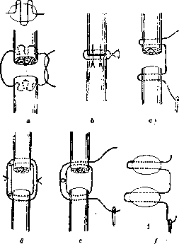
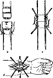
TENDON SUTURE. For restoring function to a tendon when its integrity is compromised, primary T. suture is applied. T. suture was first applied by Varicel following the method of Ambroise Pare. At the 5th Congress of German Surgeons in 1876, Küster recommended the application of sutures to tendons, at which time the participants of the congress showed great interest in the suture technique. In recent times, especially in foreign medical literature, considerable attention has been paid to the technique of T. suture. At present, there are more than 20 methods for applying T. suture. Such a large number of methods shows that in this direction there is not yet a method that would meet all requirements and give good results. T. suture must meet the following requirements. It must provide good contact between the ends of the tendon being sutured, without cutting through. When applying T. suture, necrosis of the tendon should not occur. In the subsequent period, there should be no adhesion of the tendon to adjacent tissues. The method of T. suture must be technically simple for an average surgeon to perform. The most suitable moment for applying T. suture is during the primary treatment of the wound. Only in exceptional cases with severe infection and crushing of the wound tissues is it necessary to refrain from applying a primary suture to the tendon. As material for T. suture, most authors recommend silk, and only individual authors (Jensen, Bartels) recommend catgut. Catgut does not guarantee the strength of the suture, as it dissolves very quickly and therefore cannot provide the necessary strength to the suture, especially when early movements after the operation are prescribed, as recommended by most authors as a preventive method against adhesions between the sutured tendon and surrounding tissues. After the ends of the severed tendon have been found, they should be refreshed and sutured in case of their fraying or scar changes. It is necessary to strive to apply T. suture so that the transverse cuts of the tendon are in good contact, and if possible, avoid lateral apposition of the tendon ends. The simplest method of applying T. suture is the method of Hagler (Fig. 1 b), when one suture is applied, stitching the tendon transversely, and then sutures are applied longitudinally, making the needle entry below the transverse thread; the latter thus prevents the longitudinal sutures from cutting through. A similar modification was proposed by Friedrich (Fig. 2 a). Not difficult in technique is the suture proposed by Krasin of Kazan and described by Fishman (Fig. 2 b) and Lange (Fig. 2 c and d). Krasin's method consists of splitting the ends of the severed tendon for 1 cm into equal halves, inserting a piece of fascia, specially cut from nearby, into the split ends, and fixing the ends of the tendon together with the fascia with 4 sutures at the corners. Lange's method - applying sutures to the ends of the tendon longitudinally, a mattress suture. Technically somewhat more difficult is the application of the suture according to Belfler (Fig. 1 a) and Trinka (Fig. 1 c-f). The latter applies the suture in the form of a through double loop in the transverse direction. Experimental determinations (Kimura, Malevitsch) of the strength of the suture on cadavers with hanging weights up to 3,000 g on tendons sutured by different methods showed that the strength of T. suture by the method of Wilms, Lange, Hagler is quite sufficient. It is recommended to open the tendon sheath along its lateral surface with subsequent application of thin sutures to it. Bier, Hueck do not recommend suturing the tendon sheath.--To avoid subsequent adhesion of the tendon to the skin scar, it is proposed to make the skin incision not over the tendon, but for example in the form of a flap incision. After applying T. suture, some recommend providing rest to the limb for 8-10 days (Pels-Leusden), for 2-3 weeks (Just), others advise early movements: Bier from the 8th day, Dreyer and Bayer - immediately after the operation. A bandage is usually applied with T. suture of extensors in the position of maximum extension and with T. suture of flexors in the position of maximum flexion. Müller believes that tendon healing occurs in 8 days. Nevertheless, cases have been described (Just) where rupture of the tendon occurred 30 days after applying the suture. Most authors consider the prognosis regarding the functional result after applying a primary suture to be good, other authors are pessimistic about the final result. In particular, Bier indicates a good result with T. suture of extensors, but as for flexors, especially of the fingers and hand, the final results of suturing leave much to be desired. The causes of failure lie, on the one hand, in infection of the wound, and on the other hand, in the scar adhesion of the tendon to adjacent tissues. In case of wound infection, there is a great danger of the infection spreading to the tendon sheath. It is necessary to immediately release the skin sutures and treat the wound in the usual way; sometimes the T. suture is preserved, and in individual cases recovery with preservation of function even occurs. According to Müller, out of 136 cases, there was suppuration in 46 and healing occurred. Failures due to adhesions of tendons to adjacent tissues were found in Lier's study of 450 injuries to the tendons of the fingers and hand: in 39.5% for extensors and in 53% for flexors. In individual authors, the percentage of healings after applying primary T. suture is as follows: Authors Percentage of healings Extensors Flexors Dubs 45 62 Just 22 20 Pels-Leusden 10 38 Bier 80 68 Müller 85 50 These figures show that the final result for flexors is significantly worse compared to extensors.--To avoid subsequent adhesion of the tendon to adjacent tissues, some authors (Henschen) recommend surrounding the tendon with fatty tissue, others for the same purpose advise creating a kind of sheath for the tendon from a vein (Thole), from gelatin (Lotheissen) and other means. In recent times, most authors tend to apply early movements as a method that provides the best guarantee against subsequent adhesion, and recommend starting movements on the 2nd-3rd day after the operation. Secondary T. suture should be performed after complete healing of the skin wound, but not earlier than 1 month. The prognosis for applying secondary T. suture is poor according to many authors, and therefore they advise against applying a secondary suture, especially to flexors. The statistical data of Lier and Dubs clearly show this. Authors Muscles Percentage of healings Primary suture Secondary suture Dubs Flexors 10 50 Extensors 38 61 Lier Flexors 0 10 Extensors 17 32 The reason for such a poor result of secondary T. suture, authors see firstly in the rapid atrophy of the muscle, and therefore Rehn recommends in such cases to suture the proximal end of the tendon to a healthy muscle or to an adjacent tendon; secondly, during the operation of secondary T. suture, the operation is complicated by the appearance of a large defect between the ends of the tendon after excision of scar tissue and refreshing of its ends, so that the application of secondary T. suture is often accompanied by great tension. In such cases, it is better to use tendon plastic surgery. The final result after applying secondary T. suture in most cases is unfavorable, and therefore it is necessary to strive during primary treatment of the wound to apply primary T. suture with the use of early movements after the operation.
Figure 1: a-Belfler's method; b-Hagler's; c-Trinka's.
After applying T. suture, some recommend providing rest to the limb for 8-10 days (Pels-Leusden), for 2-3 weeks (Just), others advise early movements: Bier from the 8th day, Dreyer and Bayer - immediately after the operation. A bandage is usually applied with T. suture of extensors in the position of maximum extension and with T. suture of flexors in the position of maximum flexion. Müller believes that tendon healing occurs in 8 days. Nevertheless, cases have been described (Just) where rupture of the tendon occurred 30 days after applying the suture. Most authors consider the prognosis regarding the functional result after applying a primary suture to be good, other authors are pessimistic about the final result. In particular, Bier indicates a good result with T. suture of extensors, but as for flexors, especially of the fingers and hand, the final results of suturing leave much to be desired. The causes of failure lie, on the one hand, in infection of the wound, and on the other hand, in the scar adhesion of the tendon to adjacent tissues. In case of wound infection, there is a great danger of the infection spreading to the tendon sheath. It is necessary to immediately release the skin sutures and treat the wound in the usual way; sometimes the T. suture is preserved, and in individual cases recovery with preservation of function even occurs. According to Müller, out of 136 cases, there was suppuration in 46 and healing occurred. Failures due to adhesions of tendons to adjacent tissues were found in Lier's study of 450 injuries to the tendons of the fingers and hand: in 39.5% for extensors and in 53% for flexors. In individual authors, the percentage of healings after applying primary T. suture is as follows: Authors Percentage of healings Extensors Flexors Dubs 45 62 Just 22 20 Pels-Leusden 10 38 Bier 80 68 Müller 85 50 These figures show that the final result for flexors is significantly worse compared to extensors.--To avoid subsequent adhesion of the tendon to adjacent tissues, some authors (Henschen) recommend surrounding the tendon with fatty tissue, others for the same purpose advise creating a kind of sheath for the tendon from a vein (Thole), from gelatin (Lotheissen) and other means. In recent times, most authors tend to apply early movements as a method that provides the best guarantee against subsequent adhesion, and recommend starting movements on the 2nd-3rd day after the operation. Secondary T. suture should be performed after complete healing of the skin wound, but not earlier than 1 month. The prognosis for applying secondary T. suture is poor according to many authors, and therefore they advise against applying a secondary suture, especially to flexors. The statistical data of Lier and Dubs clearly show this. Authors Muscles Percentage of healings Primary suture Secondary suture Dubs Flexors 10 50 Extensors 38 61 Lier Flexors 0 10 Extensors 17 32 The reason for such a poor result of secondary T. suture, authors see firstly in the rapid atrophy of the muscle, and therefore Rehn recommends in such cases to suture the proximal end of the tendon to a healthy muscle or to an adjacent tendon; secondly, during the operation of secondary T. suture, the operation is complicated by the appearance of a large defect between the ends of the tendon after excision of scar tissue and refreshing of its ends, so that the application of secondary T. suture is often accompanied by great tension. In such cases, it is better to use tendon plastic surgery. The final result after applying secondary T. suture in most cases is unfavorable, and therefore it is necessary to strive during primary treatment of the wound to apply primary T. suture with the use of early movements after the operation.
Figure 1: a-Friedrich's method; b-Fishman's; c and d-Lange's.
The final result after applying secondary T. suture in most cases is unfavorable, and therefore it is necessary to strive during primary treatment of the wound to apply primary T. suture with the use of early movements after the operation.
I. Minin,

Related articles
Mentioned in
Cite this page
“Tendon Suture.” Soviet Medical Encyclopedia. English translation of Bolshaya Meditsinskaya Entsiklopediya, 1st ed. (Moscow, 1928–1936), ed. N. A. Semashko. https://sovietmedicalencyclopedia.pages.dev/article/tendon-suture/