Tenon's Capsule
Historical document, translated for reference. It reflects medical knowledge of the 1920s–30s and is not medical advice.
Summary
Tenon's capsule is a complex system of fascia in the orbit that connects all its contents and envelops the eye with its appendages. It was first described in 1806 by French surgeon Tenon, after whom it is named.
Encyclopedia article (1928–1936)
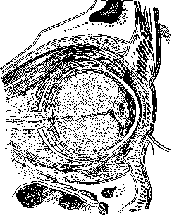
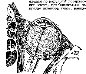
TENON'S CAPSULE (syn. Bonnet's capsule), a complex system of fascia of the orbit, closely connecting all its contents and enveloping the eye with all its appendages. It was first described in 1806 by the French surgeon Tenon, after whom it is named. This system of fascia can be traced from the very apex of the orbit, where individual bundles of it fuse with the periosteum of the latter, as well as with the fibrous sheath of the optic nerve in the area of attachment of the eye muscles and the upper eyelid. From here it extends forward, enveloping each muscle individually with its sheaths in the form of a thin layer of areolar tissue, and giving off numerous lateral branches of the same character, dividing the entire mass of surrounding orbital fat into individual fat lobules (fig. 1). The bundles of nerves and vessels located here, which supply the eyeball, in turn are enclosed between duplicatures of such thin partitions. Near the posterior pole of the eye, the muscular sheaths immediately sharply thicken, transforming from arachnoid membranes into dense elastic tissue of yellowish-white color, and that part of them which covers the inner (facing the eye) surface of the muscles, without reaching 2-3 mm to the equator of the eye, leaves the muscle and turns back onto the eye itself, spreading over its entire posterior half in the form of a continuous fibrous capsule (capsula posterior) (fig. 2). At the same time, the part of the fascia located on two layers, of which the firsttcontinues its original path along the surface of the muscles and tendons up to their scleral attachment, and then passes onto the sclera and spreads over the latter on one side to the edge of the cornea, and on the other-between the muscles, to the equator of the eye, enveloping its anterior half with a fibrous capsule (capsula anterior). Near the scleral attachments of the rectus muscles, this layer of fascia for a distance of 10-12 mm seems to recede from their surface,
Figure 1. View of Tenon's capsule with its branches and Tenon's spaces on a sagittal section of the eye and its appendages.
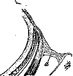
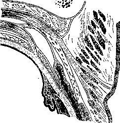
due to which here are formed cavities of an elongated shape, filled with serous fluid, or so-called 'presynovial bursae', facilitating the movements of the tendons. Between the equator of the eye and the line of muscular attachments, the anterior capsule is separated from the sclera by a capillary space, and forward it gradually fuses with the surface of the sclera, with the adhesions being most dense at the edge of the cornea. In this area it is also closely fused with the subconjunctival tissue. The second of the fascial layers transforms into dense fibrous tissue, which on one side connects with the capsule of the eye at its equator, and on the other attaches to the edge of the orbit all around its circumference, forming a kind of aponeurotic funnel, dividing the orbital cavity into two halves: anterior and posterior. In this case, those parts of this funnel that correspond to each of the muscles differ from the surrounding tissue by their special density and massiveness. French authors single them out as special anatomical formations and give them a special name 'ligamentous wings or winglets' (ailes ou ailerons ligamenteux) (fig. 3). Each of the rectus muscles has at least one such 'wing', and they are particularly well developed in the lateral and medial rectus muscles. The superior rectus, connected with m. levator palpebraethas two 'wings' extending from it to the sides (fig. 4); in addition it is connected by fascial bundles with the skin of the upper eyelid and its transition fold; a similar connection exists between the inferior rectus muscle and the lower eyelid with its transition fold. By its purpose, the aponeurotic funnel, as if suspending the eye to the edge of the orbit, fixes it in its position, and the 'ligamentous wings', when tensed during muscle contraction, protect the eye from sharp movements. Both halves of the fibrous sheath (capsula anterior et posterior), merging with each other near the equator of the eye, form around it a common sheath, or capsule, starting from the edge of the cornea and up to the optic nerve itself, onto which it passes, covering the latter with a special sheath over its own, up to foramen opticum. It is to this sheath that the name T. k. actually applies, since the author apparently considered it the central part of the entire aponeurosis of the orbit. Near the point where the optic nerve enters the eye, the T. k. is pierced by a series of ciliary nerves and vessels, receiving their own sheaths from it, thanks to which in this area it is firmly connected with the sclera; at the equator it is similarly pierced by four vorticose veins. The T. k. is separated from the sclera by a narrow cavity, which in the normal state is a capillary space filled with lymph and called Tenon's space. The anterior border of this cavity passes through the line of attachment of the muscular tendons, i.e., at a distance of 6-8 mm from the edge of the cornea, receding from the latter to 11 mm only in the area of attachment of the superior rectus muscle. Posteriorly it directly passes into the suprachoroidal space of the optic nerve and thus comes into connection with the subarachnoid space of the brain. The walls of Tenon's space are lined with a layer of endothelium and have the character of serous membranes, the cavity itself is penetrated by numerous finest fibers stretched between its walls, which contradicts the rather widespread opinion that the T. k. is for the eye a kind of joint cavity in which it can rotate freely in all directions. According to some authors, this rotation of the eye rather occurs together with the T. k. in relation to the orbital fat, filling the entire orbital cavity.
Figure 2. View of Tenon's capsule and Tenon's spaces on a horizontal section of the eye and its appendages.
Figure 3. 'Ligamentous wings' (i), or muscular processes of Tenon's capsule, connecting the anterior parts of the eye muscles with the edge of the orbit.
Figure 4. Relationships between Tenon's capsule and the superior muscles of the orbit. Pathology of the T. k. consists of its inflammatory processes or so-called tenonites, which belong to rather rare diseases and, depending on the properties of the exudate in Tenon's space, can be of serous or purulent nature. Their distinctive features are moderate protrusion of the eye straight forward (exophthalmos), edema of the eyelids and conjunctiva (chemosis) with a yellowish tint, and severe peri-orbital type pains, as well as pains on pressure on the eye and especially during its movements, which are often limited and accompanied by diplopia. Simple edema of the T. k. and adjacent adipose tissue with subsequent exophthalmos is often observed in severe diseases of the eye itself, especially in panophthalmitis. Purulent tenonites usually develop as a result of direct introduction of infection into Tenon's space, mainly in injuries, most often as a result of operations for strabismus performed with unclean instruments. All the described symptoms in purulent tenonites are more pronounced than in serous ones, and the pus accumulated in the cavity usually breaks out at the site of attachment of one of the rectus muscles. As a complication of purulent tenonites, optic neuritis is sometimes observed. Serous tenonites always develop on the basis of general infection, most often of rheumatic or grippe nature, and therefore are often bilateral and accompanied by a febrile state. The disease usually begins quite abruptly, reaches its apex within 2-3 days and under the influence of appropriate treatment passes without a trace in a few days; sometimes recurrences are also observed. According to observations by some authors, grippe can also give rise to primary purulent tenonites. The therapy of the latter consists in opening Tenon's space and draining it, and in serous tenonites in the application of heat, anti-rheumatic and diaphoretic treatment.
Related articles
Cite this page
“Tenon's Capsule.” Soviet Medical Encyclopedia. English translation of Bolshaya Meditsinskaya Entsiklopediya, 1st ed. (Moscow, 1928–1936), ed. N. A. Semashko. https://sovietmedicalencyclopedia.pages.dev/article/tenons-capsule/