Elastic Tissue
Historical document, translated for reference. It reflects medical knowledge of the 1920s–30s and is not medical advice.
Summary
Elastic tissue is a type of fibrous connective tissue characterized by elastic fibers that provide elasticity and yellow coloration. It occurs in ligaments, tendons, and elastic membranes throughout the body, allowing organs to stretch and return to their original state.
Encyclopedia article (1928–1936)
Elastic tissue is a subtype of fibrous connective tissue (see), in the intercellular substance of which elastic fibers predominate; macroscopically it is characterized by elasticity and yellow color (yellow connective tissue). Elastic fibers differ in their luster due to their ability to strongly refract light, have varying thickness (from 1-2 μ to 6-11 and even 18 μ) (Koelliker), a smooth and sharply defined contour; sometimes provided with small spiny outgrowths; often branch at acute angles, with adjacent fibers anastomosing with each other, forming networks. Elastic fibers differ in their resistance to diluted acids and alkalis, are destroyed in strong sulfuric acid and 35% caustic alkali, and are digested by pepsin and trypsin. There are a number of specific stains for elastic fibers: orcein, resorcin-fuchsin, according to Weigert (see Weigert's staining methods), resorcin-safranin, congo (see Congo red). In mechanical terms, elastic fibers differ from collagen fibers by their strong extensibility and almost perfect elasticity: they can be stretched by 100% and return to their original position; modulus of elasticity 3.8-6.3 kg per 1 cm2. Networks of elastic fibers are always mixed in greater or lesser quantities with fibrous connective tissue, loose and dense, dense networks are found in elastic cartilage. Proper elastic tissue occurs in two main modifications. The first includes ligaments and tendons, such as: yellow ligaments of the spine (ligamenta flava), nuchal ligament (lig. nuchae), certain ligaments of the larynx, lig. stilo-hyoideum, lig. suspensorium penis, tendons of the muscles of the tongue, smooth muscles of the trachea, muscle fibers of cremaster, skin muscles of birds, etc. In ligaments, elastic fibers, reaching considerable thickness, all go in one direction along the length of the ligament; they can connect with adjacent fibers by oblique anastomoses, also running along the length. Between elastic fibers there is always amorphous ground substance and bundles of loose connective tissue, which envelop the elastic fibers and bind them into a single whole, as well as stable cells of connective tissue (fig. 1). Between fibers in connective tissue, blood vessels and nerves pass in small quantities. On cross-section, the elastic fibers of the ligament appear as rounded, angular or flattened fields (fig. 2). Elastic ligaments and tendons are found where elastic transmission of movement is required. The second modification of elastic tissue—elastic membranes and plates, found in many organs, mainly in the circulatory system, where they are rolled into tubes (media of the aorta, internal and external elastic membranes of arteries, endocard), as well as in the respiratory tract, in serous membranes, in the skin at the border with subcutaneous tissue. These membranes may be formed by a dense network of elastic fibers of varying thickness, spread out in one plane and fused with amorphous intermediate substance; in other cases the fibers are very wide and merge with each other, forming continuous plates with round or oval openings, so-called fenestrated membranes (membranae fenestratae) (fig. 3); the latter occurs in vessels. Due to their elasticity, all these formations allow stretching of the organ and help bring it back to
its original state. However, the question of the elastic properties of tissues is not limited to the presence of a certain amount of elastic tissue (Sternberg). The development of elastic tissue occurs with the participation of cells similar to fibroblasts—elastoblasts (Prenant)—and proceeds simultaneously with the formation of collagen fibers, with many details remaining unclear. In the nuchal ligament of embryos, ground substance first appears between cells from collagen fibers, in which thin elastic fibers run; as development progresses, the picture changes, the elastic fibers become thicker and come to the fore; this indicates their ability to grow. According to modern views, the first appearance of elastic substance occurs in the ectoplasm of elastoblasts in the form of small droplets, which then merge into threads (Ranvier, Gardner). Sometimes in developed tissue, round grains of elastic substance are found arranged in a lattice pattern; whether they arise as a result of development or degenerative breakdown remains unclear. Pathological changes in elastic tissue are often observed in the form of fiber breaks (elastorexis), their swelling, etc. Accumulations of formless masses of elastin are often noted, for example near skin cancers. It is possible that such masses represent not previously altered preformed elastin, but a special metamorphosis of collagen. Accumulations of formless masses of elastin are sometimes designated as elastoma.
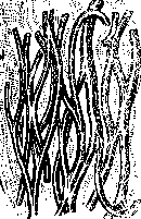
Figure 1. Elastic fibers of the yellow ligament of man with a small amount of connective tissue between them. (According to Koelliker.)
Figure of the nuchal ligament of the bull, with nuclei. Figure 2. Figure 2. Cross-section between fibers connective tissue (According to Koelliker.) Figure 3. Fenestrated elastic membrane from a. eago-tis of the horse. (According to Koelliker.)
Related articles
Mentioned in
Cite this page
“Elastic Tissue.” Soviet Medical Encyclopedia. English translation of Bolshaya Meditsinskaya Entsiklopediya, 1st ed. (Moscow, 1928–1936), ed. N. A. Semashko. https://sovietmedicalencyclopedia.pages.dev/article/elastic-tissue/