Scrotum
Historical document, translated for reference. It reflects medical knowledge of the 1920s–30s and is not medical advice.
Summary
The scrotum is a cutaneous-muscular formation that serves as a pouch for the male sex glands. It consists of skin, superficial fascia, muscle layer, and loose connective tissue, and changes size in response to temperature and nerve reflexes.
Encyclopedia article (1928–1936)
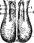
SCROTUM (from the word--moshna, Latin--scrotum), a cutaneous-muscular formation representing a pouch in which the male sex glands are located. The scrotum is an appendage to the lower abdomen, beginning its root between the penis and the perineum. The scrotum consists of two halves, separated by a partition (septum scroti); the division into two halves is also indicated on the skin by means of the raphe scroti, which on a contracted scrotum is clearly visible as a low skin ridge (Fig. 1). The scrotum consists of a) skin, which is usually more pigmented than the skin of the abdomen or thighs, differs in abundance of sweat and sebaceous glands, and is covered with hair; b) superficial subcutaneous fascia, in which there is a large amount of elastic tissue and smooth muscle fibers; this membrane is capable of contracting and relaxing the skin; it is fused to the skin by numerous bridges, and its contraction is manifested by wrinkling of the skin and the formation of transverse folds on it; the partition of the scrotum consists of the same dartos membrane; c) behind the muscular membrane there is a layer of loose connective tissue, in which the testis with its membranes and the spermatic cord lie. Since this connective tissue is a direct continuation of the superficial and Cooper's fasciae (f. superficial et f. Cooperi) of the anterior abdominal wall and the superficial fascia of the penis, the mutual connection of the processes occurring in the connective tissue of these areas and the possibility of their transition from one area to another is understandable.
Between the loose connective tissue of the scrotum and the membranes of the testis there is a connection in the form of separate delicate connective tissue bundles, through which vessels pass (anastomoses between the vessels of the scrotum and the testis). The vessels of the scrotum are supplied from the external pudendal artery (a. pudenda externa) and small anastomoses from a. spermatica interna and a. pudenda int. The veins accompany the corresponding arteries and communicate with the superficial veins of the internal thigh (v. pud. ext.), the spermatic cord (plex. pampiniformis), the anal region (v. haemorrh. inf.), and the subcutaneous veins of the abdomen. The nerves supplying the scrotum originate from two plexuses: lumbar (through n. genitocruralis) and sacral (through n. pudendus externus). The lymphatic vessels, of which the scrotum is very rich, flow into the inguinal lymphatic glands (see Femoral triangle).
The development of the scrotum occurs as part of the development of the external genital organs. The external genital parts are laid in the embryo on the eighth week, equally in both sexes, in the form of three formations: the genital tubercle, the genital folds of the skin, and the genital swelling. In male fetuses, the genital tubercle turns into the penis, the genital folds into the urethra with the cavernous body of the urethra, and the genital swelling from both sides surrounds the base of the penis, fuses along the midline and forms the scrotum in men, and in women does not fuse and forms the large labia (see the illustration to the article Hermaphroditism). Being a muscular organ, the scrotum changes its size, contracting from cold and relaxing from heat; similar changes also occur under the influence of nervous and vasomotor reflexes. In large inguinal-scrotal hernias, hydrocele, tumors of the testes, the scrotum can reach gigantic dimensions, and the penis is drawn into its thickness. The opening of the urethra is hidden in the depth of the scrotum, and on the skin of the scrotum, due to irritation by urine, eczema can develop. Congenital anomalies and malformations of the scrotum are usually not isolated, but are observed simultaneously with anomalies of other genital organs. The absence of the scrotum or half of it, more correctly-underdevelopment, is noted in cryptorchidism or monorchidism. With anomalies of the penis and urethra-hypospadias and epispadias-underdevelopment of the scrotum (partial or non-fusion-see Hermaphroditism, Epispadias and Hypospadias) and other malformations are also observed: the formation of membranes extending to the penis, etc. With significant hypospadias and epispadias, a division of the scrotum occurs (non-fusion of the genital swelling); if there is also cryptorchidism, a similarity to female genital organs is created as a result (external false hermaphroditism).
Injuries are divided into closed and open. Closed injuries (contusions) are rarely observed due to the mobility of the scrotum, which gives it the ability to escape from blows. The significance of contusions is mainly reduced to contusion of the testes, which can lead to their atrophy. In addition, contusions cause hemorrhages, sometimes very extensive due to the abundant blood supply to the scrotum. Hemorrhages can accumulate in different layers: in the skin and subcutaneous tissue in the form of flat hemorrhages; in the connective tissue surrounding the testis and spermatic cord-extravaginal hematoma; in the vaginal membrane itself-intravaginal hematoma.
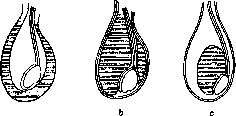
Figure 2. a-hemorrhages in the coverings; b-extravaginal hematoma; c-intravaginal hematoma.
hematoma (figure 2). The bleeding usually resolves; occasionally a thickening remains; even more rarely it suppurates. Very large hematomas cause impaired nutrition of the skin, on which blisters form under the epidermis; the process can lead to necrosis of the skin. Frequent sources of bleeding into the scrotum, besides bruises, are operations on the scrotal organs and herniotomy, when a hematoma forms in the scrotum or descends into it along the spermatic cord. Open wounds of the scrotum are the result of accidental injuries and accidents. Wounds to the scrotum, due to its good blood supply and the elasticity of the skin, usually heal well; even significant skin defects close quickly. If the scrotum or most of it is torn off, the testicles may be exposed or protrude through the defect. However, even in these cases, the skin of the scrotum contracts, and its integrity is sometimes restored without plastic surgery. Gunshot wounds to the scrotum are not uncommon and are most often accompanied by injury to the testicles, which is their significance. A unique injury to the scrotum is its removal during castration by sectarians. Inflammations of the scrotum. Acute inflammation of the scrotal skin in the form of so-called intertrigo and eczema is often observed, which is explained by the position of the scrotum, friction against the thighs and clothing, sweating, etc. Erysipelas has its own features on the skin of the scrotum. Due to the looseness of the skin and subcutaneous tissue, the skin becomes very edematous during serous inflammation. This is manifested, first, in the fact that the redness of the skin due to its edema is not pronounced and a pale color of the skin is obtained; secondly, this edema leads to the formation of blisters, pustules, etc. Phlegmons of the scrotum, i.e., purulent lesions of the deeper tissue, develop on the basis of infection penetration in injuries; other sources are infections during hernia operations and especially urinary infection (urinary infiltration, urinary accumulation) in injuries and diseases of the urethra. The anatomical structure of the scrotum creates favorable conditions for the spread of the phlegmonous process through the loose tissue, which often leads to serious consequences. Necrosis of the intestine in strangulation of an inguinal-scrotal hernia leads to a severe septic form of fecal phlegmon. The edema that forms in the loose tissue leads to thrombosis and compression of the vessels and, as a result, ischemia of the tissues; therefore, it is clear that phlegmon of the scrotum is often accompanied by gangrene of the skin and deeper tissues. In urinary accumulation, a severe septic condition develops rapidly. Another cause of gangrenous inflammation can be anaerobic infection, which easily develops in the area of the scrotum due to the proximity of the anus, from where the intestinal flora contaminates the entire area. If phlegmon of the scrotum turns into gangrene, it causes severe general symptoms and even general sepsis. Fatal outcomes are observed mainly in small children and the elderly; phlegmons can also take such a severe course at other ages. Treatment of phlegmons of the scrotum is surgical - wide incision. Phlegmons on the basis of urinary accumulation usually lead to long-unhealing fistulas of the urethra. Gangrene of the skin leads to exposure of the testicles, which subsequently requires plastic surgery, although the defect often closes on its own. Many authors isolate gangrene of the scrotum as a separate nosological unit; however, it is more correct to view it only as a complicating process. In general, the following types of gangrene of the scrotum can be distinguished: a) gangrene as a result of trauma; it affects relatively limited areas of the skin and does not tend to spread; b) inflammatory gangrene as a result of erysipelas, phlegmon of the scrotum; c) gangrene arising on the basis of general infectious diseases and general diseases (smallpox, typhoid, diabetes, etc.); d) independently occurring gangrenes of the type of putrefactive inflammations. They occur in young people, the elderly, newborns, and very exhausted patients. Tuberculosis of the scrotum is most often secondary to tuberculosis of the epididymis and manifests as the formation of infiltrates, fistulas, and ulcers on the scrotum. After removal of the affected tuberculous epididymis, tuberculous skin usually also disappears. Along with this, more common form of secondary tuberculosis of the scrotum, a primary form also rarely manifests. It exists in the form of so-called hypertrophic lupus. The skin of the scrotum thickens sharply, resembling elephantiasis. The diagnosis is not always easy to establish, and sometimes it is necessary to resort to an excision for histological examination. Treatment - see Tuberculous skin diseases. Syphilis does not play a special role in the pathology of the scrotum except for its skin lesions with primary chancre, secondary papular rashes, etc. Similarly, actinomycosis is rarely observed. Relatively often, exotic diseases such as yaws and others manifest on the skin of the scrotum.
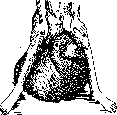
Figure 3. Scrotal elephantiasis.
vascular changes, mainly depending on the dilation of the veins of the spermatic cord. In this disease, the scrotum also changes, its skin stretches, it descends (on the left - in connection with left-sided varicocele), etc. Therefore, in the treatment of varicocele, it is sometimes necessary to also perform resection of the affected part of the scrotal skin. Diseases of the lymphatic vessels are much more important, chronic inflammation of which leads to elephantiasis (see Elephantiasis). The scrotum in tropical elephantiasis, depending on the introduction of filaria sanguinis, sometimes takes on extremely large sizes (figure 3). Elephantiasis on the basis of inflammation of the lymphatic vessels or removal of the lymphatic glands usually does not
be significant. - The
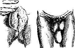
Figure 4. Dermoids along the raphe of the scrotum.
Figure 5. Lipoma of the scrotum (on the dorsum). The scrotum participates greatly in all general processes that cause tissue edema. Diseases of the heart, kidneys are often accompanied by edema of the scrotum. The latter can sometimes reach such a size that the skin becomes almost translucent. Such skin is easily susceptible to infection (erysipelas) and prone to gangrene (superficial). To reduce tension, incisions can be made in the skin or thin tubes can be inserted into the edematous tissue, through which fluid drains. Tumors of the scrotum. As benign tumors, atheromas and der

moids are observed. Atheromas are not uncommon and are located over the entire surface of the scrotum; dermoids are less common and are located strictly along the median line, along the raphe, since their very origin is associated with primordia of the epidermis that got lost during the fusion of the halves of the scrotum. Sometimes small in size dermoids are located one after another in the form of a rosary (figure 4). Benign tumors of the deeper tissues of the scrotum are rare. Cases of lipomas (figure 5), lymphangiomas and hemangiomas have been described. Fibromas and fibromyomas are somewhat more common in the scrotum, sometimes reaching very large sizes; they are dense lobular formations that completely envelop the testicle. However, there is always doubt whether the tumors originate from structures of the scrotum or from elements of the testicle (tunica vaginalis).- Atheromas can occasionally lie freely in the scrotum, being outside the testicle. Very few such cases have been described in the literature.- Of malignant tumors, only cancer (sarco- Ris- 6- part of the scrotum) is found by preference, very rarely) according to (from "chimney sweeps' warts", which has the character of either an ulcer on a dense base or a deeply penetrating node in the tissues, usually having a great tendency to disintegrate. Cancer of chimney sweeps often begins in the form of weeping eczema, with the formation of crusts. The skin thickens, warty formations appear on it; over time, these warts get a dense base, on the surface they are excoriated and sometimes lead to the formation of cancerous tumors. In view of improved working conditions in the relevant industries in recent times, these types of cancer are rare. Sometimes cancer of the scrotum is secondary, having spread from the penis. In cancer of the scrotum, the inguinal lymph glands are primarily affected. Treatment of tumors of the scrotum is surgical.
Related articles
Mentioned in
Cite this page
“Scrotum.” Soviet Medical Encyclopedia. English translation of Bolshaya Meditsinskaya Entsiklopediya, 1st ed. (Moscow, 1928–1936), ed. N. A. Semashko. https://sovietmedicalencyclopedia.pages.dev/article/scrotum/