Hypospadias
Historical document, translated for reference. It reflects medical knowledge of the 1920s–30s and is not medical advice.
Summary
Hypospadias is a congenital malformation of the urethra where the urethral opening is not located at its normal position. This article describes the embryological origins, clinical manifestations, and surgical treatments for hypospadias as understood in the 1930s.
Encyclopedia article (1928–1936)
HYPOSPADIAS, hypospadia (syn. fissura urethrae inferior congenita), a congenital malformation of the urethra in which the lower wall of the urethra is not fully formed and the external opening is not located in its usual place. Hypospadias is one of the most frequent congenital malformations. According to Bouisson, it occurs in males at a ratio of 1:300, and according to Mauo-1:350. This malformation is most commonly observed in males, as during their embryonic development it affects the tissues from which the anterior part of the urethra is formed. In females, the urethra is embryologically and anatomically equivalent to the posterior part of the male urethra, and therefore, if descriptions of this anomaly are found in females, they should rather be considered not as pure hypospadias, but as underdevelopment or an acquired defect of the urethro-vaginal septum (see Figure 1). Topographically, hypospadias can be located along the entire anterior urethra, and depending on its location, it takes on a particular appearance and topographical designation. The most common site for this defect is the glans penis, and then the external opening of the urethra opens not in the usual place, but somewhat lower and posteriorly, with the entire lower part of the glans being split into two halves. If in this case hypospadias is combined with a well-developed Cowper's valve, then two openings are observed on the glans penis: an upper one located in the usual place but leading into a blind pouch, and a lower one-the entrance to the urinary canal. The latter can be of various sizes and shapes-round, slit-shaped, crescent-shaped, etc. This location of the defect in the area of the glans is called hypospadia glandis penis. Significantly less frequently
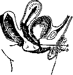
Figure 1. Hypospadias in females: a-external opening of the urethra in the vagina; b-normal location.
the external opening of the urethra opens on the remaining course of the urethra, starting from the glans to its perineal part. Accordingly, the topographical designation of this malady will bear one or another name-hypospadia penis, scrotalis, perinealis, etc. Along with this, to the described underdevelopment of the urethra, there are usually associated anatomical defects. Due to the underdevelopment or even complete absence of the urethra, as well as its cavernous bodies in front of the hypospadias, the corresponding cavernous bodies of the penis also develop insufficiently, and it acquires a downward arcuate curvature. The location of hypospadias in the scrotal or perineal part of the urethra sometimes leads to splitting of the scrotum into two halves, which gives the external genital organs a resemblance to female external genitalia (see Hermaphroditism). Hypospadias is often combined with spina bifida, cryptorchidism, etc. To clarify the origin of this malformation from an embryological point of view, it is necessary to take into account that in embryonic life, the anterior urethra is formed from a groove laid along the entire embryonic genital tubercle, along its lower surface. The edges of this groove grow somewhat stronger, deepen it, and finally fuse together, forming a closed tube. The posterior end of this tube splits and connects with the internal groove, which subsequently forms the posterior urethra. The anterior end of the tube comes into contact with the canal of the glans penis, developing completely independently in the form of a groove running toward the previously formed middle segment of the future urethra. And if at this moment the meeting of individual segments does not occur or the middle segment is not completely closed, then the mechanism of development of hypospadias becomes quite understandable- Hypospadias of the glans usually does not manifest clinically and remains unnoticed until a gonorrheal inflammatory process arises here. The latter is favored by the fact that the cleft of the glans is covered with delicate mucous membrane with clearly expressed Morgagni sinuses, into which


Figure 2.
Figure 3.
the gonococcus readily penetrates. Hypospadias of the glans can lead to disorders of urination in the form of an irregular, spraying stream and difficulty in urination due to the narrowness and low elasticity of the external opening of the urethra. All these defects in the act of urination are all the more pronounced the
closer to the bulbous part of the urethra the hypospadias is located. The urinary stream in these cases is often directed downward, wetting the skin of the scrotum and thighs, causing eczematous processes here, and at the same time creating the possibility of infection of the urinary tract. The function of the sexual apparatus is no less affected. With pronounced hypospadias, the cavernous body of the urethra is usually underdeveloped. Due to this, the cavernous tissue of the glans penis does not receive sufficient support and during erection bends downward, as a result of which during sexual intercourse the semen is not poured onto the vaginal portion of the uterus, but onto the posterior wall of the vagina. With the location of hypospadias even more posteriorly, the semen cannot enter the vagina at all and is poured onto the perineum. The treatment of hypospadias aims to restore the external opening of the urethra to its usual place and to reproduce the missing part of the urethra. As in


Figure 5.
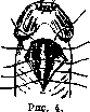
Figure 6. FIG. 7. with any plastic operations on the urethra, the first step here must be to divert urine by creating a suprapubic or perineal fistula (see Suprapubic). The second indispensable condition for the immediate postoperative and long-term cosmetic success is sufficient mobilization of the transplanted urethra; this mobility must be taken into account in all methods, keeping in mind that inevitable postoperative erections make primary suture healing impossible due to tension, and later the shortness of the urethra will give the erect penis an incorrect shape. The most common and acceptable operation to many authors for restoring the external opening of the urethra in hypospadia of the glans is the Hacker operation. An elastic bougie or catheter is inserted into the urethra, and the urethra is freed, guided by it. At the moment of freeing the urethra, its cavernous bodies must be carefully spared. The second stage of the operation is the puncture of the glans penis with the formation of a passage in it, the passage of the urethra through the formed tunnel, and its fixation at the apex of the glans (see Figures 2, 3 and 4). In cases where the lower surface of the glans is sharply split and has a deep groove, the Beck operation can be undertaken. It consists in that two flaps are previously dissected in the groove of the glans; the dissected above
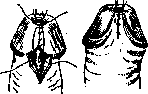
Figure 8.
Figure 9. mentioned method, the urethra is laid in the resulting bed, fixed with several sutures to the apex of the glans, and covered with the formed flaps of the glans (see Figures 5-9).-With hypospadias along the course from the glans to the perineal part, one deals not only with insufficient length of the urethra, but also with curvature of the penis, drawn downward. Therefore, the task of surgical intervention first of all includes correction of the curved penis. Most authors recommend performing the operation of straightening the penis at an early age (7-10 years). For this purpose, with the penis stretched upward, a transverse incision is made in the bands pulling the penis downward, the incision going to the tunica albuginea of the cavernous bodies of the penis; the freed urethra at this time recedes somewhat posteriorly. The suture is applied in a longitudinal direction relative to the axis of the penis. Further operations to restore the missing tube of the urethra are performed already in adulthood, when the sexual organs are fully developed. All the operations proposed for this purpose can be subdivided into
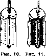

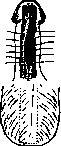

Figure 12.
Figure 13. Figure 14. Two groups: operations with plastic reconstruction of the urethra from nearby tissues and operations with free plastic surgery. The first group includes: 1) The Duplay (Birch) method. First, a canal is created in the glans, then a catheter is inserted through the formed canal and the hypospadiac opening, and parallel incisions are made in the skin of the penis at a distance of half a centimeter from the catheter, forming flaps both toward the catheter and toward the lateral surfaces of the penis. The first flaps are sutured together over the catheter with their skin surfaces inward, forming a tube for the urethra, while the second flaps cover the newly formed urethra (see figures 10 and 11). 2) The Beck method. The first step of forming the tube for the canal is the same as in Duplay's method; to cover the newly formed urethra, a flap is cut from the skin of the scrotum, which remains on a pedicle (see figures 12, 13 and 14). 3) The Rochet method. Here the urethral tube is created from the skin of the scrotum, and then a tunnel is formed under the skin of the penis using a trocar, through which the newly formed urethra is pulled and fixed in some way (see figures 15, 16 and 17). Of course, with all these methods, careful observation must be maintained to preserve the connection of the flaps with the skin to ensure adequate blood supply. To the operations with free plastic surgery can be included the method of No-ve-Josserand. In this method, a hair-free skin flap from the inner surface of the thigh, forearm, etc., 4-5 cm wide and slightly longer than the missing segment of the urethra (allowing for flap contraction) is taken to create the urethral tube. This flap is wrapped around an elastic bougie in a spiral or in the form of a tube, and the edges are sutured with thin catgut. Then two transverse skin incisions are made: directly in front of the canal opening and behind the glans, which are connected to each other by a tunnel. The tunnel is made using a thick trocar passed under the skin from one incision to the other. The prepared urethral tube is inserted through this tunnel and left under the skin along with the bougie (see figures 18 and 19). The bougie is removed on the 8th day after the operation. After this, the central end of the segment is sutured to the urethra and the glanular canal is formed. If conditions permit, i.e., there is no bifurcation of the glans, then the glanular tunnel can be performed simultaneously with the tunnel for the urethra. For free transplantation of the urethra, some authors suggest using ready-made tubes: vena saphena magna, vermiform appendix, segment of the ureter (Schmieden), etc. Some authors recommend applying the Thiersch method for H., similar to how it is applied for the upper cleft of the urethra (see Epispadias). Operations according to the method of free transplantation of the urethra, as well as operations for the formation of the urethra from skin flaps in place, are often accompanied by failures: suppuration, failure of the stitches to heal, narrowing of the newly formed urethra, necrosis, etc. Therefore, treatment always requires patience of the patient and persistence of the surgeon.

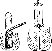
Figure 15.
Figure 16. Figure 17. but or in the form of a tube, the edges are sutured with thin catgut. Then two transverse skin incisions are made: directly in front of the canal opening and behind the glans, which are connected to each other by a tunnel. The tunnel is made using a thick trocar passed under the skin from one incision to the other. Through this tunnel, the prepared urethral tube is inserted and left under the skin along with the bougie (see figures 18 and 19). The bougie is removed on the 8th day after the operation. After this, the central end of the segment is sutured to the urethra and the glanular canal is formed. If conditions permit, i.e., there is no bifurcation of the glans, then the glanular tunnel can be performed simultaneously with the tunnel for the urethra. For free transplantation of the urethra, some authors suggest using ready-made tubes:


Figure 18.
Figure 19. vena saphena magna, vermiform appendix, segment of the ureter (Schmieden), etc. Some authors recommend applying the Thiersch method for H., similar to how it is applied for the upper cleft of the urethra (see Epispadias). Operations according to the method of free transplantation of the urethra, as well as operations for the formation of the urethra from skin flaps in place, are often accompanied by failures: suppuration, failure of the stitches to heal, narrowing of the newly formed urethra, necrosis, etc. Therefore, treatment always requires patience of the patient and persistence of the surgeon.
Related articles
Mentioned in
Cite this page
“Hypospadias.” Soviet Medical Encyclopedia. English translation of Bolshaya Meditsinskaya Entsiklopediya, 1st ed. (Moscow, 1928–1936), ed. N. A. Semashko. https://sovietmedicalencyclopedia.pages.dev/article/hypospadias/