Spina Bifida
Historical document, translated for reference. It reflects medical knowledge of the 1920s–30s and is not medical advice.
Summary
Spina bifida refers to congenital anomalies characterized by incomplete closure of the vertebral canal and defective development of the spinal cord or its membranes. The article provides a detailed classification of various forms of spina bifida and discusses their clinical significance.
Encyclopedia article (1928–1936)
SPINA BIFIDA (literally split spine), a collective name for congenital anomalies characterized by incomplete closure of the vertebral canal and defective development of the spinal cord or its membranes. The name was given by Tulpius in 1641. Path.-anat. Definition is
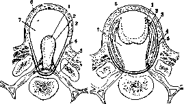
Figure 1. Complete rhachischisis: 1- medullary plate; 2-pia mater; 3-arachnoidea; 4-dura mater; 5-nerve roots. (Recklhausen, 1886). The classification currently prevailing and given below was developed by Hildebrand. Based on the most severe degrees of delay in the evolutionary process, the following types of S. b. are distinguished: I. Rhachischisis, i.e., defect of the posterior wall of the vertebral canal and absence
Figure 2. Partial rhachischisis: 1-medullary plate; 2-pia mater; 3-arachnoidea; 4-dura mater; 5-nerve roots. of closure of the neural groove into a tube. The spinal cord is spread out on the surface, uncovered behind by neither skin nor membranes (fig. 1). The cleft may extend over the entire vertebral column (rhachischisis totalis, s. holorhachischisis) and be accompanied by acrania or cranioschisis (see Cranioschisis), amyelia (see) or secondary death of brain tissue. In other cases, only a limited segment of the spine is affected, and the spinal cord, less spread out, retains the shape of a groove [rhachischisis partialis (fig. 2), s. merorhachischisis].
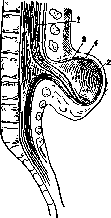
II. Cystic forms of S. b.: 1. Myelocele represents a partial rhachischisis with a watery accumulation of cerebrospinal fluid ventrally under the unclosed medullary plate, protruding posteriorly (fig. 3 and 4). The fluid accumulates between the pia mater and arachnoid membrane, nerve roots freely pass through the cyst cavity. 2. Myelomeningocele differs from the previous form only in that the hydrops developed between the layers of the arachnoid membrane. As a result, not only the medullary plate with pia mater protrudes posteriorly, but also the adjacent arachnoid leaf (fig. 5). The nerve roots are located in the thickness of the wall. A strict boundary between myelo- and myelomeningocele cannot be drawn, since in the formation of the wall of the first, the pia mater participates significantly, and it is often impossible to distinguish it from the arachnoid due to secondary degenerative and inflammatory changes. The so-called terminal myelomeningocele in the sacral region deserves special attention (fig. 6). In this form, the central spinal canal opens only at its distal end onto the surface of the tumor-like S. b. Mention can be made of ^- > Figure 3. Myelocele: 1-medullary plate; 2-pia mater; 3-arachnoidea; 4-dura mater; 5-nerve roots.
Fig. 4 Myelocele 1-medullary plate; 2-pia mater (two layers); 3-arachnoidea; 4-dura mater; 5-nerve roots. another variety of myelomeningocele depicted by de Quervain. Rhachischisis here is incomplete, i.e., the central canal is closed, but the spinal cord protrudes in a loop through a cleft in the spine (fig. 7 and 8). The protrusion is caused by a watery accumulation on the anterior (ventral) side. The protruding portion of the spinal cord is covered only on the sides by the pia mater. 3. Myelocystocele (syn. hydromyelocele, hydromyelos, syringomyelocole) is characterized by accumulation of watery fluid inside the closed central canal of the spinal cord (fig. 9). The skin over the tumor is more or less preserved, the dura mater is missing, and in the vertebral arch part a cleft gapes. Thus, the posterior wall of the cyst is formed by skin, arachnoid membrane, pia mater and the stretched, thinned dorsal half of the spinal cord. Nerve roots never lie inside the cavity, but pass in the wall of the cyst. 4. Myelomeningocystocele is a variety in which the watery accumulation formed not only inside the central canal but also in the subarachnoid space (fig. 10 and 11). Under the pressure of the fluid, the wall of the brain cyst may rupture (fig. 12)-a circumstance accelerating the death of nerve elements. If the rupture occurred on the dorsal side of the myelocyst or the latter has very insignificant dimensions and is hidden in the depth of a large meningocele, then the wall protruding - ~ 1 ,
above the skin level contains only / y
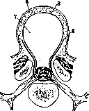
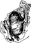
\ 4 which cases are usually taken for meningocele. 5. Meningocele in the pure sense is a hernia of the meninges, filled with cerebrospinal fluid and protruding through a cleft in the vertebral canal, most often in the sacral region. The wall is formed by skin and arachnoid membrane; dura mater is preserved only at the base of the sac, absent on the dome (fig. 13). According to Hildebrand and Marchand, hydrops may develop in the subarachnoid or subdural space, the wall of the tumor may be formed by skin, dura mater and arachnoidea or only by skin and dura mater. The spinal cord is sometimes secondarily drawn into the cyst and attached to its inner wall (fig. 14). III. Spina bifida occulta (hidden vertebral cleft) formally means only a defect in the closure of the vertebral arches without noticeable hernial protrusion of a tumor through the cleft, determinable by palpation in rare cases, usually only radiologically. However, since we are dealing with a clinical concept, its content is broader. It includes, in addition to the bone anomaly, changes in nervous tissue. IV. Spina bifida anterior differs from all the above forms in that it is not the posterior, arch parts that are split, but the bodies of the vertebrae (fig. 15). This anomaly was already known to Tulpius. Compared to the posterior cleft, it represents a much greater rarity. The clinical significance of one or another variety is greater the more frequently this form occurs and the less the anomaly itself as a developmental defect threatens viability. Newborns with rhachischisis, with myelocele and myelomeningocele are doomed to early death, as infection is inevitable
Figure 6. Terminal myelomeningocele: 1 - central canal of the spinal cord; 2-pia mater; 3-arachnoidea; 4-dura mater; 5-nerve roots; 6'- area medullo-vasculosa; 7- area epithelio-serosa.
and fatal. If the central canal of the spinal cord is covered by skin over myelo-cystocele in good condition, then by operative means, in the best case, it is possible to prevent secondary infectious and traumatic complications, but paralysis of the sphincters and extremities remains unchanged; children lead a miserable existence for a short time. Moreover, the cystic forms of S. b. are often combined with other developmental defects of the spine, with hydrocephalus (most often with myelocystocele), with defects of the abdominal wall, ectopias of the bladder, dislocation of the hips and clubfoot. Relatively favorable, with timely operation, is the prognosis for meningocele of the variety that occurs much less frequently than others. In frequency of observations, in scientific and practical interest, the first place is occupied by S. b. occulta. Finally S. b. anterior, if one does not know about the possibility of development of such cystic tumors in the pelvis, may give rise to fatal errors in gynecology.
The causal factors on which the occurrence of various forms of S. b. depends are unknown. The essence of the pathogenesis of S. b. must be sought in the primary defectiveness of the laying or development of the ectoblast. What this defectiveness is due to, i.e., the question of the etiology of S. b., remains unclear. Experimentally, a number of authors and under the influence of qualitatively very different factors'-thermal, mechanical and chemical-have succeeded in obtaining i>, РТРГч5 Figure 7. Myelomeningocele: 1 - central canal of the spinal cord; 2-pia mater; 3-arachnoidea; 4-dura mater; 5-nerve roots; 6-area medullo-vasculosa; 7-area epithelio-serosa. (According to de Quervain.)
Figure 8. Myelomeningocele: 1-central canal of the spinal cord; 2-pia mater; 3-arachnoidea; 4-dura mater; 5-nerve roots. (According to de Quervain.)
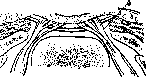
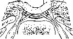
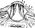
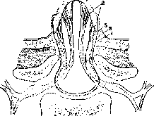
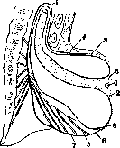
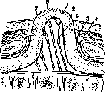
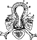
Figure 9. Myelocystocele: 1 - wall of the myelocyst; 2 - pia mater; 3 - arachnoidea; 4 - dura mater; 5 - nerve roots; 6 - skin. S. b. in animal embryos. The conclusion from this can only be one: not the stimulus, but the reaction is specific. Pathoanatomical changes are schematically outlined above in the classification of various forms of S. b. Features characteristic of each group are sometimes revealed immediately upon external examination, sometimes only during the operation and after careful examination of the specimen under the microscope. But it often happens that degenerative and inflammatory changes blur or distort the original picture. - In rachischisis, the vertebral canal is transformed into a shallow groove, to the bodies of the vertebrae rudimentary arches are attached or they are completely absent. The groove is filled with a red-brown, moist, velvety ribbon, along which sometimes a median strand with a richly developed network of blood vessels stretches. The strand represents a rudiment of the spinal cord and can be double (see Diastatomyelia). If rachischisis occupies only a certain part of the spine, then at the upper and lower ends, respectively, at the transition from the flattened to the tubular spinal cord, pits with openings, cranial and caudal, of the central canal can be found.
Myelocele differs from partial rachischisis only in that the medullary plate is protruded by fluid accumulated subarachnoidally on the ventral side, where the membranes have been preserved and are adjacent to the bodies of the vertebrae. The dorsal, surface facing the viewer of myelocele and myelomeningocele represents a tumor with, in fresh cases, three characteristic zones, named by Recklinghausen zona medullo-vasculosa, zona epithelio-serosa and zona dermatica (fig. 16). The central zone of oval or heart-shaped form has a red-brown color and corresponds to the medullary plate, whose thickness does not exceed a few millimeters, but in width and length it reaches...
Figure 10. Myelomeningocystocele: 1 - wall of the cyst of the central canal; 2 - pia mater; 3 - arachnoidea; 4 - dura mater; 5 - nerve roots; 6 - skin; 7 - subarachnoid cyst. inflammatory changes, hemorrhages, ulcerations with fibrinous deposits and purulent decay, the wall is translucent and through it the nerve roots vibrating in the cavity of the meningocele are visible. At the surface of the upper and lower poles, pinpoint openings of the central spinal canal are noticeable. The medullary zone is surrounded by a meningeal, smoother, pearl-grayish zone, formed from pia mater and epithelium growing from the edges. Maceration and hemorrhages can cause secondary changes even during intrauterine life. The sac is not covered with skin, only at its base it is bordered by the skin zona dermatica. The size of the tumor varies from a five-kopeck coin to the width of a palm, the shape is round, the base is wide.
Myelocystocele and myelomeningocystocele in size and shape, in frequent localization in the sacral-lumbar region and due to the translucent wall resemble the previous group, but differ from it essentially in that the central canal of the spinal cord is closed and the surface of the cystic tumor is always covered with skin. Under the influence of infectious and dystrophic processes, the integrity of the skin can be violated. Then the deeper layers of the wall, arachnoidea, pia and the stretched neural tube are exposed, undergoing decay. The slit in the vertebral canal can be median or lateral, wide or narrow. Cystic spina bifida, which are least common and reach the largest sizes, occur when the sac consists only of the meninges, i.e. meningocele in its pure form. Their localization is almost exclusively the sacral region. The bone slit and accordingly the pedicle of the tumor are narrow.
Figure 11. Myelomeningocystocele: 1 - wall of the cyst of the central canal; 2 - pia mater; 3 - arachnoidea; 4 - dura mater; 5 - nerve roots; 6 - skin; 7 - subarachnoid cyst.
Figure 12. Myelomeningocystocele: 1 - wall of the cyst of the central canal; 2 - pia mater; 3 - arachnoidea; 4 - dura mater; 5 - nerve roots; 6 - skin; 7 - subarachnoid cyst.
The primary involvement of the dura mater in the formation of the sac remains controversial; secondarily the cauda equina and conus medullaris can be stretched through the slit and become attached to the wall. - Spina bifida occulta should be considered pathoanatomically, as was already noted in the classification, not only as a bone, but mainly as a developmental anomaly of the spinal cord. Recklinghausen defined it as a ruptured and healed intrauterinely cystic S. b. Denuce holds the same opinion, but emphasizes that we can only be talking about myelocystocele or myelocele. In any case, various gradations of the teratological process, different times of onset of reparative changes and the corresponding variety of outcomes are possible. Natural correction of the developmental defect can create a new, secondary pathological process, which as a third element is added to the anomalies of the spinal cord and vertebral canal. Teratogenesis, as we have seen, assigns the first place in seniority to the brain defect. The bone anomaly is subordinate to it, developed later or independently of it. This explains those cases of S. b. occulta in which the severity and localization of brain disorders do not correspond to the size and location of the bone defect or the latter is not manifested in any way. However, it would be wrong to completely deny the possibility of the reverse influence, i.e. the dependence of brain disorders on the bone anomaly.
The growth of the spinal column and the spinal cord occurs disproportionately. After the 4th month of intrauterine life, the growth of the spinal cord lags behind the growth of the spine, the former shortens in comparison with the latter, gradually stretches upward from the sacral canal and by the time of birth is already at the level of the L1 vertebra. Consequently, if the spinal cord due to a developmental defect remains fixed at the bottom, it is stretched faster than the growing spine. A simple mechanical explanation, however, is also far from sufficient here.
Clinically, S. b. occulta often manifests itself only in the adolescent period, when the difference in growth between the spinal cord and the surrounding bone canal does not exceed the height of one vertebra. In addition, cases have been described (Wagner-Ribbert) in which with a typical patho-anat. picture of S. b. occulta conus medullaris was at a normal level in the spinal canal. Furthermore, it sometimes affects one-sidedly incompatible with the anomaly only of growth in length. Finally, mechanically it is impossible to explain the occurrence and action of those peculiar changes that develop in S. b. occulta on the coverings and in the soft tissues. These changes distinguish S. b. occulta essentially from S. b. aperta or cystica. They can be considered as a new moment included in the pathogenesis, if S. b. occulta developed from a spinal cord cyst that ruptured in the embryonic period. Then they are a reaction of the embryonic tissues to this rupture. The skin changes consist of navel-like or flat scars, surrounded by an area of hypertrichosis (fig. 17). In other cases, there is no local excessive growth of hair, but the skin is hypertrophied and densified, protrudes as an appendage resembling a...
Figure 13. Meningocele. Designations - see figure 10.
ducken. Angiectasias are often found. Examining the skin, one often finds a strand going from the surface to the depth, and penetrating through the bone fissure into the spinal canal to the meninges. The greatest interest is presented by changes in the nervous tissue. Unfortunately, to this day only a few cases have been published where the clinical diagnosis of S. b. occulta was supplemented by patho-anat. analysis. Not every slit in the arches represents an anomaly, a delay of the normal developmental process of the nature of aplasia. The sacral-lumbar region of man abounds in variants, as it is still in a period of rapid phylogenetic restructuring associated with the transition from horizontal to orthograde position. A. D. Spersky established that one of the manifestations of this process...
27o
SPINA BIFIDA
ft
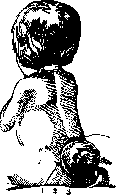
one must consider posterior clefts of the sacrum, starting from those with notches on the spinous processes and ending with a wide gaping of the sacral canal. In contrast to this regular and progressive reduction process, the bone defect in S. b. occulta is of a random, pathological character. The location of the cleft, if it occupies the thoracic or cervical region, is abnormal. The form is very diverse. The cleft is usually narrow, has an oblique direction; the spinous process is underdeveloped, absent, or displaced. In many cases in S. b. occulta, in contrast to S. b. aperta or cystica, there can be no question of aplasia of bone tissue. Both halves of the arches are not shortened, but elongated; they grew toward each other and did not fuse. It was also necessary to observe patients where, with a typical clinical picture of S. b. occulta, the arch had a normal appearance, and the ossification of the posterior wall of the sacral canal occurred excessively and asymmetrically. Denys called forms in which, instead of a fibrous plate, there is a bony closure of the former cleft, S. b. occlusa.
Symptomatology. Functional disorders in S. b. primarily depend on its localization and on the size of the defect in nervous tissue. Newborns with rachischisis and an unfused medullary tube are non-viable not due to the extent of motor paralysis, but due to the inevitability of secondary infectious complications. Deformities of the limbs, most often clubfoot, develop in utero. In other cases, on the contrary, the normal configuration of the skeleton and musculature of the limbs is affected, indicating that their development in the fetus can proceed correctly without motor impulses, due to the preservation of the spinal ganglia, which separated from the spinal cord before its formation into a tube. The deceptive appearance of such newborns can influence the decision regarding surgical help. In Myelocystocele, the greatest stretching is experienced by the dorsal wall of the brain cyst and, due to its lateral location, one half is larger than the other. Correspondingly, trophic and sensory disorders predominate, hemiplegia instead of paraplegia. Curvatures of the legs are the same as in S. b. occulta, i.e., claw-like contractures of the fingers, combinations of transverse flatfoot with pes cavus.
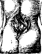
Figure 17. Spina bifida occulta with hypertrichosis. Varus and planus, ulcerative processes on the skin and bones, mixed with spastic pareses. Motor disorders may have a predominant localization in the musculature of the perineum (see Prolapse of the uterus, vagina). In pure forms of meningocele, there is usually not even that sign which is most frequently observed in S. b., namely enuresis. Basically, it must be remembered that S. b. occulta in small children does not manifest itself in any special way, that the first, unclear signs begin insidiously, by 6-10 years or even later, differing in great variety and progressive course. The diagnosis of cystic forms of S. b. presents no difficulties if on the weeping surface of the congenital tumor the 3 zones of Recklinghausen and the pinpoint openings of the central spinal canal are distinguishable. Maceration of the initially preserved skin coverings in Myelomeningocele can be misleading, all the more so since then translucence does not give positive results, and the question of the passage of nerve roots through the cavity is not resolved. In such cases, the analysis of functional disorders is more important than the data of examination. The practical significance is especially important in the diagnosis of brain hernias, in the formation of the sac of which the spinal cord does not directly participate. The absence of paralyses and other deformities, the localization in the sacral region and the significant size of the cyst speak in favor of meningocele, but the secondary involvement of the spinal cord, cauda equina or nerve roots in the sac is not excluded by this. Trial incisions or punctures are dangerous and clarify little. Recognition of S. b. occulta is impossible in small children if there are no skin signs, since the cartilaginous state of the vertebral arches on x-rays simulates clefts. The coccygeal fossa (foveola coccygea) or an unusual skin groove, longitudinal or oblique, in the sacral region is a variant only bordering on an anomaly. Typical forms of clubfoot in newborns usually have nothing in common with S. b. occulta. More convincing are the atypical curvatures of the legs in the absence of traces of amniotic bands. Only the course, namely the progressive nature of the deformation (despite correct treatment), the admixture of spastic phenomena to paretic ones and the special tendency to trophic disorders confirm the diagnosis of clubfoot on the basis of S. b. occulta. X-ray examination of patients after 10 years of age may be decisive, but the bone clefts thus found are often found without any functional disorders, local or reflected. On the other hand, the combination of a bone anomaly in the arch part of the sacral vertebrae with skin anomalies in the form of sacrococcygeal scars or capillary telangiectasias and local hypertrichosis should serve as proof of S. b. occulta. Future pathological-anatomical studies of clinical cases will show how often and in what form 'myelodysplasias' accompany the bone-skin anomaly. The prognosis of cystic forms of S. b. with the participation of the spinal cord in the formation of the wall is almost absolutely unfavorable without operation. Accompanying deformities and existing functional defects must be taken into account when assessing viability if the operative interventions aim to prevent at least infection of the brain hernia. It is impossible to influence the functional disorders, to eliminate or alleviate them by operation, since we are dealing with nervous tissue devoid of regenerative capacity. A special place in prognostic respect is occupied by meningoceles not complicated by hydromyelia of the central canal. The prognosis of these cases is even favorable without operation. As for cases of spina bifida occulta with trophic and spastic-paretic phenomena, the severing of bands and the liberation of the spinal cord from stretching or pressure may have a good result. Treatment of S. b. cystic does not achieve its purpose without careful care of the child, without prevention of secondary infectious and traumatic complications. The task of the operation is to eliminate the protrusion and to reliably close the hernial gates. The faulty development of the spinal cord remains. Only some of its consequences in the form of contractures and deformations can be more or less corrected. Cystic S. b. should be subjected to the earliest operative treatment if the child is viable at all and the condition of the coverings at the site of the tumor is satisfactory. Otherwise, one is limited to protective bandages preventing maceration of the skin and the introduction of infection, promoting epithelization. Pressure bandages, adhesive and collodion bandages, which are supposed to reduce tension and protect the cyst from rupture, do more harm than good. It is best to recommend the horizontal position on the abdomen in well-fitting plaster beds supporting the head and lower extremities. For protection during bathing, it is best to smear cystic S. b. devoid of skin with sterile petrolatum. Previously used systematic punctures of the cyst with subsequent introduction of Lugol's solution or other iodine solutions, with the aim of causing adhesive inflammation and wrinkling of the walls, are now obsolete. Similarly, ligature methods, which are even more destructive and act blindly, have been abandoned. The only rational method is the operative one, allowing reduction of the hernia without unnecessary sacrifice of preserved nerve elements and closure of the parietal defect by plastic means. The technique varies according to the type of S. b. In myelocele, a modular plate is presented, which must be disinfected, separated from the zona dermatica and reduced, then covered with normal and stable tissues, without compression of the spinal cord. There are somewhat fewer technical difficulties in myelocystocele due to the fact that the central canal is closed and the sac is not devoid of skin cover. With a flap, upwardly convex incision, the wall of the cyst is exposed, it is emptied by a median incision, i.e., where the brain tissue is most stretched and therefore functionally already unsuitable. Small cysts that are reducible without emptying are not opened. The membranes are sutured with catgut sutures. Many methods have been proposed for closing the bone cleft of the vertebral canal. In particular, Russian surgeons (Zenenko, Bobrov, Lysakov) have developed bone-plastic methods. The conditions under which one has to operate - early childhood, low tissue resistance and questionable asepsis - force one to give preference to muscle-fascial flaps from the nearest neighborhood (Bayer). Due to the narrowness of the hernial gates in meningocele, the operation in these cases can be limited to liberation of the sac, examination of its contents, isolation of the nerves from the wall and their reduction, then excision and suturing of the sac. Spina bifida anterior has no direct relation to posterior S. b. Clefts of the vertebral bodies are observed mainly in combination with rachischisis and anencephaly, with diastematomyelia and ectopias of parts of the gastro-intestinal tract. Without such deformities excluding the viability of the fetus, anterior S. b. is extremely rare. The described clinical cases can be divided into two groups. The first group consists of gynecological patients, girls and multiparous women, in whom cysts displacing the pelvic organs and fused with the sacrum were found. Punctures of these cysts through the vaginal vault or rectum led to fatal meningitis. Autopsies showed that through a defect in the sacrum a meningocele protruded into the pelvic cavity. The second group includes small girls with kyphoscoliosis of the cervico-thoracic region of the Klippel-Feil type (see Spine, developmental defects and deformities), but with clefting or even with lateral divergence of the vertebral bodies. Remarkably, in contrast to sacral forms, there is no hernial protrusion and no spinal cord phenomena at all. Sacral forms may be accompanied by clubfoot. The origin of anterior S. b. is connected by Budde with incomplete or untimely closure of the neuroenteric canal, the connection between the neural tube and the primitive intestine. In favor of this assumption are cases where the bottom of the vertebral cleft was lined with gastric mucosa or connected with loops of the intestine. The very early teratogenesis is also evidenced by the fact that in normal conditions the vertebral bodies develop from an unpaired cartilaginous primordium. Consequently, the cleft must have occurred earlier than this primordium of the vertebra differentiated from the somites of the mesoblast.
Related articles
Mentioned in
Cite this page
“Spina Bifida.” Soviet Medical Encyclopedia. English translation of Bolshaya Meditsinskaya Entsiklopediya, 1st ed. (Moscow, 1928–1936), ed. N. A. Semashko. https://sovietmedicalencyclopedia.pages.dev/article/spina-bifida/