Scoliosis
Historical document, translated for reference. It reflects medical knowledge of the 1920s–30s and is not medical advice.
Summary
Scoliosis is a lateral or frontal curvature of the spine, a common deformity particularly prevalent before modern physical education methods. This article from the 1928-1936 Soviet Medical Encyclopedia describes various types of scoliosis including congenital, rachitic, school, professional, functional, traumatic, scar, paralytic, syringomyelic, and reflexogenic scoliosis, along with their causes and anatomical changes.
Encyclopedia article (1928–1936)
SCOLIOSIS (from Greek skolios - crooked, a term introduced by Galen in the 2nd century), lateral, or frontal, curvature of the spine. A very common deformation, especially in former times, before the popularization of physical education methods for the younger generation. Thus, according to Hoffa, S. constitutes 27.6% of all orthopedic ailments, according to Döllinger - 27.9%, according to Fischer at the National Orthopedic Hospital in London - 12%, and according to Tubby later, at the same hospital - 8.1%. According to data from the Kazan Orthopedic Clinic for the period 1920-30, severe forms of S. were noted in 2.6%. The female sex is affected 3-6 times more often than the male. About 75% of all scoliotics are between the ages of 5 and 15. Depending on the height of the curvature of the spine, the following are distinguished: cervical, cervicothoracic, thoracic, or dorsal, thoracolumbar, lumbar, and lumbosacral S. In addition to the partial curvatures of the spine mentioned above, complete or total S. are also encountered. Each of the listed forms can be left-sided or right-sided. When a frontal curvature of the spine is combined with a sagittal curvature, the term kyphoscoliosis is used. Etiologically, congenital and acquired S. are distinguished. A hereditary predisposition is noted, calculated at 12-25% (Karewski, Eulenburg). Congenital S. occurs in approximately 5% of all
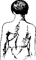
Figure 1. Congenital left-sided lumbodorsal scoliosis.
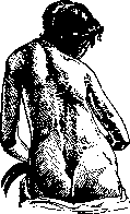
S. The mechanism of its development lies in the underdevelopment of individual vertebrae (wedge-shaped form, spina bifida) and some other anomalies of the skeleton (disease of Sprengel, cervical ribs). Congenital S. as a rule is discovered only in the second decade of life, is most often localized in the lumbosacral region, less frequently in the lumbar-thoracic region, even less frequently in the cervicothoracic region, involves a small number of vertebrae, has a small degree of curvature, is little prone to compensatory counter-curvatures and to torsion, i.e., to rotation of the spine around the vertical axis (fig. 1).-The overwhelming majority of S. of acquired origin; rachitic, school, professional, functional, traumatic, scar, paralytic, syringomyelic, reflexogenic-painful. The last three forms can be combined into a common group of neurogenic S. In the first place are rachitic and school S. In practice, they are often inseparable from each other, since their mechanism of development largely coincides. Rachitic S. occurs no less than in half of all cases of S. It is detected between the ages of 6 and 12 years, begins in the thoracic region, especially often with Dxn, has a total character, leads to compensatory counter-curvatures, is accompanied by sharply expressed torsion and is very prone to fixation (fig. 2). School S. is noted by various authors in 10-20-30% (Spitzy, Haglund, Schulthess) of all schoolchildren. In our Soviet schools, the number of scoliotics is about 10%. One of the main factors in the development of school S. is the prolonged immobility of children sitting in school. Overfatigued by prolonged static tension, the extensor muscles of the back weaken, the child becomes hunched (kyphoses), overloads the ligamentous apparatus of the spine and instinctively finds an easier position in an asymmetrical oblique posture. Over time, the posture becomes habitual. For S. to develop, it is also necessary to have reduced elastic resistance of the skeleton of the spine itself, its rachitic condition. Thus, school S. develops under the influence of four interacting agents: chronic overstrain of the back muscles, habitual asymmetrical posture, rachitic plasticity of the spine, and the resistance of the organism,
Figure 2. Rachitic scoliosis of the thoracic region with sharply expressed compensatory curvature of the cervicothoracic region.
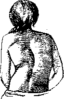
consisting in the potential energy reserve of the living tissues of the body. The severity of the scoliotic deformation depends on the degree to which the first three factors are expressed, and on the magnitude and moment of transition into a kinetic state of the body's potential energy reserves. In the early stages of school S., the spine is pliable, i.e., its pathological curvature is corrected during manual redression or during vertical traction in Glisson's loop. But then, after several years, the lateral hump takes on an increasingly fixed character due to nutritional atrophy of the muscles of the concave half of the trunk and persistent elastic changes in the ligamentous-apparatus, as well as deformation of the cartilaginous and bony tissues of the vertebrae themselves, presenting in this respect an analogy with the development of contractures. An illustration of school scoliosis can be seen in fig. 3. Professional S. is observed in teachers, persons of office and clerical work, in engravers, watchmakers, violinists, and representatives of many other professions. Here, as with school S., the basis is also a habitual asymmetrical posture with chronic unilateral overload of the back muscles. The difference is that the deforming moments in professional S. develop in persons with already completed skeletal growth. Thanks to this, the resistance of the tissues is greater, the curvature develops more slowly and does not reach such large sizes as in school S.-Functional scolioses are those that develop due to unequal length of the limbs, especially the lower ones, after unilateral amputation, after childhood paralysis, which resulted in lag in growth of one leg or arm; after a fracture of the leg, healed with shortening of the bone; after contractures of the knee and hip joints, on the basis of congenitally high position of the scapula (disease of Sprengel). Traumatic S. develops as a result of injuries to the spine and can be primary or secondary. Primary traumatic S. arises immediately after gross anatomical damage as a result of unreduced dislocation or improperly healed fracture of vertebrae. Secondary traumatic S. develops chronically on the basis of microanatomical changes in the internal structure of the spine, disrupting its nutrition and leading to its secondary deformation.-Scar S. is the result of unilateral scar contracture of the tissues of the back after burns or after severe purulent-inflammatory processes of the subcutaneous tissue and muscles. A special form of scar S. is observed after empyema of the chest cavity (retrecissement thoracique).-Paralytic S. develops mainly as a result of childhood poliomyelitis; has a total form,
Figure 3.
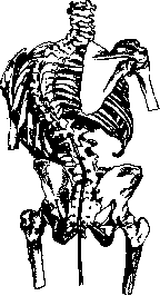
School scoliosis. ^is accompanied by a decrease in excitability of the paralyzed muscles with normal skin sensitivity. Occasionally, paralytic S. occurs due to progressive muscular atrophy and on the basis of hereditary ataxia of Friedreich.-Syringomyelic S. is one of the persistent and early symptoms of syringomyelia; is localized in the cervicothoracic vertebrae of small size; occurs due to central disorders of muscle coordination and trophic osteoporosis of the vertebrae.-Reflexogenic-painful S. is observed as a symptom of unilateral painful processes with the most diverse localization: in sciatica, in inflammation of the sacroiliac joint, in rheumatic lumbago, in appendicitis, in renal and hepatic colic. Accordingly, the following are distinguished: scoliosis ischiadica, scoliosis sacro-iliitica, scol. lumbagica, scol. appendicitica, scol. nephritica, scol. hepatica. Anatomical changes in the spine in S. consist of its lateral curvature and torsion, i.e., a spiral bend around the vertical axis similar to a grapevine. In this case, the apex of the wedge of the scoliotic vertebra faces toward the concavity and somewhat backward; the bone trabeculae of the vertebral body also have this same oblique direction. The bodies of the vertebrae rotate with their anterior parts away from the midline toward the convexity, while the spinous processes rotate toward the concavity. The transverse processes of the vertebrae follow one another like the steps of a staircase (fig. 4). The increased load on the concave side of the spine causes here plastic compaction of the bone-trabecular structure and the deposition of peripheral bony protrusions, which, in connection with the wedge-shaped atrophy of the intervertebral cartilages, leads to ankylosing synostosis of the vertebrae. The muscle-ligamentous apparatus on the convex side is stretched, weakened, on the concave side it is shortened, scar-compacted, rigid. The ribs are curved, on the convex side they protrude in the form of a hump; the intervertebral spaces on this side are widened, on the concave side, conversely, are narrowed to the point that they overlap each other like roof tiles and compress the nerves passing here. In kyphoscolioses, mainly rachitic, the chest often has a keel-like character (pectus carinatum). The disfigurement of the external shape of the chest causes corresponding displacement and disfigurement of the lungs and heart contained within it, which in turn affects their functional activity. Respiratory ventilation of the lungs
is impaired.
is weakened, in them catarrhal phenomena, emphysema, atelectasis easily develop. The heart is often dilated, hypertrophied. Bachmann, who investigated in this direction 154 cases of curvature of the spine, found in 87 cases (56.4%) hypertrophy and dilation of the right heart, in 27 (17.5%) such a lesion of the left heart, and in 40 cases (25.9%) a lesion of both halves of the heart.
The first symptom of the most common forms of S. (ricketic, school, and professional) is rapid fatigue of the back. To this is added a slight lateral curvature of the spine, especially noticeable by the end of the day. On the contrary, in the morning, when the muscles are fresh, the curvature may be completely absent. Later, pains appear, concentrated in the back or in the lumbar region, often radiating along the course of the intercostal nerves. With the years, the deformation progresses, becoming more and more fixed, so that in the end S. does not disappear even with forced attempts at correction. This moment corresponds to the rigid degeneration of the muscle-ligamentous apparatus and deforming changes in the skeleton of the spine, which is especially sharply expressed in the area of Dviii-Dx. The X-ray reveals torsion and wedge-shaped tilting of individual vertebrae, uneven density of the spongy structure, osteophyte growths. Simultaneously, compensatory counter-curvatures develop, the chest is deformed. Both halves of the shoulder girdle are set in an oblique plane: the shoulder and scapula of the convex side in S. of the thoracic department stand higher than on the concave side. Pathological disorders of respiratory and cardiac activity have been mentioned above. These disorders explain the fact that the average life expectancy of scoliotics does not exceed 40-50 years of age. The diagnosis of S. is based on the consideration of anamnestic data (age, profession, past illnesses), evaluation of clinical symptoms (fatigue of the back, pain, its localization, character), special examination of the spine with a scoliosisometer (see) and by the X-ray method. The prognosis is worst in ricketic, paralytic, as well as in all cases of old, fixed forms of S. in general. The most favorable prediction is for congenital and late-developing forms of professional S. Prevention of S. has exceptionally important significance, since it is much more effective than the treatment of this stubborn suffering. When the first signs of congenital S. are detected, the child should be provided with a physical culture regime that facilitates the work of the inadequate (convex) side of the spine: massage, mobile games, oblique sitting (on an inclined plane; in the simplest form - a book under one of the buttocks, corresponding to the convex half of lumbar S.). For ricketic weakening of the spinal column, several hours of special daily rest in the prone position, sun-air and salt baths, vitamin-rich food are indicated. The main preventive measures for school S. are: rational construction of school furniture, hourly physical culture minutes, and for younger children every half hour, based on the principle of unloading, balancing, and correcting movements; mobile games in the fresh air, swimming, rowing. The idea of harmonious development of the intellectual and physical forces of the child, implemented in our Union by the method of polytechnicalization of the school, has enormous importance for the prevention of S. and deformities of the body in general. The socio-prophylactic significance of the principles of Soviet protection and organization of labor is equally effective: general sanitary-hygienic norms of work premises, rationalization and mechanization of the most tiring production processes, wide coverage of workers with physical culture in everyday life and in production, one of the most vivid expressions of which (physical culture) is the preparation and passing of standards for the GTO badge (Ready for Labor and Defense).
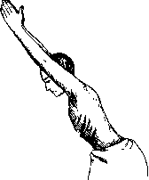
The treatment of S. is mainly based on gymnastic exercises. They are divided into general mobilizing and specifically correcting exercises. Gymnastics is conducted 2-3 times a day for 5-10-15 minute sessions. The character of gymnastic exercises (gymnastic prescription) is extremely diverse depending on the form of scoliosis and individual properties of the patient (age, general state of health). Examples of general mobilizing exercises: 1. Standing position. Arms are extended upward along the head. Bending and straightening of the trunk in the hip joint, without bending the spine (fig. 5). 2. Standing position. Hands on the waist. Active upward extension of the trunk and return to the starting position (fig. 6). 3. Hanging position, holding with hands for the bar. Pulling up and lowering of the trunk. Examples of correcting exercises for total left-sided scoliosis. 1. Standing position. Right arm is raised upward along the head. Left arm is bent at the elbow and rests with the palm on the side, closer to the back. Bending of the trunk to the left and slightly backward, then to the starting position. 2. Lying position on the stomach. Right arm is extended upward along the head. Left arm grasps the lower part of the left shin, bent at the knee. Extension of the spine and return to the starting position. 3. Standing position with the left side to a Lorenz roller. The roller is grasped with the left hand. Right arm is raised upward, bent at the elbow. Bending to the left side slightly backward, strongly pressing against the roller (fig. 7). Examples of correcting exercises for right-sided dorsal scoliosis with compensatory counter-curvature of the lumbar region. 1. Standing position. A long stick is brought behind the back. Its upper end is grasped by the right hand over the right shoulder, and the lower end of the stick is grasped by the bent elbow of the left hand. Pressing with the stick against the dorsal convexity, extend the trunk backward and to the right. 2. Lying position on the stomach. Right arm grasps the lower part of the left shin, bent at the knee. Left arm is extended upward, along the head. Extension of the trunk.
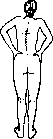
Figure 5. General mobilizing exercise.
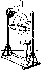
Figure 6. Active extension of the spine.
In addition to free gymnastic exercises, medical-gymnastic exercises on various apparatuses are sometimes used: vertical or on an inclined plane traction in Glisson's loop, lateral traction, exercises on Zander's, Krukenberg's and other apparatuses. Parallel to gymnastic exercises, massage is applied 1-2 times a day for 5-10 minutes: stroking, rubbing, kneading and tapping of the muscles, and for paralytic S. also vibration and faradization. The greatest attention is paid to the massage of the convex side of the back.
In severe cases of deformity, supportive or even corrective corsets are prescribed. The purpose of the former is to provide static support to the muscles until they become strong enough to do without the support of the apparatus. The supportive corset is removable, as it is only worn during the day for sedentary work. Corrective, otherwise called redressing or, as they are also called, 'active' corsets, are made of non-removable plaster. The 'active' Hoffa-Schede corset covers the pelvis and the convex side of the rib hump, which is maximally bent in the opposite direction at the time of application; sometimes the plaster also includes the thigh on the concave side. The corrective Abbott corset is also applied in the corrected position of the spine in a special frame or with the patient on all fours. The arm corresponding to the dropped shoulder is raised up, resting on a bench; the main scoliotic curvature is bent in the opposite direction. Several pieces of dense felt are applied to the scoliotically concave side of the back. Then a circular plaster cast is applied, covering the entire torso. The armpit on the concave side should be strongly supported by the corset, while the armpit on the convex side should be free, for which the edge of the plaster is cut low here. Two windows are cut in the back: opposite the concavity and opposite the convexity of S. From the window of the concave side, one layer of felt is removed and transferred to the opposite side, where it is slipped under the edge of the window. The large window corresponds to the scoliotically concave side, which here, as it should be, is hypercorrected. Finally, the windows are closed with a circular soft bandage. The transfer of subsequent layers of felt from the concave side to the convex side is done every 5-6 days. Such treatment lasts for several weeks and even months, sometimes requiring a change of corset, until stable hypercorrection is achieved. The external wedge effect of 'active' corsets is often good, but unfortunately short-lived, because this method only mechanically stretches the tissues in the desired direction, without fixing the results by strengthening the muscles and corresponding anatomical changes in the skeleton itself (Port). Therefore, in recent years, the results of preliminary correction of S. have been increasingly fixed by subsequent surgical fixation of the spine according to the Albee or Henle-Whitman method. The first method consists of autoplastically transplanting a plate of the tibia into the split of the spinous processes of the vertebrae; the second method consists of transplanting two plates, preferably a split rib, on the sides of the vertebrae, into the groove between the transverse and spinous processes, under the thickness of the muscular ridge located here. After the operation, a plaster fixing corset is applied for several months. Success, i.e., reduction and arrest of the scoliotic curvature, reaches 70% in the hands of some authors (Hibbs, Risser, Ferguson). Attempts to correct the hump by resection of the convex side (Volkmann, Hoffa and others) or by incision and excision of the concave side, expecting its subsequent straightening (Hoessly, Lange, Sauerbruch), proved to be life-threatening and ineffective. In individual cases, satisfactory results were obtained from muscle-plastic operations. Thus, Krukenberg achieved mobilization and partial correction of lumbar S. by incising the shortened aponeurosis of the t. obliq. abdom. ext. and incising the t. psoas on the concave side.
Related articles
Mentioned in
Cite this page
“Scoliosis.” Soviet Medical Encyclopedia. English translation of Bolshaya Meditsinskaya Entsiklopediya, 1st ed. (Moscow, 1928–1936), ed. N. A. Semashko. https://sovietmedicalencyclopedia.pages.dev/article/scoliosis/