Sacrum
Historical document, translated for reference. It reflects medical knowledge of the 1920s–30s and is not medical advice.
Summary
The sacrum is the terminal part of the spine that forms the posterior wall of the pelvic ring, typically consisting of five fused vertebrae. It serves as a wedge-shaped structure connecting the spine to the pelvis and plays a crucial role in weight-bearing and body mechanics.
Encyclopedia article (1928–1936)
SACRUM (sacrum) together with the coccyx forms the terminal part of the spine, which in the form of a wedge closes the pelvic ring from behind. In the vast majority of cases, the sacrum consists of 5 vertebrae, which are separated from each other only in young age, while in adults they are fused together into one common mass. However, there are also variants both toward an increase in the number of vertebrae and toward a decrease. An increase in the number of vertebrae forming the sacrum to six is more often observed in men (Braus). This increase can occur from the upper (anterior) end of the sacrum, when the lower lumbar vertebra partially or completely fuses with the sacrum (sacralization); or
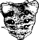
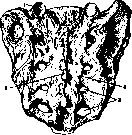
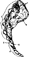
Figure 1. Sacrum from the front: 1- superior articular process; 2- anterior sacral foramen; however, numerical increase is also noted from the caudal end of the sacrum. Much less frequently, the sacral bone consists of four fused vertebrae. Quantitative and qualitative variants of the spine in the lumbar-sacral region often lead to deformity of the trunk (scoliosis). The bodies of the vertebrae and the lateral parts (rudiments of ribs) of the sacrum, fusing together, decrease in size from top to bottom, from which the sacral bone has the shape of a triangle, the apex of which connects with the coccyx, and the base with the last lumbar vertebra (fig. 1 and 2). The lateral parts of the sacrum through the articular surfaces connect with the corresponding surfaces of the ilium, forming sacroiliac joints (fig. 3). The anterior surface of the sacrum, facing the pelvic cavity (pelvic surface), appears slightly curved, creating a larger volume for accommodating pelvic organs. The four anterior sacral foramina on the anterior surface serve for the passage of large nerve plexuses. The upper end of the sacrum at the junction with Ly, on the contrary, protrudes forward significantly in the form of a promontory (promontorium). The posterior convex surface of the sacrum (dorsal surface) in the middle has a well-expressed ridge (median sacral crest) from the spinous processes and two lateral ridges (articular sacral crest), which are fused articular processes. The superior processes (superior articular processes) articulate with Lv; the inferior ones, in a rudimentary form (sacral horns), symmetrically limit the entrance to the sacral canal. The latter has a triangular shape and curves according to the curvature of the sacral bone. The superior opening of the sacral canal, located at the base of the sacrum, serves as a direct continuation of the vertebral canal. The inferior opening (sacral hiatus) is covered by the outer (periosteal) lamina and the posterior sacrococcygeal ligament. Due to the fact that the conus medullaris ends at the level of S2, below, to the sacral hiatus, there remains a space of 6-9 cm, which is called the extradural or epidural cavity. The opening closing the sacral canal from below can be of considerable size, which depends on the non-closure of the rudimentary arches of the sacral vertebrae.
Figure 2. Sacrum from behind: 1- superior articular process; 2- median sacral crest; 3- posterior sacral foramen; 4- articular sacral crest.
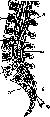
Through the transversely running intervertebral (intersacral) foramina, the sacral canal communicates with the anterior and posterior sacral foramina, which serve for the passage of nerve trunks. Puncture through the sacral hiatus is usually used for sacral anesthesia. The sacral horns serve as identifying points, which limit the lower opening of the canal from both sides. The injection is made in the middle with a needle, which is first set at an angle of 20° to the longitudinal line of the body, then advanced 4-5 cm along the line of the body (fig. 4). The four openings (posterior sacral foramina) located on both sides of the lateral sacral crest serve for the passage of posterior sacral nerves. From the anterior and posterior surfaces of the sacrum to the pelvic bones, a series of ligaments (sacrotuberosum, sacrospinosum, anterior sacroiliac, posterior sacroiliac longum et breve, etc.) go, which hold the bones forming the pelvic ring in a strong connection. From the muscles, the piriformis and coccygeus muscles are connected to the inner surface of the sacral bone; behind the sacrum is covered by the tendinous ends of the long back muscles (sacrospinalis). The synergistic action of individual groups of muscles (back, abdominal, and gluteal) and the strength of the ligamentous apparatus hold the movable part of the spine (above the sacrum) in relation to the sacrum in a certain position, advantageous from the point of view of mechanics and statics of the spine. The lower immobile part of the spine, including the sacrum and coccyx, is located at a certain angle of inclination to the horizontal. This angle usually approaches 60° or 67°, according to Fick (pelvic inclination). In accordance with this angle, the physiological curves (lordosis and kyphosis) of various parts of the movable part of the spine fluctuate (fig. 5). From the point of view of mechanics, the sacrum can be considered as a balancing object on a transverse axis through the sacroiliac joints. The weight of the trunk presses the sacrum forward and downward. When the trunk is inclined and at the same time a load is lifted, the pressure force increases, as a result of which excessive tension is created in the ligaments holding the sacroiliac joint. Under the influence of a rough turn with excessive tension and chronic stretching of the ligamentous apparatus, rotational displacement can occur in the sacrum (for more details, see Sacroiliac joint). Fractures of the sacrum also occur in severe pelvic injuries. Fractures of the sacrum with a longitudinal line of fracture have been observed, and such a fracture in itself does not cause gross functional disorders or causes
Figure 3. Lateral surface of the sacrum and coccyx: 1-sacrum; 2- median sacral crest; 3- coccygeal horn; 4- coccyx; 5- first coccygeal vertebra; 6- auricular surface; 7- tuberosity.
them to a much lesser degree than, for example, fractures in the region of L5. Transverse fractures of the sacrum occur more often from direct application of force and are observed mostly below the sacroiliac joint. Of the congenital anomalies of development of the skeleton (dysplasia) of this part of the spine, besides the non-closure of the posterior wall of the sacral canal, sacralization has practical significance, which can be in the form of a symmetrical connection of the transverse processes of L5 with the sacrum, and then it is clinically manifested to a lesser degree; if sacralization-
L ~
of the lower part of the spine: 1- XII rib; 2- rarely between the transverse process of L5 and the sacrum, a lumbar vertebra develops; 5 and 6- sacral horns; 7- sacral hiatus; 8- coccygeal canal; 9- conus medullaris; 10- cauda equina; 11- posterior sacral foramina; 12- anterior sacral foramina; 13- median sacral crest; 14- articular sacral crest; 15- sacral canal; 16- base of the sacrum; 17- apex of the sacrum; 18- coccyx; 19- first coccygeal vertebra; 20- second coccygeal vertebra; 21- third coccygeal vertebra; 22- fourth coccygeal vertebra; 23- fifth coccygeal vertebra.
one half, painful phenomena are more often observed. Tumors of the sacrum and coccyx region have a very diverse structure. Besides pure forms of myelocystocele and myelomeningocele, teratomas and congenital lipomas are also found here. Along with simple and complex dermoids, sarcomas and subcutaneous parasites giving a picture of a tumor are also observed. Tilman (Tillmann) in 110 pelvic sarcomas notes localization on the sacrum in 29 cases. More favorably progressing exostoses and enchondromas, often located at the border of the sacrum and ilium, are also described. The sacrococcygeal region presents many interesting features in terms of embryogenesis and phylogenesis, and therefore tumors that are usually not observed in other places are sometimes found here. Such are, for example, chordomas, arising from the notochord. Such tumors according to the classification of Cohnen (Cohnen) can be cranial, vertebral, and caudal. Caudal or sacrococcygeal chordomas can be presacral, retrosacral, and central sacral. Melnikov collected in the literature 36 cases of caudal chordomas, to which he added his 37th case. Surgical intervention for tumors of the sacrum and sacrococcygeal region should be radical in relation to most tumors, which with palliative interventions tend to recur,
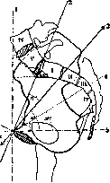
Figure 5. Diagram of inclination angles: 1- vertical line; 2- horizontal line; 3- anatomical conjugate; 4- line of inclination; 5- iliac line; 6- normal conjugate.
giving an unfavorable prognosis. Radical removal of the tumor in most cases must be combined with resection of the coccyx and partial or complete resection of the sacrum. Among inflammatory diseases of the sacrum, tuberculous osteomyelitis is most frequently noted. - Osteomyelitis of the sacral bone, like other pelvic bones, proceeds severely, requires rapid recognition and radical intervention. Denier observed 3 cases, which ended in death with symptoms of general septicopyemia. Out of 8 cases, Klemm had 4 recoveries thanks to early and radical intervention. Tuberculous lesions of the sacrum proceed more slowly and favorably. An infiltrate often arises near the sacroiliac joint, but destruction involves a larger or smaller part of the sacrum. In this case, such severe disorders are not observed as in osteomyelitis: patients often continue to work for a long time. Abscesses arise almost exclusively on the posterior surface of the sacrum and extremely rarely break forward. Therapy, in contrast to osteomyelitis, should be conservative. Abscesses are punctured according to the type of cold abscesses. Fistulas heal persistently under the influence of heliotherapy. Systemic injections of a mixture of Calmette also have a favorable effect. Syphilitic lesions of the sacrum in the form of tertiary gummous osteitis are observed more rarely. In individual cases, along with specific treatment, removal of necrotic foci by surgical means is indicated. - The sacrum together with the coccyx is also an object of surgical intervention in diseases of pelvic organs, especially for access to the rectum. Such is, for example, the posterior approach according to Kraske. In recent years, anomalies of the spine and especially the 'transitional' fifth lumbar vertebra in combination with the sacrum have been thoroughly studied and explain the causes of many unclear 'lumbar pains', as well as the causes of deformity formation of the spine. In the study of spinal anomalies, scientists of the USSR also contributed a certain share of work, among which the works from Turner's clinic (N. I. Chirkin) and from Vreden's clinic (A. A. Kozlovsky) occupy a prominent place.
Related articles
Mentioned in
- Achondroplasia
- Asynclitic Presentation
- Bandages (a198)
- Breech Presentation
- Center of Gravity
- Coccygodynia
- Coccyx
- Coitus
- Douglas Space
- Electrodiagnosis
- Gaffky and Haff Disease
- Gluteal Region
- Haematometra
- Hirsutism (hypertrichosis, hypertrichia, polytrichia,)
- Iliac Region
- Innominate Bone
- Leyden Type of Dystrophy
- Lordosis
- Lumbar Region
- Magnus-Kleijn Reflexes
Cite this page
“Sacrum.” Soviet Medical Encyclopedia. English translation of Bolshaya Meditsinskaya Entsiklopediya, 1st ed. (Moscow, 1928–1936), ed. N. A. Semashko. https://sovietmedicalencyclopedia.pages.dev/article/sacrum/