Gluteal Region
Historical document, translated for reference. It reflects medical knowledge of the 1920s–30s and is not medical advice.
Summary
This article from the 1928–1936 Soviet Medical Encyclopedia describes the anatomy, topography, and phylogenetic development of the gluteal region. It details the muscular, ligamentous, and vascular structures, as well as the clinical significance of the region's fat layers and fascial spaces.
Encyclopedia article (1928–1936)
GLUTEAL REGION (synonym sciatic), regio glutaea (synonym nates, clunes). Boundaries of the region: superior—iliac crest (crista ossis ilii), inferior—gluteal fold (plica natium), medial—sacrum and coccyx, lateral—a line extending from the anterior superior iliac spine (spina iliaca anterior superior) to the greater trochanter (trochanter major).
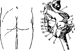
Figure 1.
Figure 2.
Figure 1. Gluteal fold and the lower edge of the m. glutaeus max., the edges of which are indicated by a dotted line. Figure 2. Ligamentous apparatus of the female pelvis from the side: 1—crista iliaca; 2—spina iliaca ant. sup.; 3—spina iliaca ant. inf.; 4—ram. sup. ossis pubis; 5—symphysis; 6—ram. inf. ossis pubis; 7—for. obturatum; 8—tuber ischiadicum; 9—trochanter major; 10—lig. sacro-tuberosum; 11—for. ischiad. min.; 12—incisura ischiad. min.; 13—lig. sacro-spinosum; 14—for. ischiad. major; 15—incisura ischiad. major; 16—spina iliaca post. inf.; 17—spina iliaca post. sup. (Figs. 1 and 2). The rounded shape of the gluteal region, determined by the muscular-skeletal foundation, varies to a greater degree than other regions due to the deposition of fatty masses, especially in women (see below). The downward extent of the gluteal region does not correspond to the boundaries of its muscles (the edge of the gluteus maximus), but is determined by the edge of the fascial sheath enclosing the fat pad (Fig. 1). The gluteal region is one of the largest regions of the human body. Its length ranges from 25 to 27 cm (from the middle of the crest to the gluteal fold), and its width from 20 to 24 cm (from the middle of the posterior surface of the sacrum to the middle of the line connecting the anterior superior spine with the greater trochanter). The gluteal region is phylogenetically connected with the appearance and development of the lower limbs—organs of support and locomotion. In fish, the lower limbs (hind fins) produce elementarily simple movements that only facilitate a change in direction and form a weakly developed pelvic girdle, which is why one cannot properly speak of a specifically gluteal musculature in them, as the movements of the lower limbs are performed by the muscles of the trunk. In monkeys, the gluteus maximus muscle is less developed than in humans, which is closely related to the vertical position of the latter's body. In lemurs, the middle and large gluteal muscles represent a single muscle. In Old World monkeys, the sciatic musculature is not voluminous. The m. glutaeus maximus in lower monkeys reaches the lower part of the thigh or even the upper part of the lower leg. In chimpanzees and gorillas, the attachment of the glut. maximus reaches the lateral condyle of the femur and can merge with the short head of the biceps; in the orangutan, it reaches the middle of the thigh, and in the gibbon, it attaches to its upper third. The latter type of attachment variation is also caused by a change in statics—the transition of the trunk to a vertical position. For ontogenesis, see Pelvis and Hip joint. For the function of the muscles of the gluteal region, see Muscles of man; it should only be added here that the large gluteal muscles, according to Frosche, contracting simultaneously, can act as a sphincter ani externus, which acquires great practical importance in the use of this muscle as an artificial sphincter during surgical interventions. The osteo-ligamentous foundation of the gluteal region is the external (posterior) surface of the ilium (os ilii), the ischium (os ischii), the posterior surface of the sacrum, the sacro-spinous (lig. sacro-spinosum) and sacro-tuberal (lig. sacro-tuberosum) ligaments, the hip joint capsule, the neck of the femur, and the greater trochanter (Fig. 2). The sacro-spinous ligament turns the greater sciatic notch into the greater sciatic foramen. The sacro-tuberal ligament, together with the previous one, turns the lesser sciatic notch into the lesser sciatic foramen. It is necessary to emphasize that the center of the region is the hip
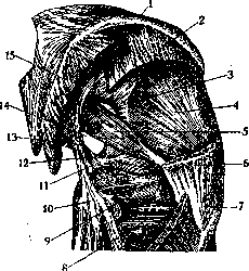
Figure 3. Musculature of the gluteal region: 1—crista iliaca; 2—m. glutaeus med. (for. suprapiriforme); 3—upper part of for. ischiad. major; 4—m. glutaeus min.; 5—m. piriformis; 6—m. glutaeus med.; 7—trochanter major; 8—m. quadratus femoris; 9—m. obturat. int. and mm. gemelli; 10—tuber ischiadicum; 11—for. ischiad. min.; 12—spina ischiad. and lig. sacro-spinosum; 13—lig. sacro-tuberosum; 14—lower part of for. ischiad. major (for. infrapiriforme); 15—m. glutaeus max.
joint. Stretched over this bony foundation are the muscle masses (for their innervation and attachment, see the table Muscles of man): the piriformis muscle (m. piriformis), the internal obturator muscle (m. obturator internus), the external obturator (m. obturator externus), the gemelli (mm. gemellus sup. and gemellus inf.) (Fig. 3). The entire mass of the region is covered from above by the gluteus maximus muscle. Variants of the gluteal muscles include
both an increase in their number—glutaeus quartus (s. scansorius), splitting of the gluteus maximus into two layers, and a decrease in the mass of individual muscles, or their fusion—atavism. The main vessels of the region arise from the hypogastric artery (a. hypogastrica)—the superior and inferior gluteal arteries. The nerves are branches of the sacral plexus (plexus sacralis).
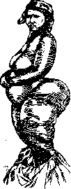
Topography of the gluteal region. The subcutaneous tissue (tela subcutanea), approximately 2 cm thick, contains grape-like arranged fat lobules. The next layer of fat, characteristic only of humans and not observed even in anthropoid apes, is the fat pad (corpus adiposum). The usual thickness of the fat pad is about 4 cm. Variations in the positive direction have received the name steatopygia in anthropology. In children, this pad is poorly developed. In women of certain tribes of South Africa, especially after puberty, a grandiose amount of fat is deposited in these places (Fig. 4).
The fat pad in question lies between the superficial and deep layers of the superficial fascia (fascia superficialis). The third fat layer of this region, named by Charpy "le coussinet graisseux lombofessier" and by Stromberg "massa adiposa lumbo-glutaealis," located between the deep layer of the superficial fascia and the muscular fascia, continues into the lumbar region. In the gluteal region, this layer descends to its middle third. Through this tissue, parametritis (Stromberg) and parametrial hemorrhages (Richter) can descend into the gluteal region. In the fat layer run the nerves innervating the skin (nn. cutanei clunium sup.): the superior gluteal—branches of the lumbar plexus, the middle gluteal branches—from the sacral plexus, and the inferior—from the posterior cutaneous nerve of the thigh (Fig. 5). The next layer is the gluteal fascia (aponeurosis)—a closed sheath for the gluteus maximus muscle. Hyrtl considers it a continuation of the fascia lata, which splits at the lower edge and embraces the gluteus maximus muscle; at the upper edge of the latter, the layers of the fascia rejoin and attach to the iliac crest and the sacrum. Above the edge of the gluteus maximus muscle, this fascia passes over the gluteus medius muscle in the form of a free, triangular, shiny plate. Under the fascial sheath of the gluteus maximus muscle lies a layer of fatty tissue that does not disappear even in high degrees of starvation. This layer of fat covers the deep part of the gluteal region—the site of development of deep abscesses (compare subpectoral abscess). When this layer is injected, a kind of pocket is formed, the upper boundary of which is the upper edge of the gluteus maximus muscle, and the lower is the popliteal fossa; therefore, pus can pass from the deep sections of the gluteal region into the posterior compartment of the thigh and into the popliteal fossa. Between the middle and small gluteal muscles lies the deepest interfascial space. The piriformis muscle passes through the greater sciatic foramen and forms two slits: the supra- and infrapiriform foramina (foramen supra- et infrapiriforme). Both slits are very tightly filled with the structures passing through them; this is evident from Starkov's experiment: "during injections into the parametrium, in 2 cases out of 15, the mass penetrated into the gluteal region." Through the suprapiriform foramen pass the superior gluteal artery and vein and the superior gluteal nerve. In the foramen itself lie lymph glands (Bourgery and Jacob) (Fig. 6). The superior gluteal artery (the largest branch of the iliac artery), upon appearing in the gluteal region, immediately divides into a number of branches anastomosing with the lumbar arteries (aa. lumbales), the iliolumbar (a. ilio-lumbalis), the lateral sacral (a. sacralis lat.), the inferior gluteal (a. glutaea inf.), and the lateral circumflex femoral artery (a. circumflexa femoris lat.). These anastomoses ensure collateral circulation of the gluteal region during ligation of the external iliac.
iliac and femoral arteries. Through the infrapiriform foramen (f. infrapiriforme) pass the inferior gluteal artery and vein, the sciatic nerve (n. ischiadicus), the posterior cutaneous nerve of the thigh (n. cutaneus femoris post.), the inferior gluteal nerve (n. glutaeus inf.), the internal pudendal artery and vein (v. et a. pudenda interna) (Fig. 6). The inferior gluteal artery lies between the sciatic nerve and the internal pudendal artery, gives branches to the muscles and a separate branch accompanying the sciatic nerve (a. comes n. ischiadici), and anastomoses with the arteries surrounding the thigh (aa. circumflexae femoris) and the perforating branches of the deep artery of the thigh (rami perforantes a. profundae femoris) (see Thigh). The development of the inferior gluteal artery is subject to numerous variations, partly in connection with changes in the power of the gluteus maximus muscle; the artery accompanying the sciatic nerve also varies sharply. This has practical significance in high amputations of the thigh. The shortness of the gluteal arteries makes their ligation in situ difficult (see below). Upon exiting the pelvic cavity, the sciatic nerve lies on the posterior surface of the muscles: the internal obturator, the gemelli, and the quadratus femoris.
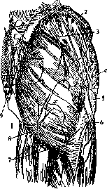
Figure 5. Gluteal region. Fascia and superficial nerves: 1-nn. clunium sup.; 2-crista iliaca; 3-upper edge of m. glutaeus max.; 4-collum femoris; 5-trochanter major; 6-n. cutaneus femoris lat.; 7-nn. clunium inf.; 8-lower edge of m. glutaeus max.; 9-nn. clunium medii; 10-spina iliaca post. sup.
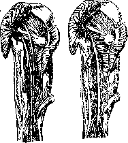
At the lower edge of the gluteus maximus muscle covering it, the nerve lies more superficially—a site for its easy surgical exposure. Of practical importance is the high division of the sciatic nerve, in which the common peroneal nerve exits either above the piriformis muscle or by splitting it (Fig. 7). The internal pudendal vessels and the pudendal nerve, upon exiting the infrapiriform foramen, curve around the ischial spine and re-enter the pelvis through the lesser sciatic foramen (Fig. 6). Mucous bursae of the Gluteal Region. The ischial bursa of the gluteus maximus (bursa ischiadica m. glutaei magni) is on the posterior surface of the ischial tuberosity; it occurs very frequently; the trochanteric bursa of the gluteus maximus (bursa trochanterica m. glutaei maximi) is found constantly on the posterior and external surface of the greater trochanter. Gluteofemoral bursae (bursa glutaeo-femoralis), two
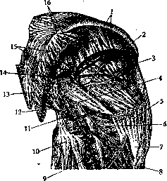
Figure 6. Topography of the right gluteal region: 1-crista iliaca and m. glutaeus med.; 2-rami inf. a. glut. sup.; 3-m. glutaeus minimus; 4-n. ischiadicus and a. glut. inf.; 5-m. glutaeus med.; 6-m. glutaeus inf.; 7-trochanter major; 8-n. ischiad.; 9-n. cutaneus fem. post. and a. glut. inf.; 10-tuber ischiadicum; 11-a. and n. pudendus at for. ischiad. min.; 12-lig. sacro-tuberosum; 13-m. piriformis and n. pudendus; 14-a. and n. glutaeus sup.; 15-sacrum and m. glutaeus max.; 16-branches of a. glutaei sup. to m. glut. max. (ram. sup.).
or three, are located on the femur, at the attachment site of the gluteus maximus (medially or laterally); the anterior trochanteric bursa of the gluteus medius (bursa trochanterica m. glutaei medii ant.) is between the tendon of the gluteus medius and the apex of the greater trochanter; the posterior trochanteric bursa of the gluteus medius (bursa trochanterica m. glutaei medii post.) is between the tendons of the gluteus medius and piriformis muscles. Bursae of the piriformis muscle (bursae m. piriformis)—below the tendon of the piriformis muscle, between the latter, the bone, and the gemellus muscle. The bursa of the internal obturator (bursa m. obturatorii interni) is between the muscle and the lesser sciatic notch. Lymphatic pathways of the Gluteal Region are divided into superficial, carrying their lymph to the subinguinal nodes (lgl. subinguinales), and deep, the first station of which are the nodes located in the greater sciatic foramen below and above the piriformis muscle along the course of the vascular bundles. The next station of the deep pathways is the pelvic nodes, with which the superficial inguinal nodes also anastomose, forming a pelvic lymphatic ring.
AB
Figure 7. Types of the sciatic nerve: A-dispersed, high division of the sciatic nerve; B-main, low division. (see Pelvis). The projection of the most important parts of the gluteal region onto the surface is presented in Fig. 8. The Roser-Nélaton line is of great practical importance (see Roser-Nélaton line). In the clinic of the Gluteal Region, congenital tumors of the sacrum and coccyx, which often occupy the entire Gluteal Region as well, acquire special significance (see Sacrum). These tumors represent remnants of a second fetus (inclusio foetalis) (see Pelvis, Sacrum, Coccyx). All kinds of tumors are encountered in the gluteal region, especially sarcomas, which sometimes reach an enormous size. Among other tumors, atheromas, lipomas, soft fibromas, neurofibromas, and also neurosarcomas. In acute injuries of the Gluteal Region, wounds of the superior and inferior gluteal arteries, as well as the sciatic nerve, acquire special significance. Surgical approach to these formations is hindered by two circumstances: the thickness of the gluteus maximus muscle and the shortness of the arteries. Previously proposed methods
l
Figure 8. Finding the aa. glut. sup. and inf., a. pudenda int., and n. ischiadicus: 1-spina iliaca post. sup.; 2-a. glut. sup. in for. suprapiriforme; 3-m. piriformis; 4-a. glut. inf. and a. pud. int. in for. infrapiriforme; 5-troch. major; 6-troch. minor; 7-for. ischiad. minus; 8-lig. sacro-tuberosum. Methods of ligation of the superior and inferior arteries after separating the fibers of the gluteus maximus muscle must be considered inconvenient; the trauma is great, the muscle is denervated (Fig. 6), there is little space, and in case of severe bleeding, it is practically impossible to reach the vessels. Therefore, the incision proposed by A. L. Radziyevsky for approaching the damaged sciatic nerve (wounds, scars)—transection of the attachment of the gluteus maximus muscle with reflecting it medially—is particularly convenient. When approaching the vessels for their ligation within the pelvic foramina themselves, it is also necessary to perform disinsertion of the piriformis muscle or partial resection of the bony edge (French authors). Upon completion of the operation, the muscles are sutured, the gluteal starting from below. Many authors recommend not wasting time on ligation of the gluteal arteries from the side of the Gluteal Region, which often cannot be ligated, but to go into the pelvic cavity to ligate the hypogastric artery. Aneurysms of the gluteal arteries and nerve injuries are treated according to general rules. Purulent processes of the Gluteal Region include parametrial and paravesical processes passing through the greater and lesser sciatic foramina, then cold abscesses descending here, and finally abscesses and phlegmons arising after all kinds of injections; the latter should be performed only in the lateral quadrants of the region (see the arrangement of vessels and nerves). Phlegmons from infection of the mucous bursae are also encountered, especially from the large mucous bursa of the ischial tuberosity. More often than others, inflammation and suppuration of the mucous bursae between the skin and the posterior surface of the sacrum are observed. In these processes, incisions should also not be made by the "classical method"—through the thickness of the gluteal muscle. Anatomically justified are 3 incisions for the Gluteal Region, which often have to be made simultaneously: 1st—in the tendinous part of the gluteus maximus parallel to the greater trochanter, 2nd—in the posterior region of the thigh below the gluteal fold, and 3rd—along the upper edge of the gluteus maximus muscle. In severe cases, it is necessary to perform temporary detachment of the gluteus maximus muscle. In the differential diagnosis of purulent processes of the Gluteal Region, it is necessary to pay attention to diseases and lesions of the hip joint, the purulent processes of which often spread to the Gluteal Region. Lesions of the bones of the Gluteal Region—see Pelvis.
Related articles
Mentioned in
Cite this page
“Gluteal Region.” Soviet Medical Encyclopedia. English translation of Bolshaya Meditsinskaya Entsiklopediya, 1st ed. (Moscow, 1928–1936), ed. N. A. Semashko. https://sovietmedicalencyclopedia.pages.dev/article/gluteal-region/