Sacroiliac Joint
Historical document, translated for reference. It reflects medical knowledge of the 1920s–30s and is not medical advice.
Summary
The sacroiliac joint is a strong connection between the articular surface of the ilium and the lateral part of the sacrum. It is a true synovial joint with limited mobility but high stability, strengthened by multiple ligaments.
Encyclopedia article (1928–1936)
SACROILIAC JOINT (articulatio sacro-iliaca), a rather strong connection between the articular surface of the ilium and the lateral part of the sacrum (fig. 1). The slightly protruding part of the articular surface of the ilium is tightly wedged into the depression of the articular surface of the sacrum corresponding to the I, II, and III sacral vertebrae. According to Petersen's data, based on the study of 280 joints, the I, II, and III sacral vertebrae participated in the articulation 242 times; participation of only the I and II vertebrae was noted in 21 cases. Both the size of the articular surfaces and their shape vary considerably in different individuals. In children, the articular surfaces are smoother, while in adults they have bends. In structure, the sacroiliac joint represents a true joint (amphiarthrosis) with a synovial membrane and a small amount of synovial fluid. The articular surfaces are covered with fibrocartilage, with the cartilage on the sacrum being significantly thicker, covered with fibrocartilage on the surface and having a layer of hyaline cartilage in the depth. In rare cases, the articular surface is covered not with cartilage but with connective tissue. In the posterior part of the joint, Rudolf Fick, and even earlier Henle, established the presence of connective tissue bundles extending from one articular surface to another. Fick also notes the frequent presence of a narrow cartilaginous bridge between both articular surfaces. The joint space is clearly defined in most cases and is well visible on an X-ray. The joint cavity is bounded by a strong capsule, which arises directly at the cartilaginous edge in the sulcus paraglenoidalis. It is very tightly stretched throughout the joint and serves as a continuation of the periosteum of the ilium and sacrum. Inside the joint, there is a small amount of synovial fluid. The joint space, according to various authors, forms at different periods of life. While Luschka found a clearly defined space already in a 20-week embryo, Barkow did not find a space in an 8-month embryo and believes that it does not even exist in newborn infants. The articular surface of the ilium at this time is not yet covered with cartilage but is connected to the cartilaginous surface of the sacrum by connective tissue fibers. Barkow found a clearly defined joint space in a 7-year-old girl and believes that only from this age does it exist constantly. Such studies, however, not based on large material, do not give an accurate picture of the period of formation of the joint space. But what is important practically is that the space with all elements of the joint is already present in childhood and always in adults, and consequently the joint can undergo inflammatory changes and traumatic injuries, like other large joints of the body. Due to its special structure, movements in the sacroiliac joint occur within very limited limits. Such joints are adapted not so much for mobility as for stability. The stability and strength of the joint, in addition to the anatomical relationships of the joint, are also determined by the strength of the ligaments that strengthen the capsule. The capsule and joint are directly strengthened in front by the lig. sacro-iliaca anteriora, in the back by the lig. sacro-iliacum posterius longum, *
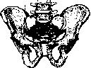
Figure 1. Sacroiliac joint (i).
lig. sacro-iliacum posterius breve, lig. sacro-iliaca interossea (fig. 2, 3, 4 and 5). The anterior ligaments are usually poorly developed, located on the pelvic surface of the joint capsule, strengthening it with their fibers. The posterior long ligament, starting from the edge of the sacrum in the area SII-IV, goes steeply upward to the spina iliaca post. sup. The lateral edge of this ligament intertwines with the fibers of the lig. sacro-tuberosum, and the medial edge merges with the fascia lumbodorsalis. The lig. sacro-iliacum posterius breve is located medial to the posterior long ligament and is slightly covered by it. The short posterior ligament starts from the crista sacralis lat. and attaches to the spina iliaca post. inf. The fibers of the ligament extend over the foramina sacralia post., partially covering them. The lig. sacro-iliaca interossea form a deep fossa behind between the tuberositas iliaca and tuberositas sacralis. These ligaments, in the form of short strong fibers, run transversely, intertwining with each other. In the strengthening of the sacroiliac joint, the following ligaments also play an important role: lig. ilio-lumbale, which arises from the transverse processes of the last two lumbar vertebrae and fans out over the anterior and posterior surfaces of the ilium. Lig. sacro-tuberosum arises from the lateral edge of the sacrum and coccyx, attaches to the
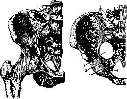
Figure 2. Sacroiliac joint: 1 - lig. ilio-lumbale.
Figure 3. Ligaments: 1 - lig. ilio-lumbale; 2 - lig. sacro-spinos.; 3 - lig. sacro-tuberos.; 4 and 5 - for. ischiad. majus et min.
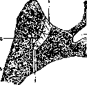
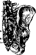
Figure 4. Frontal section: 1 - lig. sacro-iliac. post.; 2 - can. sacr.; 3 - os sacr.; 4 - lig. sacro-iliaca ant.; 5 - os ilium; 6 - lig. sacro-iliaca interossea.
Figure 5. Ligaments of the sacroiliac joint behind: 1 - lig. ilio-lumbale; 2 - lig. inter-spinale; 3 - lig. sacro-il. interossea; 4 - lig. sacro-il. post. breve; 5 - lig. sacro-il. post. long.; 6 - lig. sacro-tuber.; 7 - lig. sacro-coccyg. laterale.
326 325
Sacroiliac joint. The ligament sacrospinosum begins at the ischial spine, runs horizontally, crosses the ligament sacrotuberosum, and attaches to the lateral edge of the lower part of the sacrum and coccyx. The stability of the joint is also achieved by the constant tone of the surrounding muscles, especially the long muscles of the back, the straight and oblique muscles of the abdomen. Due to the significant reinforcement of the joint, access to it from behind is difficult. Albee, analyzing the anatomical data regarding the sacroiliac joint, notes the large disagreements that exist among the greatest anatomists regarding the structure of this joint. Based on his research on 50 corpses, Albee concludes that the sacroiliac joint has all the elements of a joint: movements in it occur especially during childbirth. The blood supply to the joint is provided by branches of the hypogastric artery. Branches of the superior and inferior gluteal arteries penetrate into the sacroiliac joint from the pelvic side and from the outside. The joint receives a significant branch from the iliolumbar artery (Fick) and smaller branches from the lateral sacral arteries. The innervation of the sacroiliac joint has enormous practical importance due to the variety of pain points in inflammatory diseases and injuries of this area. Articulatio sacroiliaca, according to Morris (Morris), is supplied by branches of the superior gluteal nerve, the fourth and fifth lumbar nerves, and the fifth sacral nerve, as well as by the posterior branches of the first and second sacral nerves. The fourth and fifth lumbar nerves together with the obturator nerve go in front of the joint, and the lumbar branches are in close connection with the sacroiliac joint. This explains the circumstance that when this joint is affected, pain is experienced in the sacral region (upper sacral nerve) and the buttock (gluteal nerve). Often the pain radiates to the hip and knee joints, as well as along the inner surface of the thigh (obturator nerve). According to the latest research by English and American authors in connection with traumatic injuries to the sacroiliac joint, the statics and mechanics of this joint present much that is unique and valuable for clinical practice. The sacroiliac joint together with the lumbosacral region are areas that are most subject to static overstrain as places of connection between the fixed and movable parts of the spine. It is known that in the standing position of a person, the articular surface of the ilium is located almost horizontally, while the sacrum is inclined to the horizon usually at an angle of 60-67°, according to Fick. In the position of the body that a person assumes during work, the sacrum is positioned more horizontally, and the sacroiliac joint receives less support from the bony structures that make up the joint. Although older authors (Sapreus) almost completely deny movement in the sacroiliac joint, according to the latest research by Meyer, Fick, and especially Goldthwait, they are absolutely proven. One type of movement in the sacroiliac joint, according to Meyer, occurs around a transverse axis through the sacrum. Another type of movement also goes around a transverse axis, which is located behind the articular surface of the sacrum and passes through the interosseous ligaments. The movements are of a rocking nature. The range of motion varies individually depending on the structure of the articular surface of the sacrum. By sawing through the pelvis while preserving the ligaments, Goldthwait proved on fresh corpses that movements in the sacroiliac joint do exist and that the joint can be dislocated. Movements in the sacroiliac joint occur depending not on the action of muscles, but mainly on changes in the position of the trunk and spine. The muscles themselves, although they surround the joint in large numbers, have little influence on the movements of this joint. Only the straight and oblique muscles of the abdomen can indirectly influence the known mobility of this joint, as they tend to bring the trunk closer to the pelvic ring. On the contrary, the weight of the trunk when it is moved plays a serious role in the mechanics and statics of the sacroiliac joint. In the upright position of the trunk, the line of gravity passes in front of the axis of rotation of the joint. Therefore, when the trunk is straightened, the sacroiliac joint also indirectly participates with its movements. For the same reason, in the standing position the posterior ligaments of the sacroiliac joint are tense, and when sitting they are relaxed. When the mobility of the sacroiliac joint changes depending on pathological conditions, the nature of gait also changes significantly: it becomes somewhat constrained, the patient does not take large, free steps as a healthy person does. Anatomical-physiological and especially mechanical data explain many pathological phenomena observed in connection with traumatic or inflammatory changes in the sacroiliac joint. The range of mobility of the joint plays an enormous role not only in the mechanics of movements of the entire body, in the nature of gait, but is also important in obstetrics, influencing to a certain extent the development of the pelvis. Clinic. Like other joints, the sacroiliac joint is subject to inflammatory diseases and various types of traumatic injuries. Due to the peculiarities of the structure and mechanics of this joint, as well as due to the deep location of the anterior (pelvic) part of this joint, the clinical symptomatology sometimes presents much that is unclear, especially in traumatic injuries to the sacroiliac joint. A number of pain symptoms after injury to the lumbar region, which are sometimes taken for 'contusion' or 'sciatica', often have deeper causes, especially if the pains are prolonged and do not disappear after usual manipulations. Rupture of the posterior ligaments in traumatic injuries or their chronic stretching in an unfavorable professional position of the trunk can be accompanied by a pain syndrome. In recent years, American authors have also proven the possibility of a true subluxation of the sacroiliac joint. Even older authors (Rooge) note the possibility of subluxation of this joint and attribute it to the act of childbirth. Edwards and Kerr in 1889 described in detail a subluxation of the sacroiliac joint in a pregnant woman, which arose suddenly when she rose from a sitting position. Jewett mentions pathological instability of the pelvic joints and connects this with rickets, syphilis, tuberculosis, a large fetal head, or abnormal births. However, at present, inflammatory changes of tuberculous or luetic (more rarely) origin are strictly distinguished from true traumatic subluxations. The clarification of the latter question belongs to the classic work of Goldthwait, based on extensive material (500 hospital cases) and detailed anatomical research. Cox (Cox) in his anatomical research also established movements in the sacroiliac joint ranging from 0.5 to 3 mm. He managed to cause actual displacement in the joint, more often displacement of the ilium forward in relation to the sacrum. There are two distinct types of traumatic subluxation of the sacroiliac joint. The first is associated with a specific sudden trauma, such as a fall, lifting a heavy weight, suddenly rising from a sitting position, or a sharp lateral turn of the trunk. The second type of subluxation is observed with prolonged incorrect position of the trunk depending on professional peculiarities (chronic trauma). Cases of subluxation in pregnant or parturient women have also been described by individual authors. In the last months of pregnancy, there is hyperextension of the spine due to lordosis. The range of motion in the pelvic joints increases, a certain instability of the joint arises. This circumstance together with a minor trauma can serve as an important etiological factor for the occurrence of subluxation in the sacroiliac joint. From the analysis of the mechanism of subluxation, according to Cox, most often there is a slipping of the ilium forward in relation to the sacrum; the sacrum remains unchanged. Subluxation in most cases is unilateral. In the mechanism of acute subluxation, two forces acting in opposite directions play a role. The first force is the hyperextension of the spine that a person suddenly produces, for example when stumbling; the second force is the sudden forward pull exerted on the ilium by the action of a group of muscles originating from the anterior superior and inferior iliac spines (rectus femoris, sartorius, tensor fasciae latae). These two forces, fixing the sacrum, cause displacement of the ilium forward in the upper part of the sacroiliac joint. Symptomatology of acute subluxation. Lumbar pains, usually unilateral, arise suddenly after trauma, lifting heavy weights, a sharp turn of the trunk, etc. The patient with difficulty can straighten his back. Any movement or position of the body associated with movements of the sacral part causes sharp pain. Thus, raising the leg upward with the knee straight causes pain corresponding to the joint and can simulate sciatica. There is a Goldthwait test for testing subluxation of the sacroiliac joint. The subject stands on the leg of the affected side, bending the hip with the leg straightened. The surgeon places one hand on the suspicious joint and the other on the symphysis. In the presence of stretching or subluxation in the sacroiliac joint, the symphysis should move with every movement of the leg. There is also a test that Steindler attributes to Goldthwait: the hip of the affected side is sharply bent, the knee is extended. This test causes tension of the flexors of the leg and pain in the affected sacroiliac joint. Gaenslen proposed the following test for testing the sacroiliac joint.
The thigh and knee of the affected side are brought into a position of sharp flexion so that the limb is pressed against the chest. With hyperextension of the other thigh, pain is noted in the corresponding sacroiliac joint of the affected side. In the acute period, a more pronounced protrusion of the sacrum, rigidity of the back muscles, and disappearance of the lumbar curvature of the spine are observed due to the forward displacement of the ilium, with lateral curvature of the spine in the opposite direction. Pressure on the iliac crest causes severe pain. Particularly important signs are marked tenderness upon pressing on the sacroiliac joint from behind and the inability to bend the trunk forward without pain with the knees straight. In chronic cases of subluxation, the onset is gradual, lumbar pains are more diffuse, affecting both joints and of lesser intensity. The patient feels better with a pillow placed under the lower back. It is important to establish a connection with occupational peculiarities. In individuals of delicate constitution who work in uncomfortable positions, habitual stretching of the ligaments is a precursor of chronic sacroiliac dislocation. Referred pain may occur along the course of the sciatic nerve and often simulate sciatica or lumbago. According to Gault, many cases of chronic sciatica are due to unrecognized subluxation of the sacroiliac joint. A more detailed analysis of the mechanism of injury, localized tenderness, and the signs mentioned above allow for clarification of the diagnosis. Unfortunately, a radiograph in the standard projection does not indicate displacement, as according to Gault, rotational displacement occurs which cannot be demonstrated on such plates. A stereoscopic photograph provides more clarity. Treatment of sacroiliac subluxation in the acute period should consist of reduction and subsequent fixation. Cox recommends the following reduction method. The patient is placed on the table face down, his weight is supported on the elbows and abdomen, and he firmly grasps the end of the table with his hands. The surgeon, standing on a box near the patient's feet, firmly grasps his ankle joint and lifts the body off the table so that the entire body rests only on the elbows. Thus, the trunk is held for several minutes with the legs apart, and constant strong traction is applied to the leg of the affected side, while an assistant applies vigorous pressure to the sacrum. The body is lifted up and down during the traction. Sudden relief is usually noted when the bone slips back into place, and the lumbar curvature is immediately restored. After reduction, tight bandaging with dense material (or plaster bandage) is performed so that the turns of the bandage go from the front of the ilium around in the form of a belt, enclosing the sacrum and the lower part of the lumbar region below the greater trochanter. Cox places a firm pillow on the sacrum to maintain constant pressure. This bandage is applied for 6-8 days, during which time the patient is kept in bed. In acute cases, this method of reduction and fixation is considered quite reliable. In chronic cases, chronic arthritis often develops in addition to the subluxation. Resting position on the back for 2-4 weeks with a firm pillow under the sacrum, traction on the leg of the affected side, or a circular bandage in the position of abduction relieve pain. In severe cases where conservative therapy does not achieve the desired result, it is necessary to resort to arthrodesis of the sacroiliac joint of the affected side. Among inflammatory diseases of the sacroiliac joint, tuberculosis is in the first place. Tuberculous involvement of this joint (sacro-coxi-tis tuberc.) occurs more often than is generally thought. Since the sacroiliac joint has all the components of a joint, tuberculosis can develop here as in other large joints, in synovial or osseous form. In practice, the osseous form is mainly encountered, and according to van Hook, localization of the inflammatory process in the anterior part of the joint occurs in 61.8%, in the posterior part in 38.2%. The course of the tuberculous process in this joint is extremely slow, not accompanied by serious disturbance of function, and patients can perform their duties for a long time, as observed by Chaklin in 2 cases. All manipulations involving movements in the affected sacroiliac joint cause pain; movements of the spine are slightly limited only in the lower lumbar region. A positive Trendelenburg phenomenon is often observed. Tenderness is experienced when lifting the leg with the knee straight and especially when pressing from behind along the line of the sacroiliac joint. Pressure on the lateral parts of the pelvis causes less pain. In women, some tenderness is noted during vaginal examination. In contrast to subluxation, in tuberculous involvement, a good radiograph clarifies the diagnosis even in the period of hidden, indefinite pains. Uncertainty in the outline of the articular surfaces, presence of erosion, often at the lower edge of the joint, and osseous atrophy of the adjacent parts of the pelvic bones in later stages of the disease provide valuable assistance for diagnosis. Referred pain along the course of the sciatic nerve often leads to a diagnosis of 'sciatica' until a thickening appears along the line of the joint from behind. When the tuberculous process is localized closer to the posterior part of the joint, infiltration is soon followed by an abscess, which does not reach significant size and soon ruptures. The diagnosis at this stage is not difficult. The task is much more difficult when the process is localized in the anterior part of the joint. In the absence of clinical data of involvement of the hip joint and the fifth lumbar vertebra and in persistent pain in the sacral region and along the course of the sciatic nerve with a slight elevation of temperature, serious attention should be paid to the sacroiliac joint, avoiding too frequent and indefinite diagnosis of 'sciatica'.- Treatment of tuberculous sacrocoxitis. Since the disease of the sacroiliac joint comparatively little disturbs the patient's usual life, there is no need for early fixation, and in only a few cases is bed rest indicated. Unloading of the joint can be achieved by giving the patient crutches and a high sole under the healthy leg. In significant exacerbation of pain-fixation of the pelvis with a belt-like bandage. Patients continue their usual work if it does not involve heavy physical exertion. When an infiltrate appears from behind and then an abscess, the latter is usually punctured and 8-10 cm3 of Calot's fluid is injected. If a fistula develops, the same fluid is systematically introduced into it every 3-4 days during the winter period. In summer, the sun has a beneficial effect on such fistulas. Incision of abscesses and curettage of the focus are not recommended, as this increases the number of fistulas and often worsens the course of the process. On the contrary, in the recognition of closed tuberculosis, surgical interventions such as resection or arthrodesis (see below) have a more reliable effect. They should not be resorted to in the presence of tuberculosis of the lungs or intestines, or in far-advanced cases of joint and pelvic bone involvement. In addition to local therapy, general treatment for tuberculosis of bones and joints is also indicated. Osteomyelitis may involve the sacroiliac joint when a primary inflammatory focus arises in one of the pelvic bones near the joint. Osteomyelitis proceeds more acutely than tuberculosis, is accompanied by a relatively rapid development of an abscess, and often a severe general condition of the patient. Surgical intervention in suspected osteomyelitis should be radical and timely. In primary chronic osteomyelitis of the sacroiliac joint area (e.g., of typhoid or paratyphoid origin), the clinical picture with its sluggish course may resemble tuberculosis. When an abscess develops, culture and inoculation of pus into a guinea pig are decisive factors in diagnosis. A characteristic feature of abscesses arising from the pelvic bones is their direct location on the bone, usually near the primary focus.-Syphilitic involvement of the pelvic bones and in particular the sacroiliac joint area is extremely rare. In individual cases, localized chronic osteomyelitis with sluggish granulations may give rise to suspicion of gumma. On the contrary, chronic hypertrophic arthritis of the sacroiliac joint occurs more frequently on the basis of constant cooling, chronic trauma, and disturbance of normal static conditions. In hypertrophic arthritis of this joint, persistent limitation of mobility is noted, causing the gait to be somewhat stiff, the patient being forced to take small steps, especially during periods of arthritis exacerbation. A radiograph may show proliferative growths of bone tissue. Decrease in function in hypertrophic arthritis, together with constant primary irritation, causes muscular atrophy, which is often marked in such types of arthritis. Therapeutically, in these cases, dry-air baths, mud therapy, irritating manipulations in the area of the joint, and massage are effective.-Some authors also note the possibility of gonorrheal involvement of this joint, with the inflammatory process usually involving the entire joint.-Various types of tumors can also arise in the sacroiliac joint area. Near this joint, exostoses and enchondromas often occur, with exostoses containing cartilaginous tissue, which probably connects their origin with the primary cartilaginous anlage.
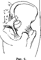
Treatment of such tumors is surgical. Surgical approaches to the sacroiliac joint. In the presence of an abscess, an incision according to Delbet is made along the greatest diameter of the abscess. Along the course of the fistula, sequestra and necrotic areas of bone are removed from behind, then the joint is trepanned, providing access to the anterior portion. For partial resection of the joint, the method of Oilier-Bardenheuer is used, in which the incision is made in the shape of the Roman numeral I. In cases of significant tuberculous involvement of the sacroiliac joint, extensive resection of the pelvis according to Rieder is used in rarer cases. Good drainage of the joint is also achieved with the approach to the sacroiliac joint according to Piquet. In recent years, American orthopedists have introduced a number of methods for arthrodesis of the sacroiliac joint. The main indications for this type of intervention are tuberculosis, chronic subluxation with rare pains, and osteomyelitis. The most developed methods are those of Smith-Petersen and Albright. The technique of arthrodesis according to Smith-Petersen (Fig. 6): an arcuate incision from the posterior superior iliac spine along the iliac crest, extending forward 2/3 of the distance to the anterior spine. The incision is made through the muscles and periosteum; sparing the superior gluteal nerve and artery. The periosteal-muscular flap is reflected outward. The sacroiliac joint is located 2 1/3 cm above the upper edge of the sciatic notch and 2 7/8 cm anterior to the posterior inferior iliac spine. Here a window is sawn out or chiseled out, including a portion of the ilium and sacrum. Through it, access to the joint is gained and the cartilage is removed. Then the window is again used to cover the opening made. Suture of the soft tissues. Ordinary bandage. Bed rest for 4 weeks.
Albright for arthrodesis of the sacroiliac joint uses a bone autograft, which he fixes between the first spinous process of the sacral vertebra and the posterior wing of the ilium. It is important to thoroughly scarify the bone over a considerable area where the graft will be applied, so that it will take well and not be subject to resorption. The second modification of arthrodesis according to Albright consists in exposing the joint through the posterior ligaments. A groove is made, a graft of appropriate size (its usual length is 7 cm and width about 1 cm) is taken, which is firmly hammered into the prepared bed. If it is necessary to arthrodese the lower part of the spine at the same time, Albright uses two grafts (Fig. 7): one longitudinal through the spinous processes of the lumbar vertebrae, and another, adjacent to it, goes at an angle, involving the sacrum and ilium.
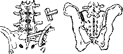
Figure 7.
Arthrodesis of the sacroiliac joint according to Smith-Petersen is relatively simple in technique and is especially applicable in traumatic injuries of this joint when it is necessary to eliminate movements in it that cause pain. The technique of arthrodesis according to Albright is applicable in tuberculous involvement. In the study of the anatomy and mechanics of the sacroiliac joint, some progress has been made in recent years, especially in connection with traumatic and professional injuries of the joint. In connection with this, the diagnosis of unclear 'lumbar pains' has been clarified and new operative methods have been introduced, mainly by an American group of orthopedists.
Related articles
Mentioned in
Cite this page
“Sacroiliac Joint.” Soviet Medical Encyclopedia. English translation of Bolshaya Meditsinskaya Entsiklopediya, 1st ed. (Moscow, 1928–1936), ed. N. A. Semashko. https://sovietmedicalencyclopedia.pages.dev/article/sacroiliac-joint/