Movements
Historical document, translated for reference. It reflects medical knowledge of the 1920s–30s and is not medical advice.
Summary
This article from the first edition of the Great Medical Encyclopedia (1928–1936) provides a biomechanical analysis of human movement. It defines the geometry, kinematics, and connectivity of the human motor apparatus, categorizing joints by their degrees of freedom and mobility.
Encyclopedia article (1928–1936)
MOVEMENTS. Contents: Geometry of movements....................452 Kinematics of movements...................456 Dynamics of movements....................461 Motor mechanisms............465 Methods of studying human movements.........471 Pathology of human movements............. 474 The movements and mobility of the human organism and its parts are so diverse and numerous that to survey them in an exhaustive manner is an impossible task. It can be asserted that there is not a single process during a human's life that would not contain within itself elements of movement. For any concrete approach to the question of human movements, it is necessary a) to isolate the basic, essential aspects inherent in any movement in general, and b) to consider each of these aspects in relation to human movements, noting and clarifying, firstly, that which is invariably inherent in any human movement, and secondly, that which is of the greatest practical interest. The signs of any movement in general are as follows: firstly, it occurs in space and represents a series of changes in time; secondly, it is always conditioned by a combination of forces, the sources of origin of which can be very diverse. Accordingly, the overview of human movements is presented in accordance with these signs.
Geometry of movements. The human organism is mobile as a whole, i.e., it can move from place to place (locomotor mobility, locomotion): walk, run, swim, etc.; furthermore, it possesses mutual mobility of individual parts (deformational mobility). Locomotion, if it is performed by the forces of the organism itself, is impossible without deformations; therefore, first of all, one should review the latter. The locomotor mobility of the human organism is unlimited, whereas the deformational mobility of the organism has limits depending on the structure and connection of various parts of the body. Connectivity of the parts of the human motor apparatus. The human organism possesses connectivity of two kinds: 1) kinematic connectivity, i.e., the inability to undergo deformations accompanied by the separation of some parts of the organism from others (in this, the human organism is similar to the majority of machines, except for projectile ones, such as a bow, an artillery piece, a weaving loom); 2) molecular connectivity, i.e., the absence of parts that would be connected to the organism otherwise than by forces of molecular cohesion of particles. In this respect, the organism differs from the vast majority of machines, not having at its disposal parts capable of performing a full rotation around an axis. This limitation of deformational possibilities is compensated in the organism by the enormous variety of partial deformations available to it. Being, by virtue of kinematic connectivity, capable only of limited translational deformations, and by virtue of molecular connectivity, only of limited rotational deformations, the human organism nevertheless, in the variety of its mobility, far exceeds the majority of artificial mechanisms created to date. Measure of mobility and measure of connectivity. The mutual mobility of the parts of the human organism is determined by two signs: a) the measure (or freedom) of mobility and b) the boundaries of mobility. Let us mentally divide the human organism into such parts that can, in a first approximation, be considered as non-deformable (shoulder, thigh, lower leg, lower jaw). We will henceforth call elements of this kind links. It is necessary to determine first of all what freedom of mobility (otherwise measure of mobility, Bewegungsfreiheit) represents. Mechanics considers every body located outside of connection with any bodies as possessing maximum freedom of mobility, since it can be unlimitedly displaced wherever and however one likes. The mobility of a body connected by rigid links to other bodies will be to one degree or another limited by this connection; the degree of this limitation is called the measure of connectivity. It can be said that the measure of mobility of a partially connected body will be determined if one subtracts the measure of its connectivity from the measure of maximum mobility (inherent in a body connected to nothing). The position of an invariable body in space is determined completely by the positions of three of its points not lying on one straight line; therefore, if one fixes immovably any three points of a body not lying on one straight line, then the body will lose all mobility, and the measure of its connectivity will be maximum. The measure of connectivity will be less if not three, but only two points of the body are fixed; under these conditions, it will already acquire some minimal freedom of mobility. If only one point of the body is fixed, its freedom of mobility will increase, while the measure of connectivity will decrease further. One can determine quantitatively what the freedom of mobility and the measure of connectivity of bodies fixed by a different number of points are equal to in the following way. Each point of a completely free body can move in space along three dimensions; the geometry of movements expresses this by saying that each point of a free body has three degrees of freedom of mobility. Therefore, if one fixes one point of an unconnected body, one can deprive it of three degrees of free mobility, in other words, create for it 3 degrees of connectivity. If one point of an invariable body is fixed, then its other point can move around the fixed point along a spherical surface, on which its position is determined by two coordinates (e.g., latitude and longitude); consequently, by fixing the second point of such a body, one gives it two more degrees of connectivity or deprives it of two more degrees of mobility. Two fixed points preserve for the body only one possibility of movement—rotatability around an axis passing through the two fixed points, whereby each unfixed point not lying on the axis will move along one possible line—a circle, on which the position of such a point will be determined by one coordinate (for example, ascension). By fixing such a third point, one adds one more degree of connectivity to the body and, as was already noted above, thereby deprives it of all mobility. Thus, the total number of degrees of connectivity possible for an invariable body is equal to 3 + 2 + 1 = 6; the number of degrees of mobility of an unconnected body in space is the same. The indicated relationships are more clearly visible from table 1.
Freedom of mobility of human joints. Since the human organism possesses ubiquitous connectivity, each movable rigid link of it has at least one point fixed to an adjacent link; therefore, the relative mobility of rigid links cannot exceed three degrees of connectivity (the lower jaw relative to the head, the shoulder and hip relative to the girdles). Freedom of mobility table 1. Number of fixed points: 0, 1, 2, 3. Number of degrees of connectivity: 0, 3, 5, 6. Number of remaining degrees of freedom of mobility: 6, 3, 1, 0. Number of degrees of freedom of mobility of an individual point: 3. The soft parts of the body (tongue, heart) cannot, of course, be expressed by a number of degrees, because due to the compliance of their connections, one cannot speak of the fixing of one of their points relative to others. The measure of connectivity (and consequently the measure of mobility of rigid links) depends on the different structure of the joints connecting them. Below is a summary of the measure of mobility and connectivity of the main joints of the human skeleton. 3 degrees of freedom of mobility (3 degrees of connectivity): intermaxillary joint, scapulohumeral joint, metacarpophalangeal joints, hip joint, acromioclavicular joint. 2 degrees of freedom of mobility (4 degrees of connectivity): atlanto-occipital joint, humeroradial joint, radiocarpal joint, carpometacarpal joint of the thumb, knee joint in flexed position. 1 degree of freedom of mobility (5 degrees of connectivity): atlanto-axial joint, humeroulnar joint, radioulnar joint, interphalangeal joints of the fingers and toes, sacroiliac joint, knee joint in extended position, talocrural joint, subtalar joint. 0 degrees of freedom of mobility (6 degrees of connectivity): cranial sutures. Boundaries of mobility of human joints (see figure 1). The boundaries of mobility of links relative to each other are conditioned not by the geometric properties of the joints, but by the structure and compliance of the ligamentous apparatus and the shape of the apophyseal ends of the bones; thus, three-degree joints with extremely narrow boundaries of mobility are possible (acromioclavicular joint) and, conversely, one-degree joints with very wide boundaries of mobility (humeroulnar joint). In general, the boundaries of mobility not only nowhere allow a rotation of a link by 360°, as indicated above, but do not even reach 180°, constituting a maximum of 170° (knee joint). The greatest ranges of mobility in individual joints on average are given in table 2 on p. 455. Table 2. Knee joint, passive.......... 15°. Carpometacarpal joint of the middle finger ... 8°. Freedom of deformation of parts of the human body. If a movable link is connected by a partial connection to a completely immovable body (e.g., a wing
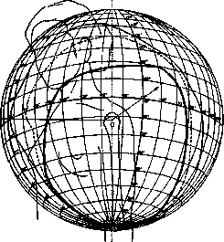
Figure 1. Limits of shoulder mobility in the scapulohumeral joint. The inner, bold curve represents the limits of shoulder mobility with a fixed scapula; the dashed curve represents the same with a mobile scapula; the outer solid curve represents the same with mobile scapula and clavicle. (According to Braune and Fischer.)
semaphore with a post), then the measure of its connectivity fully determines the measure of its mobility. If, however, two mobile links are connected by a partial link (e.g., the legs of a compass), then the measure of their connectivity determines only their relative mutual mobility, and not the absolute mobility of each link individually. The measure of the relative mutual mobility of two articulated links is called the freedom of deformation of a two-link kinematic chain. This freedom is obviously equal to the freedom of mobility of one of its links relative to the second, if this second one is made fixed. It is also obvious that the freedom of mobility, for example, of the thigh relative to the pelvis (assumed to be fixed) is equal to the freedom of mobility of the pelvis relative to the thigh, and both are equal to the freedom of deformation of the 'thigh-pelvis' two-link chain. The freedom of deformation of more complex kinematic chains is determined by the variety of shape changes accessible to these chains. The freedom of deformation of an open multi-link kinematic chain (i.e., a chain that breaks into two disconnected parts upon the cutting of any of its joints, such as 'pelvis-thigh-lower leg-talus-calcaneus') is equal to the sum of the degrees of relative mobility of all pairs of adjacent links of which it consists. The freedom of deformation of a closed kinematic chain (i.e., a chain that does not break into disconnected parts upon the cutting of one of the joints, such as the 'shoulder-ulna-radius' chain) is less than such a sum. Thus, the open kinematic chain 'pelvis-thigh-lower leg' possesses 3 degrees of freedom of deformation due to the hip joint + 2 degrees due to the knee joint. Below is a summary of the freedoms of deformation of the main systems of the human body. Table 3. Finger of the hand... 2. Hand... 33. Forearm + hand... 36. Entire arm from the shoulder... 37. Entire arm from the scapula... 40. Arm and shoulder girdle... 43. Entire body... 191. Head and neck... 21. Head with lower jaw... 3. Head and 2 upper vertebrae... 11. Foot... 13. Lower leg + foot... 15. Entire leg from the thigh... 18. It should be added that the kinematic chains of the vast majority of machines are closed kinematic chains, and therefore their freedom of deformation in most cases is significantly less than the freedom of deformation of the human body. For comparison, the freedoms of deformation of some machines are given. Table 4. Bicycle... 5. 4-axle locomotive without leading wheels... 10. 5-axle locomotive with a swivel bogie... 15. Automobile engine... 1. Rotary printing press... 1. 'Underwood' typewriter... about 60. Concert grand piano... 480. The active mobility of the human organism is everywhere less than the passive one. This is caused either by the fact that the muscles of a given joint are too weak to fully utilize its mobility (knee flexion, hyperextension of fingers), or by the fact that there are no suitable muscles for performing a passively possible movement (e.g., rotation of fingers in the metacarpophalangeal joints) or suitable innervation (e.g., rotation of the hand in the wrist joint), or finally by the presence of insurmountable synergies (see below), i.e., associated innervations that make it impossible to actively perform certain complex systemic movements separately. Thus, few people are capable of flexing the distal phalanges separately from the middle ones; the passive mobility of each vertebra relative to the neighbor has three degrees, whereas actively we are not in a state to perform movements in any one isolated intervertebral joint, etc. Even less is the freedom of deformations for habitual movements, which, as indicated below, almost entirely consist of complex synergies. Kinematics of movements. Form of human movements. Even the simplest direct observation shows that the human organism does not use all the possibilities of mobility that it possesses: a kinematic chain with two degrees of freedom of deformation, fixed at one end, is capable of rectilinear movement of the other end; meanwhile, approximately rectilinear movements of the ends of such a free system as the arm (43 degrees of freedom of deformation) are a rarity, and strictly rectilinear ones are not encountered at all. Just as rare are breaks in movement trajectories at an angle. As a rule, human movements possess to a greater or lesser degree smoothness and roundness. One could assume that rectilinear movements are absent precisely because they require complex synergy and that, by virtue of the rotational structure of most joints, arcs of circles should predominate in human movements, but precise observations show that arcs of circles are no less a rarity in human movements than straight lines. (For the kinematics of the most important motor syndromes, see below, as well as the corresponding words.) Regarding the questions of the form of movement trajectories, there exists an extremely small number of quantitative studies. Qualitative observations cannot bring much benefit in this area, since, first of all, they are extremely inaccurate and subjective. Furthermore, the following must be pointed out. Only during rotational movement around a fixed axis (such movements the human organism never performs) and during translational displacement (unrealizable for the organism) do all points of a moving system perform movement of exactly the same form. For any other movement, the forms of the trajectories of all points of the moving system differ from one another, and therefore, if one does not make precise quantitative measurements, it is easy to overlook points whose movement is in some respect regular, even if such points exist in the system. Some general propositions can, however, be established already now. As already stated, the variety of active movements of individual systems of the human organism is incomparably less than the variety of those passive movements that are accessible to this system according to the freedom of its mobility. If one compares the freedom of mobility of a link or system of the human body to a large plain, then the active trajectories accessible to this system will be similar to highways on this plain: the place occupied by them will be vanishingly small compared to the entire surface of the plain (see Figure 2). Continuing this comparison, it will be possible to say that the 'plains' of the freedoms of mobility of various systems of the human body will be saturated with the 'highways' of the trajectories of active movements to a very different degree. Although between the vastness of the anatomical mobility of one or another system or link and the variety...
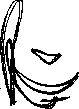
There is a certain parallelism between the anatomical mobility of a system and the active movements available to it (active mobility in the shoulder joint is richer than that in the talocrural joint), but the variety of active movements possible in a given system is determined mainly by the innervation development of the motor centers of this system. With almost identical anatomical mobility, the shoulder is incomparably more diverse in its active mobility than the hip; the fingers of the right and left hands are anatomically mobile to the same degree, and yet the variety of their active mobility is very different for one hand versus the other. The anatomical mobility of the toes is almost no less than that of the fingers (the same freedom of deformation and only slightly smaller boundaries of mobility), yet the active movements of the toes, except for the hallux, are reduced only to the possibility of simultaneous flexion or extension of them all at once in all phalanges. The variety of active mobility does not depend noticeably on the number and variety of muscles surrounding a given link; the muscular fund of the hip joint is greater in the number of muscles and more diverse in their arrangement than the muscular fund of the shoulder joint, the active mobility of which is significantly greater than that of the hip. The kinematic complexity of any given trajectory of movement of a person, i.e., the complexity of its spatial form, is conditioned again not by the measure of mobility of a given point of the moving system, but by the magnitude of the mass concentrated at this point: as a rule, the greater the latter, the simpler the form of movement (see Figures 3 and 4). Hence, the colossal mobility of the hand and fingers, which are rich in both degrees of freedom of deformation and highly articulated innervations, and at the same time poor in mass, is understandable. [Figure 5 shows a cyclogram of reloading a rifle (from the author's photograph)—an example of an automated movement of high complexity.] Both the variety of movement trajectories and the complexity of the latter decrease significantly even in richly innervated systems under the influence of the automation of human movements; the high degree to which the uniformity of successive automated movements can reach is shown in Figures 2 and 6.
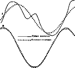
Speeds of movements. The speeds of movements along trajectories have also been studied very little. The greatest speeds of movements of parts of the human organism are achieved by the distal ends of the limbs during swinging movement from the proximal joint: by the hand during movement from the shoulder girdle and by the foot during movement from the hip joint. The hand can reach speeds of up to 20 m per second (72 km per hour) during a throwing movement; the foot gives slightly lower figures during fast running. During rhythmic movements, the speeds are lower, and they depend not on the tempo of the movements, but on their range: with an increase in the tempo of movement, its range usually decreases, and in this case, the speeds do not increase much. Under normal physiological conditions, the speeds of the hand and foot of a healthy person often reach 5-6 m per second. The maximum tempo of movements depends directly on the mass and moment of inertia of the moving part. The highest tempo (up to 8-10 movements per second) is possessed by the movements of the fingers, the lowest—by the trunk when swinging it in the hip joints. Rhythmic movements of a person. A completely general kinematic law has been successfully formulated to date only for rhythmic movements. An exact exposition of this law contains some mathematical difficulties; therefore, only the most general concept of it is given here. Any oscillatory rhythmic movement can represent either 1) a simple pendulum-like (sinusoidal) oscillation or 2) a sum of simple pendulum-like oscillations of different frequencies, simultaneously performed by the oscillating body. In order for such a sum of oscillations to still be rhythmic, all its component oscillations must have a common rhythm; in other words, the durations of the complete cycles of each of the component oscillations (or, as they say, the periods of oscillations) must relate to each other as 1:1/2:1/3:1/4, etc. In the form of such a sum of sinusoidal oscillations, of course, every rhythmic movement of a person can be represented, more precisely—the movement of every point of the moving organ. Such movements include: walking, running, turning a handle, numerous work movements (filing, blacksmith and locksmith striking, movements of the hand on the piano), pathological movements (tremors, clonus), etc. In all such movements, the basic rhythm is represented by the so-called fundamental oscillation, the period of which is equal to unity; additional oscillations with periods of 1/2, 1/3, 1/4, etc., are layered onto it. The ranges, or amplitudes, of the component oscillations can, of course, be very diverse; it is obvious that the more insignificant the amplitudes of the additional oscillations are in comparison with the amplitude of the fundamental oscillation, the simpler the structure of the movement.
The kinematic law of the progression of rhythmic movements of a person, which was mentioned above, states that the amplitudes of the additional components have different values relative to the amplitude of the fundamental oscillation for different points of the moving human organ and for different cases of movement, namely, that they are smaller (and consequently the movement is simpler): 1) the greater the mass and the greater the moment of inertia concentrated in a given moving point and 2) the higher the tempo of the movement. Thus, for a large moving system (e.g., the leg during walking), the movements of the centers of gravity of the parts of the system will be simpler, closer to a pendulum-like oscillation, than the movements of the joints of the same system, and the movement of the center of gravity of the entire system will be simpler than the movements of the centers of gravity of its parts (see Figure 7). Let us clarify this with examples. During normal walking, the amplitudes of the component movements of various points of the leg (referred to the fundamental component, taken as 100%) are equal to: Table 5. Moving point. Amplitudes of 1st component, 2nd, 3rd, 4th. Center of gravity of the knee joint: 100%, 26.1%, 20.6%, 22.5%. Tip of the foot: 100%, 4.1%, 8.2%, 2.1%, 2.8%. Center of gravity of the lower leg: 100%, 20.0%, 4.7%, 3.8%, 2.1%. Center of gravity of the entire leg: 100%, 0.7%, 0.7%, 1.6%, 0.3%, 0.5%. With an accelerating tempo of piano octave movements, there are the ratios indicated in Table 6 for the center of gravity of the hand (performer—a great virtuoso).
MOVEMENTS
Thus, the mechanical structure of rhythmic movement is, as a rule, simpler the faster the movement is and the greater the moments of inertia of the moving masses are. (Regarding the kinematics of the most important motor syndromes - see Running, Speech.) The study of the kinematics of human movements in normal and pathological cases is of enormous diagnostic interest (see below). Let us mention for now that, as clinical experiment shows, the finest deviations of human movements from the norm at the onset of any disease, deviations completely inaccessible to the naked eye, are clearly revealed by recording these movements using a sensitive method, which should in the near future make the study of the kinematics of human movements one of the important tools of clinical work. Dynamics of movements. Nowhere, perhaps, have so many mistakes and hasty judgments been made as in the questions of the dynamics of human movements. This is understandable: it was pointed out above that the motor system of the human body belongs to the number of the most complex, immeasurably diverse, and free mechanical systems, for which a satisfactory mechanical interpretation is generally impossible. For a mechanic, the human organism is a tangle of complex and insoluble problems. It is natural that in this field (which is maximally difficult for a mathematician, but out of ignorance seems simple to a physician), the physician is not insured against gross errors. Therefore, one should examine here with particular care the basic facts of the dynamics of human movements, leaving aside more complex phenomena of a secondary order. External and internal forces. The body of a mobile organism is constantly under the action of external and internal forces. External forces include the following: the force of gravity, the force of resistance of the medium (air, water when swimming, etc.), the forces of support reactions, and finally, all kinds of inconstant forces acting from the outside (jolts, impacts, etc.). Internal forces include: molecular forces of cohesion of particles, elastic forces arising during muscle excitation, stretching of ligaments, compression and bending of cartilage and bone, and friction forces arising during movement in all moving parts of the organism. If a prolonged equilibrium is observed between all these forces, the organism is at rest. If, however, there is no such equilibrium of forces, i.e., if the resultant of all forces and its moment are not equal to zero, then the organism begins to move, which depends entirely on the magnitude, direction, and moment of the resultant. Since all the forces listed above (except only the force of gravity) represent variable forces, each of them, by changing, can cause the beginning or a change in the movement of the organism. From the point of view of the mechanics of movement, it is essential only whether a given force or resultant is an internal or external force in relation to the organism or to a given part of it (kinematic chain), regardless of the source and origin of this force. Therefore, it is necessary to define what is an internal and an external force in relation to a given kinematic chain. According to the third principle of mechanics, the action of each force is balanced by an equal force counteraction directed along the same straight line as the given force, but in the opposite direction. If the point of application of the force and the point of application of the counterforce are inside the given chain, then the force is considered internal in relation to the chain. Thus, for the chain "the entire arm from the shoulder joint," the tension force of the m. brachialis interni is an internal force, and the tension force of the m. pectoralis major is an external force; in relation to the "forearm-hand" system, however, the tension force of the m. brachialis interni is already an external force. However, the force of the m. brachialis, acting on the humerus, is transmitted along it to the ligaments of the shoulder joint, i.e., to points external in relation to the system "the entire arm." The counterforce for this transmitted force is applied at the center of gravity of the kinematic chain suspended from the shoulder joint. Thus, any force that is internal in relation to a given suspended chain also creates an external force, but always of the same order: a force directed along the straight line connecting the center of gravity of the chain and its point of suspension. In other words, no internal muscle of a given kinematic chain can act on the center of gravity of the chain otherwise than by pulling or pushing it along the straight direction connecting it with the point of suspension of the chain. The moment of a force passing through a given point is equal to zero in relation to this point; thus, one arrives at the fundamental theorem of muscle dynamics: the moment of a muscle force, internal in relation to a given system, is not equal to zero for all internal points of the system and is equal to zero for all points lying outside it. Muscle moment. Action of a muscle external and internal in relation to the system. The above-mentioned theorem of muscle dynamics provides an unmistakable path to determining whether the muscles of a given joint participate in the movement or position we are interested in and to what extent. It is sufficient to determine the force moments of the moving system in relation to each joint: if the moment in relation to a certain joint is equal to zero, then its muscles do not participate in the given act; if it differs from zero, then its magnitude directly characterizes the measure of the efforts of the muscles of the given joint. For the entire human body as a whole, each muscle is internal; consequently, the moment of each muscle of the body individually, and therefore the moment of all of them taken together, in relation to an external point of support is equal to zero. Therefore, with one external point of support, no muscle of the entire body can shift the center of gravity of the body otherwise than along the straight line connecting it with the point of support; in other words, with one point of support, a person can move from their place only at the expense of an external force (gravity, a jolt, etc.). If there are more than one point of support, then, of course, the internal forces of the body will possess a moment different from zero, at least in relation to one of the points of support, and then any displacements are possible. Parametric, tonic, and contractional forces. From the point of view of the physiology of movement, the forces acting in a given kinematic chain should be subdivided differently. According to their physiological significance, these forces fall into three groups: 1) forces arising outside the organism, i.e., forces whose presence and value do not depend on the organism in any way—independent forces (forces of gravity, wind, external jolts, etc.); 2) forces arising in the organism and completely depending on the position and state of movement of its parts—parametric forces (forces of tension of ligaments and tendons, elastic stresses of bones and cartilage, internal friction of muscles, inertia forces of links, etc.); finally, 3) forces arising within the organism itself, but capable of changing independently of the positions and states of movement of its parts—these are muscle forces (tonic and contractional). At each given state of movement of an organ, the organism does not have access to influence either independent forces, the sources of which lie outside it, or parametric forces, for each of which only one unchangeable curve of successive values is possible during a given movement, and only the forces of muscle tension are accessible to constant control and regulation by the organism. Thus, physiologically, a muscle is not only not the only engine of the organism, but not even its main engine, but only the only controllable engine. The motor system of a person can be likened to a sailing ship with an auxiliary steam engine—a collection of muscles. Refining this comparison, it must be said that the "steam engine" of the organism itself is not at all weak and in most cases is quite sufficient for moving the ship against any wind and storm, but that the entire evolution of motor mechanisms, both in general ontogenesis and in the acquisition of any new motor skills, boils down to a continuous growth in the ability to use the wind and sails and to an ever greater economy of fuel. Studying the physiology of the act of walking in a healthy person, one can see with extraordinary clarity how widely "free" forces are used for walking and to what a minimum active muscle activity is reduced. Relationship between tension, muscle contraction, and movement. The dynamic action of a muscle depends on its tension, but, as should be clear from the previous exposition, this tension does not at all have to be caused by muscle excitation. An unexcited muscle is the same elastically-tensioned formation as a ligament, fascia, or bone, only with different characteristics of extensibility and elasticity. The main difference between the function of a muscle and the function of a ligament lies only in the fact that for a ligament, each given stretch corresponds always to one definite tension, whereas for a muscle, at one and the same stretch, the tension can be different, depending on the measure of its excitation. Thus, muscle excitation is a path not to causing tension, but to changing it.
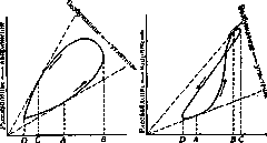
A change in muscle tension causes a change in the resultant of all the numerous forces (independent, parametric, and muscular) acting on a given kinematic chain (the 1st causal link), and a change in the resultant causes a change in the movement of the chain in the order described above (the 2nd causal link). The movement of the kinematic chain changes, generally speaking, the distances between the attachment points of the muscles located on this chain. If these points move apart, the muscle is stretched; if they move closer together, the muscle, as an elastic formation, shortens; but since, given the extremely complex interaction of forces acting on the system, it is completely impossible to say in a general form how an increase in the tension of a given muscle will affect the movement of the entire system, it is also impossible to predict whether the muscle will stretch or shorten as a result of this movement. Thus, under physiological conditions, the "contraction" of a muscle has no direct connection with an increase in its tension and is merely one of the possible consequences of the system's movement, just as possible as the stretching of this muscle or its remaining at the same length. Situations in which muscle contraction is the cause of movement do not exist; situations in which muscle contraction is an obligatory and unambiguous consequence of movement caused by its own tension relate only to the simplest, maximally schematized cases (a muscle in a myograph, isolated local stimulation of a muscle by faradization, etc.). Indicator diagram of muscle work. Errors arising from the failure to distinguish between excitation, tension, and contraction of a muscle depend on the confusion of the concepts of force and work of a muscle. Perhaps Figure 8 will best explain the actual relationships of all these quantities. If one plots the changes in muscle length (contraction-stretching) on the abscissa axis and the changes in its tension (tension-relaxation) on the ordinate axis, then each muscular process will be represented on such an "indicator diagram" by a curve; if, after some time, the muscle returns to the same state of length and tension from which it began the given process, then the curve will be closed, as in the figure. The area bounded by this curve represents the work of the muscle: work performed by the muscle if the curve goes around this area counterclockwise (see Figure 8, 9—the lower ring of the figure eight), and work absorbed by the muscle if the curve goes clockwise (see Figure 9—the upper ring of the figure eight). The measure of muscle excitation is characterized by the ratio of muscle tension to its corresponding state of stretching (since both excited and unexcited muscles
Stretching-stretching
Contraction-stretching
Figure 8. Indicator diagram of work Figure 9. The same-more
...a complex case. Muscles tense upon stretching and relax upon contraction): the more strongly a muscle is excited, the greater this ratio is. Therefore, on a graph, excitation will be characterized by an increase in the slope of the straight line connecting a given point on the graph with the origin of coordinates; inhibition or calming will correspond to a decrease in this slope. In Fig. 8, at ordinate D, the length and tension of the muscle are at their minimum; from D to A, the muscle is stretched, tension increases, and the measure of excitation falls; from A to B, all three values increase, i.e., the muscle is excited and tenses, but is stretched; from B to C, the measure of muscle excitation continues to increase, the muscle contracts, tension falls; from C to D, excitation and tension fall, while the muscle contracts. A more complex case is depicted in Fig. 9. Here, the increase in excitation occurs in the segment AB, during continuous stretching of the muscle; starting from B, excitation begins to fall, while the muscle is still being stretched until C, and contraction occurs from C to D, with a continuous fall in both tension and the measure of excitation. (This case is borrowed from experiments with hand movements during a piano strike.) Thus, muscle excitation can begin and end during the stretching phase, whereby all the energy of muscle excitation will pass into the potential elastic energy of the stretched muscle, and this converted energy will then, in the contraction phase, i.e., a few fractions of a second later, pass into the mechanical work of the muscle. Motor mechanisms. Muscle excitation depends only on the activity of the central nervous system and in no degree depends on the state of the motor organ; but muscle tension inevitably depends on the state of its stretching, i.e., on the movement of the organ to which it belongs. Thus, the tensions of the muscles of a given organ are, on one hand, the cause of its movement, and on the other, the consequence of that same movement. Thus, in human movement, a closed chain of interactions takes place between efforts (muscle tensions) and the positions of the organ: the first influences the second, and the second influences the first. Such chains of interactions are expressed in mechanics by second-order differential equations, but their consideration is very complex. One can only point to the most basic properties of such equations, which determine human movement. Every equation contains coefficients, or parameters; for example, in an ordinary quadratic equation x2+px+q=0, such parameters are the values p and q. The parameters of the equations determining human movement will be the constant values of the moments of inertia of the links, the coefficients of elasticity of ligaments, friction of parts, etc.; therefore, the forces arising in the centers of gravity of the links, in ligaments, rubbing parts, sarcoplasm, etc., were called parametric forces above. The difference between a differential equation and a simple algebraic one, like the one cited above, consists in the fact that an algebraic equation has one or several separate solutions (e.g., a quadratic equation has two solutions), whereas a differential equation has an infinite number of possible solutions. Which of these countless solutions will take place, in other words, what movement will occur given a certain change in muscle excitations, will depend not on the form of the equation and not on its parameters, but on the so-called initial conditions of the movement. For the differential equation of the movement of a part of the human body, such initial conditions can serve, for example, as the initial position and initial velocity of this part. A change in the initial conditions can completely change the entire kinematic effect, the entire external physiognomy of the movement, even if the differential equation determining it, and consequently the law of change of muscle tensions, remains exactly the same. Muscle-force scheme and initial conditions of movement. Proprioceptive mechanisms and proprioceptive coordination. Knowledge of movement ensures, according to what has been said, knowledge of its muscle-force scheme; but the reverse transition is impossible: knowledge of the muscle-force scheme is insufficient to predict the movement, because one and the same scheme, depending on the initial conditions, can produce a multitude of different movements. One can say that the diversity of movements is greater than the diversity of muscle-force schemes. By virtue of this, the organism, if it were limited to the innervation-based activation of any specific muscle-force scheme, could not have any guarantee that this scheme would produce exactly the spatio-temporal movement that it needs at a given moment. For the performance of a kinematically defined movement, once-and-for-all defined centrifugal impulses are insufficient; control centripetal impulses are also necessary, conditioned by the already mentioned initial conditions and capable of introducing corrections into the initially activated innervation scheme depending on the concrete spatio-motor situation. Such impulses are the so-called proprioceptive impulses, arising in the nerve endings of muscle tendons, articular capsules, deep layers of the skin, etc. Thanks to proprioceptive reflexes, along with the aforementioned mechanical interaction between muscle tensions and positions, another analogous interaction arises, localized already in the central nervous system; central motor impulses cause changes in the movement of the kinematic chain, and these changes, with the help of proprioceptive reflexes, in turn change and correct the central motor impulses. And here, consequently, the interaction must be characterized by a set of differential equations of no lower than the second order, but in contrast to the first case, the real construction of these equations, given the current state of our physiological knowledge, does not yet appear possible. The activity of proprioceptive reflexes conditions the possibility of so-called proprioceptive coordination of a single movement (the term coordination represents a kind of Sammeltopf [catch-all], where the most diverse motor mechanisms are placed). This type of coordination takes place in all movements of a normal person, starting from the moment of myelination of the posterior columns; it is partially impaired in diseases of the posterior columns (ataxia), but the complete exclusion of this coordination conditions not ataxia, but the complete impossibility of any movements whatsoever. The course of this coordination is best studied at the present time for rhythmic movements (for example, the scratching reflex). Spatial shortening of one muscle causes reflex inhibition of its antagonists (see below) and excitation of the protagonists (see below); innervation of one muscle group is thus accompanied by denervation of the opposite group. Conversely, sharp stretching of a muscle excites it itself (muscle recoil, Rückstoss). Thus, rapid flexion of a finger, causing stretching of the finger extensors, leads to an extensor recoil (Lewy). How proprioceptive coordination proceeds during non-rhythmic movements is as yet very little known. Systemic nature of motor innervations. Just as little is known to the present time about the place of origin and the structure of the innervation muscle scheme of movement itself. Whether by virtue of the wide irradiation of proprioceptive impulses or by virtue of the structure of the innervation scheme itself, under no conditions in a healthy state does isolated excitation of one muscle or even one group of adjacent muscles take place. The possibility of isolated innervation, undoubted for spinal motor cells, has not been proven either for the motor layers of the cerebral cortex or for subcortical nuclei with motor functions (striatum, pallidum, nucleus ruber). Central excitations are always systemic, simultaneously encompassing vast muscle aggregates. This fact has received the name of muscle synergy. Like coordination, this term is as yet devoid of any physiological and localizational specificity. Synergies. Some types of synergy are apparently innate and universal human systemic innervations (e.g., extension of the wrist when clenching fingers into a fist), inevitably occurring upon innervation of a given muscle group; other types obviously arise gradually, as a given motor mechanism is developed, and do not figure in excitations of the same muscle groups, but in other constellations (e.g., the synergy of the pronators and supinators of the forearm with the flexor-extensor system of the fingers, functioning during writing). -In this group of synergies, one rightly sees the source of all possible motor skills. A special position among synergies is occupied by so-called associated movements, arising in adjacent kinematic chains during the purposeful movement of one of them.
In a significant number of cases, associated movements represent rudiments of once-purposeful extensive synergies (e.g., swinging the arms while walking, which has been preserved from the times of walking on four limbs); sometimes the results of motor-innervation irradiation of excitation during the acquisition of an unfamiliar motor skill are also classified as such (sticking out the tongue in children while drawing and writing, the inability of beginners learning to play the piano to make different movements with two hands due to associated movements, etc.). Among the most extensive purposeful synergies, encompassing all the muscles of the body, are locomotor movements—walking, running, swimming, etc. Static synergies, which ensure the possibility of standing and sitting, should be attributed to equally extensive synergies. Indirect clinical indications allow for the presumptive localization of synergies in the subcortical centers of the brain. Rhythmic innervations. The temporal sequence of systemic innervations is also determined by the activity of the central nervous system. For the simplest rhythmic sequences, the temporal alternation of innervations and denervations is regulated, as we have already seen, by the activity of simple proprioceptive reflexes. For more complex rhythmic sequences encompassing large groups of muscles, the mechanism of rhythm regulation is still very unclear, although there are grounds to assume that rhythmic coordination is also determined to a significant extent by subcortical (and specifically pallidal) activity. Innervation structure of human movements. The central nervous system has at its disposal, for the execution of movements, a very finely articulated musculoskeletal apparatus, characterized above. Its connection with this apparatus is carried out through peripheral centrifugal nerves running from the anterior roots of the spinal cord and from sympathetic ganglia to the neuromuscular junctions, and through centripetal nerve fibers extending from articular surfaces, tendons, and the muscle perimysium to the intervertebral ganglia. Through this connection, all movements accessible to the human organism are carried out by the central nervous system, regardless of which parts of the nervous system they originate from. Thus, in the majority of complex human movements, controlled simultaneously by a whole series of motor divisions of the central nervous system, very complex impulses flow along the peripheral nerves, arising as a result of the superposition of numerous central impulses of various origins. Spinal motor reflexes possess the simplest structure, the motor impulse of which arises in the cells of the anterior horns of the spinal cord in response to excitation arriving from the periphery of the body. However, all these reflexes encompass more than one muscle with excitation; only hypothetical local reflexes (Eigenreflexe, Spiegel) are apparently limited to the involvement of one single muscle (tendon reflex of the quadriceps extensor of the knee, the triceps of the arm, etc.). Spinal reflexes, as a rule, already encompass an entire group of muscles of associated action. From the point of view of the participation of individual muscles in the complex act of human movement, the muscles participating in the movement are endowed with special names. Muscles similar in their function to a given muscle are called agonists of that muscle (e.g., m. brachialis internus is an agonist of the biceps brachii for elbow flexion). Muscles that are dissimilar in their function to a given muscle but perform a role associated with it during a certain movement are called protagonists, synergists, or synergetics of that muscle. For example, when raising the arm (elevation of the arm), the muscles of the scapula (m. serratus ant. major, m. trapezius, etc.) work as synergists for the deltoid muscle, although their general motor function is completely different from that of the deltoid. Finally, muscles that perform a function opposite to the function of a given muscle are called antagonists of that muscle. It should be emphasized that both protagonism and antagonism are not, in the vast majority of cases, constant functions of a muscle in relation to a given one; for each movement, the distribution of roles between muscles will be different. Thus, for lateral abduction of the shoulder, the m. pectoralis major is an antagonist to the m. deltoideus; however, for raising the shoulder forward medially, both of these muscles are mutual protagonists. For elbow flexion, the m. brachialis internus and m. biceps brachii are agonists, for supination of the forearm they are protagonists, and for light flexor pronation, they may turn out to be antagonists. Spinal motor reflexes usually encompass an entire system of agonists and protagonists simultaneously. Thus, the reflex of withdrawing the hand from pain already encompasses an entire system of muscles. It is even more important to note that one and the same irritation from the periphery is capable of causing a pain reflex of a very different motor structure depending on the initial position of the reacting limb. Upon pain stimulation of the fingers of the hand with an induction current, flexion of the shoulder will occur if the hand was in front, and extension if it was behind. In lower vertebrates, spinal reflexes can also produce a rhythmic form of alternation of impulses between agonists and antagonists (the wiping reflex in a decerebrated frog). Whether the same alternation of purely spinal origin is possible in humans is difficult to say. More complex human movements are performed in a healthy state mostly with the participation of the motor zone of the cerebral cortex by means of impulses transmitted to the peripheral neuron through the pyramidal tract. However, there is not a single movement that would be performed only by the pyramidal nervous system; the cortex never acts in movement in isolation from subcortical motor centers connected to the spinal cord by means of extrapyramidal tracts. The measure and degree of participation of one or the other system are very different in different cases of movement, but it is now certain that, on average, the measure of participation of the extrapyramidal system in normal movement is greater than the measure of participation of the cerebral cortex: the existence of movements performed by the extrapyramidal system alone without the participation of the cortex is very likely. Movements that include pyramidal innervations are still often referred to as voluntary or volitional movements, although both of these names, due to their subjectivity, should have been removed from use long ago. It would be more accurate to speak of cortical movements, in which the extrapyramidal system and the cortex participate, in contrast to subcortical or extrapyramidal ones, which are innervated only by the striopallidal apparatus. Cortical movements arise in humans only a few months after birth, in connection with the delayed myelination of the pyramidal tract, and the infant in the first months innervates its movements only extrapyramidally. In an adult, three forms of interaction between the cortex and the subcortical apparatus can be distinguished. 1. Clearly cortical movements (according to the old terminology—voluntary). All single complex impulses and chain movements consisting of a series of non-identical complex impulses should be attributed to such movements. These are: the movements of an artist painting a picture, the movements of disassembling and assembling some mechanism, complex sorting, the movements of a surgeon during an operation, etc. In these movements, the participation of the cortex is undoubtedly predominant, and the extrapyramidal innervations constitute only a general background and foundation. Movements during periods of acquiring a new motor skill—walking, speech, writing, etc.—also belong to this group. 2. Movements with a predominance of extrapyramidal innervations (automated or habitual movements). The overwhelming majority of human movements belong to this group: walking, writing, speech, habitual professional movements, etc. In these movements, the participation of the cortex is so reduced that the individual constituent impulses are not perceived by the subject at all, and psychologically these movements are perceived as a direct transition from an image (visual image of a letter, auditory image of a word, etc.) to its reproduction. All movements of this kind are not innate but are developed by more or less prolonged exercise, leading to their automation. It is interesting to note one property characteristic of this group of movements and distinguishing all its representatives from other groups of movements. It is precisely in these (and only in these) movements that the individual manner of movement of a given subject is manifested. Walking is colored by the sign of gait, writing by handwriting, speech by accent, piano playing by touch. In movements of this kind, it is possible to observe constitutional differences in the manner of movement. A greater degree of participation of the subcortical apparatus manifests itself in the smoothness, gracefulness of movements, and a tendency toward extensive and well-coordinated synergies, etc. The predominance of the cortex leads to angular, sharp, precise, but less elegant movements. 3. Movements in which the cortex apparently does not participate at all in a normal state, although it can participate. These movements are called automatisms. Such movements include: breathing, blinking, yawning, stretching, defecation, etc. Movements of this group are innate; they exist in a child from the first day of its life. Automatisms differ from reflexes partly by their greater complexity, and mainly by the fact that they are excited not from the periphery, but from the center due to causes arising within the organism itself.
In diseases of the extrapyramidal system, new pathological automatisms may appear: tics, athetosis, chorea, etc. (see below—pathology of Movements). Methods of studying human movements. In a brief overview, it is impossible to cover in any detail the numerous methods that have been and are being used to study human Movements. Therefore, only the most important of them, and those of significance for clinical practice, will be presented here. - Pre-photographic methods. Among the pre-photographic methods of studying human Movements, the methods of pneumatic registration using the so-called Marey capsule have retained their significance to the present time. It is difficult to name movements to which one has not attempted to apply pneumatic registration. For recording muscle tension, in particular, the method developed by Bruzhes in the USSR and Johnen in Germany is significant: the limb segment being studied is encircled by a pneumatic cuff under pressure (as in measuring blood pressure), connected by a tube to a Marey capsule. The thickening of the tensing muscles increases the pressure in the tube and thereby moves the pen of the Marey capsule. No less numerous are the methods of recording by means of mechanical transmission of Movements to a recording device. The Weber brothers studied knee flexion during walking by connecting the foot to the greater trochanter of the femur with a taut tape and recording the change in distance between both points. Isserlin, and later Lewy, studied finger movements by connecting the fingertip with a thread passed over a pulley to a pen writing on a drum. A similar technique was used by Fischer and Wodak (M. Fischer, Wodak) for recording arm displacements during reflex changes in tone; recently, this same method has been used for recording the knee reflex. Sommer built a so-called three-dimensional apparatus (see Figure 10), which allows, by means of a lever transmission, the recording of the displacements of the fingertip (or the tip of the foot) along all three coordinates of space. - In great use in
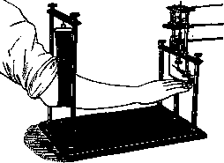
Figure 10. Sommer's three-dimensional apparatus.
clinical practice are methods in which the part of the body being studied draws its own movement without any transmission whatsoever. This includes recording gait by obtaining imprints of coated soles on a paper track, as well as the method of cephalography, i.e., recording head displacements while standing by means of a point fixed on the crown of the head and drawing on a smoked sheet spread over the head. The method of recording changes in joint angles, being developed by Filimonov, promises to yield valuable results: a wooden compass is attached to the joint area, which folds and unfolds during flexion and extension in the joint; the angles of the compass opening are registered by means of electrical contacts. Cinematography and rapid-cinematography. The emergence of photography opened up new, immense possibilities for the study of human Movements. Even at the dawn of instantaneous photography, Muybridge in America, Anschütz in Germany, and Marey and Demeny in France made series of instantaneous snapshots of human and animal movements. From the experiments of Marey and Demeny arose modern cinematography, which continues to remain a most valuable means for studying movements, especially where visual clarity is more important than high precision. A motion picture camera is now an indispensable accessory of a well-equipped neurological, psychiatric, orthopedic, or ophthalmic clinic. In the post-war period, the so-called rapid-cinematography, or time magnifier (Lehmann, Labrelie), became widespread abroad, making up to 250-300 shots per second instead of the 16-18 characteristic of a standard movie camera, and thereby providing the possibility of a most detailed study of rapid movements (Ascher, Nouneberg). For the movements of small animals, the same principle of rapid-cinema was applied even earlier by Bull, who obtained amazing shooting frequencies—up to 10,000 shots per second. Chronophotography and cyclography. Cinema methods, besides their high cost and complexity of application, are unsuitable for obtaining high precision. Therefore, in parallel with them, another group of techniques developed from the 1880s, known as the methods of chronophotography (Marey, Braune, Fischer, Fremont) and chronocyclography (Gilbreth, Thun, Tikhonov, Kekcheyev). The peculiarity of these methods consists in the fact that the photographing of a moving object is performed in the form of a series of rapidly following instantaneous snapshots, not on successive pieces of running film, as in cinema, but all on one and the same stationary photographic plate. This method, while giving a visual image of the trajectory of movement, greatly clutters the snapshot; therefore, it is preferred to photograph not the entire object, but selected lines (see Figure 11) or points of the object, which is achieved by placing brightly lit braids or electric light bulbs on the limbs of a body dressed in dark clothing. With such a modification of the method, the snapshot gains very great clarity and becomes suitable for the most precise measurements. Its drawback is that only for locomotor movements, when the object continuously moves in the field of view of the camera, are the snapshots obtained impeccably legible; for small, complex, and repetitive movements, the trajectories of which are constantly returning to the same place, these methods are unsuitable. Kymocyclography. Recently, Bernstein has developed the method of kymocyclography, which eliminates these shortcomings. In this method, the stationary plate is replaced by a slowly and uniformly moving film, unlike cinema, where the film moves rapidly and jerkily. (The snapshots in Figs. 5 and 6 of this article were made using the kymocyclographic method.) This method allows for filming any small and rapid movements with a frequency of up to 600 shots and higher per second and with the possibility of very precise measurements (down to tenths of a millimeter and to hundred-thousandths of a second). The kymocyclographic setup is shown in Fig. 12. It consists of a camera with a photographic lens and a device for the uniform movement of the film; a rotating shutter-obturator; a distribution box providing current to the light bulbs attached to the object, and straps with light bulbs. The light bul-
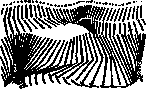
Figure 11. Cyclogram of walking, executed by Marey.
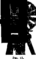
bs have a diameter of 2 mm and a length of 6-7 mm, so that they do not burden the subject in the least with their presence. The kymocyclographic method is also suitable for studying three-dimensional movements in space (and not just on a plane, like cinema snapshots). For this purpose, Bernstein has proposed a method of mirror photography, which makes it possible to determine the spatial coordinates of a moving point in the simplest way. Cyclographic and kymocyclographic methods may in the future play a very noticeable role in the clinic. Cyclography provides very valuable materials in the clinical study of pathological gaits. The kymocyclographic method is currently applied to the registration of tremors, adiadochokineses, tonic reactions of the arms, the knee reflex, etc. The applicability of precise measurements to the described methods makes it possible to calculate the forces acting during a given human Movement, and thereby to proceed to the study of muscle-force schemes and differential equations of human Movement (see above). For these purposes, methods of cyclogrammetry (Fischer, Bernstein) are being developed, allowing one to determine the muscle dynamics of a human from the measurement data of the Movement on the snapshot.
Electromyography and electrical methods of recording Movements. One should also touch here upon the electrical methods of recording the processes of human Movements. This includes, first of all, the method of electromyography (Einthoven, Yudin, Samoylov), i.e., the recording of action currents of an excited muscle using a string galvanometer. This method, to the present time, is the only one allowing one to judge with certainty the course of muscle excitation in an intact organism. Among other methods of electrical recording, the method of Popov, which has not yet received a definite name, deserves the most serious attention of the clinician. The device consists of a high-frequency oscillatory circuit, implemented using cathode tubes, as in radio receivers. The capacitance of this circuit is connected to a plate placed in front of the subject. Minute approaches or withdrawals of the studied part of the body in relation to the plate change its capacitance, which immediately affects the current strength in the circuit. Through a cathode amplifier, these changes are transmitted to a recording galvanometer. The sensitivity of Popov's device is colossal and is calculated in thousandths of a millimeter.
N. Bernstein. Pathology of human movement. The motor functions of the organism are constructed very complexly. For their normal execution, the integrity of the osteoarticular apparatus, the muscular system, the peripheral nervous apparatus, and the very complex central motor systems, as well as the so-called coordinative apparatuses regulating movement, is necessary. In accordance with the complexity of the structure, disturbances of motor functions are distinguished by great variety. In general, the character of the disturbances is determined on one hand by the localization of the anatomical lesion, and on the other hand by its intensity and extent. Disturbances of the integrity of joints, ankylosing and deforming processes in them must obviously change the motor function, disturbing the range of movements, changing their direction, and changing the place of application of the acting forces. Just as understandable are those disturbances of movements which arise as a result of lesions of the muscular system. Atrophies of muscles of one origin or another, dystrophic processes in them, subsequent cicatricial contractions due to the replacement of dead muscle fibers with connective tissue lead to a decrease or complete disappearance of the strength of the corresponding movements, to a limitation of their range, to a distortion of their direction. The analysis of such disturbances, given knowledge of the anatomy and physiology of the muscular system, does not present particular difficulties. The question of changes in motor functions caused by lesions of the nervous system is significantly more complex. But even here there is a localization of the lesion which leads to comparatively elementary motor disorders, namely, localization in the region of the peripheral motor neurons: motor cells of the brain stem and spinal cord (cells of the anterior horns)—anterior roots—motor nerves. A lesion of the peripheral motor neuron gives complete paralysis of the corresponding muscle. The motor function drops out completely, as in a primary lesion of the muscle itself: both voluntary and associated and reflex movements drop out. The second essential sign of peripheral paralysis is a complete inability to compensate. The spinal motor center (cells of the anterior horn) is absolutely necessary for the motor function of the corresponding muscle; no bypass innervation pathways can correct the defect if the destruction of the center is irreparable. Thus, paralysis of the biceps muscle of the arm will never smooth out or improve if its spinal innervation center is destroyed. Partially, the corresponding function can of course improve due to compensatory strengthening of the function of synergists, i.e., muscles working in approximately the same direction (in this example: m. brachialis internus, m. supinator longus), but the function of the m. biceps itself drops out forever. The third characteristic feature of peripheral paralysis is deep changes in the trophics of the corresponding muscles, accompanied by typical changes in electrical excitability. The entire uncomplicated semiotics of peripheral paralysis essentially boils down to these three main signs. Central paralyses differ significantly from peripheral paralyses, i.e., paralyses arising from lesions of central motor systems, in other words, those motor apparatuses which are located above the spinal motor centers and carry out their influence through these latter. The number of central motor systems—centers and conducting pathways—is very significant. The most well-studied of them is the pyramidal system. Upon its lesion, a so-called pyramidal paralysis arises, characterized both by the peculiarities of its distribution and by the peculiarities of the disturbance of motor functions it presents. The distribution of paralyses corresponds to the so-called selective Wernicke-Mann type: the muscles of the neck and trunk remain relatively spared; on the upper limb, the abductor muscles of the shoulder, the extensors and supinators of the forearm, and the extensors of the wrist and fingers suffer particularly; on the lower limb—the flexors and abductors of the thigh, the flexors of the lower leg, and the dorsal flexors of the foot and toes. As for the paralysis itself, it is deeply different from peripheral paralysis in its very essence. While in peripheral paralysis the motor function drops out completely, in pyramidal paralysis its dissociation, its splitting, takes place: only the higher part of the complex motor function drops out, while its elementary component part, carried out by spinal centers, not only does not suffer damage but even strengthens its action. Thus, in pyramidal paralysis, one observes not a fading of reflexes and not a drop in tone, as in peripheral paralysis, but an increase in reflexes and hypertonia. The presence of this increase in reflex excitability very typically influences the form of movement in a pyramidal lesion. In normal movement, in a certain phase of it, tension of the antagonists arises by reflex means, i.e., muscles exerting resistance to the given movement. Thanks to this tension, the movement is inhibited in its final phase, and upon its completion, the limb tends toward a reverse movement, toward reaching the initial position. This phenomenon, which has very great significance in the doctrine of normal and pathologically altered movement, is known as the "rebound" phenomenon. In pyramidal paralysis, the rebound is always more or less sharply increased. The tension of the antagonists, firstly, arises in an earlier phase of the movement than in the norm, and secondly, it is stronger than there. Let us take as an example the extension of the forearm in a pyramidal lesion. Initially, the movement may proceed at a normal or almost normal tempo, then very early (approximately around 90°) a sudden, "jerky" tension of the flexors of the forearm arises, which completely changes the character of the curve of velocities and accelerations. In a deep lesion of the pyramidal system, this tension can become insurmountable and fix the limb in a certain position. In such cases, one speaks of pyramidal contractures. Motor disorders in extrapyramidal lesions, the most frequently encountered example of which are disorders in paralysis agitans and in parkinsonism, have a completely different character. Here, the tension of the antagonists arises already in the very initial phase of the movement. And in this case, there is essentially an exaggeration of the phenomenon observed already in the norm: normal movement also begins with the tension of the antagonists, but in the norm, this tension quickly disappears and is replaced by the tension of the agonists, which realizes the movement. In parkinsonism, the normal relaxation of the initial tension of the antagonists does not occur; during the entire time of the movement, they remain tense more or less evenly. Thanks to this, the movement turns out to be evenly slowed down, the curve of velocities acquires a typical evenly flattened character. As a result—a characteristic feature of the motor skills of such patients, the extreme slowness of all their movements, known by the name of bradykinesia. The second feature of parkinsonians is the so-called poverty of movements, expressed by the dropping out of all side movements in the given motor act. The latter is maximally simplified and reduced to the minimum of movements required for the set goal. This also includes the decrease in the ability to simultaneously perform several motor operations. A direct opposite to pyramidal movement disorders are movement disorders in hypotonia. While there is an increase in normal inhibition on the part of the antagonists there, here this inhibition is decreased. Thanks to the decrease in inhibition, movements acquire an excessive, "overshooting" character. Furthermore, disturbances of the coordinative apparatuses (conductors of deep sensitivity, cerebellum) are reflected to a strong degree on the form of movements. Every movement, even the simplest, is performed essentially by a whole group of muscles: both agonists, i.e., muscles directly realizing the given movement, and synergists, i.e., muscles helping the agonists, and antagonists, i.e., muscles inhibiting the action of the agonists and synergists in one phase of movement or another. In order for the movement to have a normal character and reach the precisely set goal, it is necessary that the innervation impulses be distributed between these muscle groups in a completely definite way, and be dosed completely accurately. If there is no such correct distribution, then the movement, despite the normal strength of the corresponding muscles, will be performed incorrectly, will be pathologically altered. These changes are most clearly revealed in the classic tests of hitting the tip of the nose with the index finger and the kneecap of the other leg with the heel of one foot with eyes closed. They constitute the symptom complex of motor ataxia. But such changes also take place during standing—standing requires the innervation of a large number of muscles, and this innervation must be distributed in a very precise way. Otherwise, so-called static ataxia arises: the subject, when standing with feet together and eyes closed, loses balance (Romberg's sign).
Lesions of the cerebellum, the most important coordination center in the nervous system, manifest themselves also with other, more specific symptoms of coordination disorder: asynergy, i.e., the inability to perform movements in the correct combination; adiadochokinesia, i.e., the inability to quickly and correctly (in tempo and volume) perform alternating movements, e.g., rotation of the upper extremities inward and outward, flexion and extension of both forearms or shins, etc.; dysmetria, i.e., excessive, overshooting the set goal, volume of movements. Great changes are introduced into the form of movements by hyperkineses, or excessive movements (tremor, athetosis, chorea, etc.). These excessive movements, arising spontaneously, involuntarily, are woven into voluntary movements, interrupt them, change their direction, etc. The relationships here can, however, be very different. Thus, tremor in paralysis agitans weakens or completely disappears during voluntary movement, thus having no significant influence on the movement. But for the most part, hyperkinesis intensifies during movement or does not change in its intensity. Thus, the so-called intention tremor in multiple sclerosis sharply intensifies during voluntary movement. Barely noticeable or even absent at rest, during movement it brings the corresponding limb into a state of sweeping oscillations, completely changing this movement and posing a great obstacle to achieving the goals set by it. Athetosis, characterized by slow spastic tension of either agonists or antagonists and expressed by excessive tonic extension and abduction of the fingers, torsional grotesque movements of the limbs and trunk, etc., strongly interferes with the correct execution of movement. No less serious disturbances are introduced into movement by choreatic hyperkinesis—voluminous violent movements of a rapid, jerky tempo. In tempo, and in similarity to voluntary grimaces, choreatic hyperkinesis closely resembles tics, which differ from it mainly by their systematic nature. One of the most frequent forms of tic is stuttering, which deeply disturbs speech motor skills. Pains have a huge influence on the form of movement, limiting their volume or making them completely impossible, forcing patients to give the pain-affected limbs a special forced position ('antalgic postures'), etc. The symptomatology of such motor disturbances is naturally extremely motley and does not lend itself to precise description. It is also very difficult to give such a description to motor disturbances of psychogenic origin. The clinical picture of hysterical paralyses, pareses, etc., is extremely whimsical and diverse, and this diversity and discrepancy with the basic types of organic motor disorders described above constitutes its particularly characteristic sign. The distinction from organic motor disorders can, however, in some cases present very great difficulties. This applies especially to hysterical hyperkineses. For a comparatively recent time, neuroses, i.e., diseases without a definite anatomical substrate, included such hyperkinetic disturbances that are now recognized as unconditionally organic. Undoubtedly, the process of separating organic hyperkineses from the chaos of hysterical motor disturbances is not fully completed even at the present time, and we are still, to a certain extent, deprived of proper reference points in this area when making a differential diagnosis. On the other hand, in no other area does hysteria simulate an organic lesion as closely as in the area of hyperkineses. The diagnosis of hysterical paralysis is much easier. Especially helpful here for the diagnosis is the presence of characteristic changes in reflexes in organic lesions (so-called pathological reflexes). Of the disturbances of motor functions in mental illnesses, of particularly great interest are the disturbances characterizing the catatonic symptom complex, which from the outside presents a great similarity to the disorders described above in parkinsonism and paralysis agitans (the same poverty of movements, sometimes reaching extreme limits here, bradykinesia, flexibilitas cerea). A characteristic sign of schizophrenia is further a special affectation, mannerism of movements. Depressive states (circular psychosis, etc.) are characterized by lethargy of motor reactions; manic states, on the contrary, by motor restlessness, excessive richness of movements. In detail, disturbances of movement in psychoses were studied by Kraepelin (study of writing by means of a special apparatus, the so-called 'Schriftwage') and Isserlin (study of movements of the index finger with the help of Weiler's apparatus).
Related articles
Mentioned in
Cite this page
“Movements.” Soviet Medical Encyclopedia. English translation of Bolshaya Meditsinskaya Entsiklopediya, 1st ed. (Moscow, 1928–1936), ed. N. A. Semashko. https://sovietmedicalencyclopedia.pages.dev/article/movements/