Sciatic Nerve
Historical document, translated for reference. It reflects medical knowledge of the 1920s–30s and is not medical advice.
Summary
This article from the 1928–1936 Soviet Great Medical Encyclopedia provides a detailed anatomical description of the sciatic nerve, its origins in the sacral plexus, and its pathway through the lower limb. It outlines the nerve's division into the tibial and common peroneal nerves and details the innervation of the muscles and skin of the leg and foot.
Encyclopedia article (1928–1936)
ISCHIADICUS HERVUS (sciatic nerve) belongs to the nerves of the lower limb and is the longest and largest (1.5 cm in diameter) of all peripheral nerves. It originates from the roots of the sacral plexus (plexus sacralis, LIV-SIII), of which it serves as a continuation (Figure 1). It forms near the sciatic foramen, through which it leaves the pelvic cavity, passing between the tuber ischiadicum and the trochanter major, posterior to the muscles that rotate the thigh (m. piriformis); it is covered by the m. gluteus maximus. Further downward, it runs along the posterior surface of the thigh, where it is situated along the midline on the m. adductor magnus, covered posteriorly by the muscles that flex the lower leg (mm. biceps, semitendinosus, semimembranosus), and in this way reaches the popliteal fossa, where it lies superior and lateral to the a. et v. popliteae. The fascia poplitea is located above it. In most cases, in the upper corner of the popliteal fossa, it divides into two terminal branches (Figure 4): one, thicker, which acts as its continuation, the n. tibialis, and the other, thinner, the n. peronaeus communis; sometimes this division occurs halfway down the thigh, or even both branches can be separated from one another at the place where the nerve exits the plexus. The sciatic nerve innervates the surrounding muscles of the pelvic cavity and the posterior surface of the thigh [see separate table (Vol. III, art. 103-104), Figs. 2 and 3] and gives off branches to the capsule of the hip joint. The n. tibialis (LIV-V-SI-III), the tibial nerve, continues the path of the sciatic nerve (Figure 2); in the popliteal fossa, it lies posterior and lateral to the vasa poplitea; then it descends along the posterior surface of the lower leg, situated together with the a. et v. tibiales posticae between the deep and superficial layers of the calf muscles. In the lower parts of the lower leg, it deviates medially together with the vessels and reaches the medial side of the ankle joint, where it divides into its terminal branches: nn. plantares lateralis et medialis, which head under the lig. laciniatum to the sole. On its path, the n. tibialis innervates the muscles of the posterior surface of the lower leg (see diagram), the knee and ankle joints, the tibia, and gives off sensory branches to the skin of the lower leg and foot: n. cutaneus surae medialis and rami cutanei mediales. The first begins in the popliteal fossa and, together with one of the branches of the n. peronaeus, forms the n. suralis, which innervates the outer surface of the ankle joint, the outer edge of the foot, and the outer part of the fifth toe. The latter innervate the skin of the inner side of the heel and the posterior part of the sole.
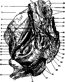
Figure 1. Nerves of the right half of the pelvis from the left: 1-gangl. lumbal.; 2-corp. vert. lumb. IV; 3-a. iliaca comm. dex.; 4-fascia iliaca; 5-m. psoas major; 6 and 32-tr. lumbo-sacralis; 7-a. hypog.; 8-n. cutan. fem. lat.; 9 and 13-a. et v. iliaca ext.; 10 and 12-n. et a. obtur.; 11-plexus sacralis; 14-m. obtur. int.; 15-m. levat. ani; 16-symph. os. pubis; 17 and 18-n. dors. penis (dex. et sin.); 19-nn. perin.; 20-n. pudend.; 21-m. coccyg.; 22-pl. coccyg.; 23-rami viscerales; 24-ram. muscul.; 25-pl. pudend.; 26-a. glut. inf.; 27-gangl. sacralia; 28-rami musculares; 29-m. piriform.; 30-ram. ant. n. sacral. I; 31-promont.; 33-ram. ant. n. lumb. V; 34-pl. lumbalis; 35-ram. ant. n. lumbal. IV; 36-pr. spin. vert. lumb. IV. (According to Spalteholz.)
The larger terminal branch of the n. tibialis, the n. plantaris medialis, having reached the sole, divides into two terminal branches: medial and lateral (Figure 5). Even before its division, it gives off branches to the muscles: m. abductor hallucis and m. flexor digitorum brevis. The medial terminal branch innervates the skin of the inner part of the foot and gives off muscular branches to the inner head of the m. flexor hallucis brevis; the n. plantaris medialis ends in the skin of the inner side of the big toe (n. plantaris hallucis medialis). The lateral terminal branch divides into three branches—nn. digitales plantares communes of the I, II, and III toes. At the anterior end of each interosseous space, each of these branches splits into 2 nn. digitales plantares proprii for the facing sides of the I-IV toes. The second terminal branch of the nervus tibialis, the n. plantaris lateralis, together with the a. plantaris lateralis, heads outward and forward, describing an arc, lies under the m. quadratus plantae and m. flexor digitorum brevis, under which it divides into two terminal branches to the muscles—r. superficialis et profundus. Before its division, it gives off branches to the muscles near which it passes. The ramus superficialis in turn is subdivided into two branches: the n. plantaris lateralis digiti quinti for the outer side of the V toe and the n. digitalis plantaris communis IV, which innervates the inner side of the little toe and the outer side of the IV toe. The first branch also innervates the muscles of the V toe and the mm. interossei IV; the second branch innervates the mm. lumbricales III-IV. The ramus profundus forms an arc with the convexity forward and outward; from this convexity, nerves depart for the first three mm. interossei and for the muscles of the big toe; sometimes the muscles of the little toe and the mm. interossei IV of the interosseous space also receive branches from it. The n. peronaeus communis (LIV-SII) heads to the outer corner of the popliteal fossa and, along the neck of the fibula, emerges onto its outer surface, where it divides into two branches (Figure 3): a predominantly sensory one—nervus peronaeus superficialis, and a predominantly motor one—n. peronaeus profundus. Before the place of division into terminal branches, branches depart from it to the knee joint, to the joints of the tibia and fibula, to the short head of the m. bic. femoris, and a sensory nerve (nervus cutaneus surae lateralis), which innervates the outer half of the anterior and posterior surface of the lower leg. The n. peronaeus superficialis heads down along the anterolateral surface of the lower leg, initially between the m. peronaeus and m. extensor digitorum communis; in the lower third of the lower leg, the nerve divides into two terminal branches—n. cutaneus dorsalis medialis and n. cutaneus dorsalis intermedius, which pass through the fascia cruris under the skin, run under the skin along the anterior surface of the foot, and divide into medial and lateral branches, which innervate the anterior surface of the foot and the toes. Before dividing, the n. peronaeus superficialis gives off branches for the mm. peronaeus brevis et longus. The n. peronaeus profundus, crossing the outer surface of the lower leg, heads forward, reaches the anterior tibial vessels, together with which it descends initially between the mm. tibialis anticus et extensor digitorum communis, and then between the first muscle and the m. extensor hallucis longus; together with this muscle, the nerve passes under the lig. cruciatum pedis onto the anterior surface of the foot, where it divides into medial and lateral branches, which, together with the branches of the n. peronaeus superficialis, innervate the skin of the foot and toes, the mm. interossei dorsales (nervi interossei pedis), and then other muscles and joints passing here. On the lower leg, the n. peronaeus profundus gives off motor nerves to the muscles— M. quadratus femoris, Mm. gemelli, M. obturator internus, M. adductor magnus, M. gastrocnemius, M. soleus, M. plantaris, M. popliteus, M. flex. halluc. long., M. tibialis posterior, M. flex. digit. commun. long., M. flex. digit. commun. brev., N. cutan. surae med. (sensory branch), Rami cutan. medial (sensory branch).
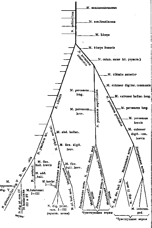
M. abduct. dig. V, M. quadrat. plantae, M. flex. dig. V. [Sensory]. Diagram of the branching of the n. ischiadici.
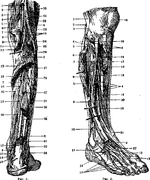
Figure 2. Nerves and muscles of the right lower leg from behind: 1-sciatic n.; 2-popliteal v.; 3-popliteal a.; 4-vastus medialis m.; 5-adductor magnus m.; 6-articular branch; 7-tibial n.; 8-semimembranosus m.; 9 and 10-gastrocnemius m. (medial and lateral head); 11-popliteus m.; 12-soleus m.; 13-posterior tibial a.; 14 and 27-muscular branches; 15-flexor digitorum longus m.; 16-posterior tibial m.; 17-calcaneal (Achilles) tendon; 18-medial calcaneal branches; 19-laciniate ligament; 20-biceps femoris m.; 21-medial sural cutaneous n.; 22-common peroneal n.; 23-lateral sural cutaneous n.; 24-plantaris m.; 25-head of fibula; 26-interosseous nerve of the leg; 28-peroneal a.; 29 and 31-flexor hallucis longus m.; 30-peroneus longus m.; 32-sural n.; 33-lateral calcaneal branches. Figure 3. Nerves of the right lower leg from the front and outside; 1-biceps femoris m.; 2-common peroneal n.; 3-head of fibula; 4-muscular branches; 5-superficial peroneal n.; 6-peroneus longus m.; 7-anterior intermuscular septum; 8-brevis m.; 9-medial dorsal cutaneous n. of the foot; 10-intermediate dorsal cutaneous n. of the foot; 11-lateral dorsal cutaneous n. of the foot; 12-extensor digitorum brevis m.; 13-dorsal digital nerves of the foot; 14-patella; 15-deep peroneal n.; 16-anterior tibial a.; 17-interosseous nerve of the leg; 18-tibialis anterior m.; 19-extensor digitorum longus m.; 20-extensor hallucis longus m.; 21-cruciate ligament; 22-extensor hallucis brevis m. (From Spalteholz.)
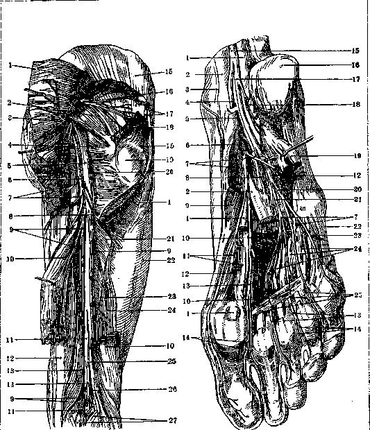
Figure 4.
Figure 5.
Figure 4. Nerves of the right thigh from behind: 1-gluteus maximus m.; 2-piriformis m.; 3-inferior gluteal n.; 4-obturator internus and gemelli mm.; 5-sciatic n.; 6-posterior femoral cutaneous n.; 7-perineal branch; 8-inferior cluneal nn.; 9-muscular branch; 10-biceps femoris m.; 11-tibial n.; 12-semitendinosus m.; 13-semimembranosus m.; 14-medial sural cutaneous n.; 15-gluteus medius m.; 16-superior gluteal n.; 17 and 18-gluteus minimus m.; 19-greater trochanter; 20-quadratus femoris m.; 21-adductor magnus m.; 22-iliotibial tract (Maissiat); 23-articular branch; 24-biceps femoris m. (short head); 25-common peroneal n.; 26-lateral sural cutaneous n.; 27-gastrocnemius m. Figure 5. Deep nerves of the right sole: 1-flexor hallucis longus m.; 2-flexor digitorum longus m.; 3-tibialis posterior m.; 4 and 6-medial plantar n.; 5-laciniate ligament; 7-muscular branches; 8-quadratus plantae m.; 9-abductor hallucis m.; 10-adductor hallucis m. (oblique and transverse heads); 11-flexor hallucis brevis m.; 12-articular branch; 13-common plantar digital nn.; 14-proper plantar digital nn.; 15-tibial n.; 16-calcaneal tuberosity; 17-lateral plantar n.; 18-muscular branches; 19-flexor digitorum brevis m.; 20 and 21-superficial and deep branches of the lateral plantar n.; 22-opponens digiti V m.; 23-flexor digiti V brevis m.; 24-plantar interossei mm.; 25-lumbrical mm. Motor function of the sciatic nerve and its branches: Sciatic nerve. Muscles: Semimembranosus m., Semitendinosus m., Biceps m., Quadratus femoris m., Gemelli mm., Obturator internus m., Adductor magnus m. (partly). Function: Flex the lower leg. Rotate the thigh outward. Adduction of the thigh. Gastrocnemius m., Soleus m., Plantaris m., Popliteus m. I. Tibial nerve. 1) Lateral plantar nerve. Produce plantar extension of the foot. Tibialis posterior m. Flexes the lower leg, rotates it inward. Adducts the foot inward, lifting the inner edge upward. Flexor digitorum communis longus m. Flexes the last phalanx of the toes, partly the second. Flexor digitorum communis brevis m. Flexes the II phalanx of the big toe. Abductor digiti V m., Quadratus plantae m. A) Superficial branch. Flexor digiti V m. B) Deep branch. Interossei externi et interni mm. Opponens digiti V pedis m. 2) Medial plantar nerve. A) Lateral branch. B) Medial branch. Adductor hallucis m. Abducts the little toe from the other toes. Plantar flexion of the I phalanx of the I-V toes. Abductor hallucis m. Flexes the I phalanx of the little toe, extends the II phalanx. Flex the I phalanx of the toes, extend the II and III; interossei externi mm. abduct the toes, and interossei interni mm. adduct them. Adducts the big toe to the others. Abducts the I toe from the others. Flexor digitorum communis brevis m. Function-see above. Lumbricales mm. Flexor hallucis brevis m. Flex the I phalanx, extend the II and III. Flexes the I phalanx of the big toe, extends the I and II. Common peroneal nerve. Biceps femoris m., Tibialis anterior m., Extensor hallucis longus m. 1) Deep peroneal nerve. 2) Superficial peroneal nerve. Function-see above. Extensor digitorum communis longus m. Flexes the foot on the dorsum, lifts its inner edge. Extends the I phalanx of the big toe. Extends the I phalanx of the toes, produces dorsal flexion of the foot. Extensor digitorum communis brevis m. (Extends the I phalanx of the toes). Peroneus longus m., Peroneus brevis m. Peroneus longus m., Peroneus brevis m. Abduct the foot outward, lifting its outer edge. Tibialis anterior, extensor digitorum communis, extensor hallucis longus and to the joints of the foot. As can be seen from the table, the sciatic nerve innervates some muscles of the hip joint, the posterior muscles of the thigh, and all muscles of the lower leg and foot. It rotates the thigh outward, flexes the lower leg, rotates it outward, and produces all movements of the lower leg and foot. Due to the great length of the sciatic nerve and its connection with many surrounding structures, disease of the sciatic nerve is one of the most frequent in neuropathology. The sciatic nerve becomes diseased not only primarily from a process directly acting on it, but also often suffers during disease of the surrounding tissues. At its formation, it lies in the cavity of the lesser pelvis in immediate proximity (as well as the plexus giving rise to it) to the internal organs of the lesser pelvis. Above, its roots have a close connection with the membranes of the brain, with the spine; below, the sciatic nerve is surrounded by muscles, in some places directly touching bone, joints, so that any process developing in one of these places can simultaneously involve the nerve. As primary processes, infections, intoxications, tumors, and traumatic injuries are observed. The clinical picture is the same for primary and secondary diseases of the sciatic nerve; it varies depending on complete or partial damage, as well as on the localization of the process (for details on diseases of the sciatic nerve, see Sciatica).
L. Brusilovsky. Surgery of the sciatic nerve. Exposure of the sciatic nerve is performed for the purpose of suturing a damaged nerve, releasing it from adhesions formed as a result of its injury, traction, or other manipulations undertaken for the surgical treatment of neuralgia (see Sciatica), etc. To expose the nerve below the gluteal fold, an incision (with the patient in the prone position) is started at the lower edge of the gluteus maximus m. and led downward along the easily palpable long head of the biceps femoris m. An incision 8-10 cm long is thus located slightly medial to the line connecting the greater trochanter and the ischial tuberosity, and extends toward the middle of the popliteal fossa. After incision of the fascia lata, the inner edge of the biceps m. is exposed, pulled outward, the gluteus maximus muscle is pulled upward, after which the thick trunk of the nerve appears in the thickness of the loose connective tissue. Lower down, it is located behind the long head of the biceps m. in the middle of the thigh-between the biceps m. and the semimembranosus m. For more highly located injuries of the sciatic nerve, it is necessary (according to Guleke) to detach the gluteus maximus m. from its attachment to the greater trochanter, after which it is pulled upward, thus exposing the nerve. König and Iselin for this purpose do not stop even at cutting the broad tendons of the muscle, by which better access is achieved. Suturing of the sciatic nerve in its injuries gives good functional results only in fresh cases. Lit.: Anatomy.-Villiger E., Peripheral Innervation, M., 1917; Shamburov D., Sciatica, M.-L., 1928. Surgery.-Iselin H., Desinsertion der Muskeln zur Freilegung der grossen Nervenstämme an Schulter u. Hüfte, Bruns kriegschirurgische Hefte, 1917, No. 40; Klapp E., Die Operationen an der unteren Extremität (Chirurgische Operationslehre, hrsg. v. A. Bier, H. Braun u. H. Kümmell, B. V., p. 338, Lpz., 1923); König F., Die Freilegung des Nervus ischiadicus in seinem obersten Teile, Zentralblatt für Chirurgie, 1916, No. 52.
Related articles
Mentioned in
Cite this page
“Sciatic Nerve.” Soviet Medical Encyclopedia. English translation of Bolshaya Meditsinskaya Entsiklopediya, 1st ed. (Moscow, 1928–1936), ed. N. A. Semashko. https://sovietmedicalencyclopedia.pages.dev/article/sciatic-nerve/