Popliteal Fossa
Historical document, translated for reference. It reflects medical knowledge of the 1920s–30s and is not medical advice.
Summary
This article from the 1st edition of the Great Medical Encyclopedia (1928–1936) details the anatomy, boundaries, contents (vessels and nerves), and surgical topography of the popliteal fossa.
Encyclopedia article (1928–1936)
POPLITEAL FOSSA (fossa poplitea) occupies the posterior region of the knee joint, has a rhombus shape, the sides of which are formed: on the inside by the clearly protruding tendons of the semimembranosus and semitendinosus muscles, on the outside by the tendon of the biceps femoris muscle, from below by the two protruding bellies of the gastrocnemius muscle; the floor of the fossa is formed 1) by the posterior surface of the femur (planum popliteum ossis femoris) and its condyles, 2) by the posterior part of the joint capsule and the oblique popliteal ligament, and 3) by the tibia covered by the popliteus muscle (see plate 3). The length of the fossa in an adult is 12–14 cm, of which 9 cm belongs to the thigh and 3–5 cm to the leg. The appearance of the popliteal fossa (Figure 1) varies depending on the position of the leg relative to the thigh. Examination of the popliteal depression is best conducted in the position of passive flexion. The skin in the region of the popliteal fossa is smooth, rich in sweat and sebaceous glands, and possesses significant mobility and extensibility. Superficial blood and lymphatic vessels are embedded in the layer of adipose tissue; the latter are partly a continuation of the lymphatic pathways of the leg, and partly pathways of the popliteal fossa proper; the lymphatic vessels pass along the medial surface of the thigh and empty into the superficial inguinal lymph nodes. The superficial layers of the popliteal fossa are innervated by branches of the posterior femoral cutaneous nerve, from the medial edge by branches of the saphenous nerve, and from the lateral edge by branches of the peroneal nerve (Figure 2). The aponeurosis of the popliteal fossa is part of the common aponeurotic sheath of the leg; the fibers of the aponeurosis run obliquely from below and inwards upwards and outwards; in the middle, there are openings for vessels and nerves; from the aponeurosis deep into the popliteal fossa go fibrous plates (Figure 3) that cover the medioposterior surfaces of the muscles and attach to the V-diverging linea aspera of the femur here. Numerous fibrous cords reinforcing it run from the tendons to the aponeurosis, as a result of which the muscles of the popliteal fossa tense the aponeurosis upon contraction. By the named fibrous septa and aponeurosis, the entire popliteal fossa is turned into a fibrous sac filled with adipose tissue (Pirogov). A thin, loose fibrous plate covering the muscles is located beneath the aponeurosis. The quadrangular space of the popliteal fossa is divided by a transverse line into two triangles with bases facing each other—the upper, larger femoral triangle, and the lower, smaller tibial triangle. Figure 1. External appearance of the right popliteal fossa: 1—posterior surface of the thigh; 2—relief of the semitendinosus muscle; 3—popliteal fossa; 4—transverse fold; 5—posterior surface of the leg; 6—relief of the head of the fibula; 7—relief of the biceps muscle (After Testut-Jacob).
the popliteal adipose tissue communicates above with the posterior surface of the thigh, and through an opening (hiatus adductorius) in the adductor magnus muscle with the anterior and medial surface of the thigh; along the neurovascular bundle, the popliteal fossa communicates with the posterior
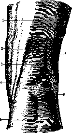
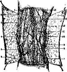
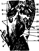
Figure 2. Superficial layers of the popliteal fossa: 1, 4, 12, and 15—branches of the peroneal nerve; 2—anastomosis between the small saphenous vein (9) and great saphenous vein (3); 5 and 11—cutaneous arteries; 6 and 14—skin; 7 and 13—aponeurosis; 8—lateral sural cutaneous nerve; 10—superficial vein (After Testut-Jacob).
posterior, anterior, and lateral surfaces of the leg. Nerves lie most superficially in it, deeper lies the popliteal vein, and still deeper lies the popliteal artery (Figure 4). The main nerve trunks of the popliteal fossa are the tibial nerve (nervus tibialis) and the common fibular nerve (nervus peronaeus communis). Both nerves are a continuation of the sciatic nerve. The tibial nerve passes from the upper corner of the popliteal fossa to the lower, disappearing between the heads of the gastrocnemius muscle, and goes onto the leg through the tendinous arch of the soleus muscle. The tibial nerve continues the direction of the sciatic nerve. To expose the tibial nerve, one should make the same incision as for ligating the popliteal vein or artery. The nerve lies immediately beneath the aponeurosis and is lateral to the popliteal vein lying beneath it; it gives off branches to the knee joint, gastrocnemius, plantaris, and popliteus muscles and
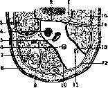
Figure 3. Cross section of the popliteal fossa: 1—thigh; 2—aponeurosis with septa (14) separating the popliteal fossa from the anterior region of the knee with its musculature (3 and 15); 4 and 5—popliteal artery and vein; 6—tibial nerve; 7—adipose tissue of the popliteal fossa; 8—skin; 9 and 12—aponeurosis; 10 and 13—aponeurotic septa; 11—common fibular nerve (After Testut-Jacob).
to the soleus muscle. Approximately in the middle of the upper part of the popliteal fossa, the nerve gives off a large cutaneous branch (medial sural cutaneous nerve) directed into the furrow between
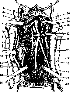
Figure 4. Contents of the popliteal fossa: 1—semimembranosus muscle; 2—semitendinosus muscle; 3—tendon of the adductor magnus muscle (4); 5 and 21—superior genicular arteries; 6, 10, and 18—glands; 7—serous bursa of the semimembranosus muscle; 8—middle genicular artery; 9—opening communicating the serous bursa with the joint; 11—saphenous vein; 12 and 17—inferior genicular arteries; 13 and 16—gastrocnemius muscle; 14 and 15—skin; 19—muscular branch; 20 and 22—popliteal artery; 23—common fibular nerve; 24—tibial nerve; 25—popliteal vein; 26—biceps femoris muscle (After Testut-Jacob).
the heads of the gastrocnemius muscle and downwards onto the leg along with the small saphenous vein. The second nerve, the common fibular nerve, runs along the medial edge of the biceps femoris muscle in the same oblique direction as the muscle itself, then along the posterior surface of the lateral head of the gastrocnemius muscle and, winding around the head of the fibula, goes into the lateral region of the leg. In the upper region of the popliteal fossa, the common fibular nerve gives off a cutaneous branch (lateral sural cutaneous nerve) directed toward the middle of the popliteal fossa, and branches to the knee joint (articular branches). The second layer, lying deeper than the first and separated from it by adipose tissue, contains the popliteal vessels—the artery and vein. The popliteal vein is normally single, but many find a second vein; in this case, the popliteal artery is located between the veins (Pirogov, Testut). The popliteal vein runs lateral and slightly posterior to the artery, covering its lateral edge. The pathway of the vein runs from the tendinous arch of the soleus muscle to the adductor hiatus, where it continues as the femoral vein. The popliteal vein and artery are closely connected by a thin layer of strong connective tissue, as a result of which simultaneous damage to both vessels is possible. The vessels are clothed in a common connective tissue sheath, in which numerous small arteries (vasa vasorum) run in downward and upward directions, forming a rich network of anastomoses. These anatomical relationships make one extremely cautious when ligating the popliteal vessels and especially when denudating the artery (periarterial sympathectomy), which is wiser to abandon here. The popliteal vein receives branches, paired veins accompanying the articular and muscular branches of the popliteal artery; the small saphenous vein also empties into it (though not always). The popliteal artery is a continuation of the femoral artery and has a length of 17–18 cm; cases are encountered where the popliteal artery is a continuation not of the femoral artery, but of the nerve-accompanying sciatic artery or the profunda femoris artery. The final division of the popliteal artery into the anterior and posterior tibial arteries occurs either above its passage through the soleus muscle, i.e., in the popliteal fossa itself, in rare cases at the femoral condyles (dispersed type), or below the ring in the soleus muscle, i.e., already on the leg (stem type). The popliteal artery occupies the deepest and most medial position in the popliteal fossa. The artery lies adjacent to the skeleton of the knee, separated from it by the joint capsule and an insignificant layer of fat, so that all movements in it unquestionably affect the position of the artery; thus, for example, with an extended leg, it bends with concavity forward. On its path in the popliteal fossa, the popliteal artery lies adjacent to: the posterior surface of the femur, the posterior wall of the joint capsule, and lower down to the popliteus muscle separating the artery from the tibia, which makes it necessary to be very cautious during surgical intervention on the femur and tibia in this area. Lateral to the artery are located: above—the lateral condyle of the femur and the lateral head of the gastrocnemius muscle; on the inside—the semimembranosus muscle, the medial condyle of the femur, and the medial head of the gastrocnemius muscle. Posteriorly, the popliteal artery is successively covered from the surface inwards by the skin with adipose tissue, the aponeurosis (containing the small saphenous vein), the fat layer and lymph nodes, the tibial and common fibular (above) nerves, and the popliteal vein; right here around the artery are located several deep [nodes].
lymph glands and vessels (see Knee joint). Deeper than all, according to Pirogov, lie the side branches of the popliteal artery (see Blood vessels, table of arteries), of which there are usually seven, counting the larger ones: five for the knee joint (aa. articulares) and two for the triceps muscle of the calf (aa. surales). This number is inconstant and more often increases. The sites of origin of the branches of the popliteal artery are grouped approximately on the lower two-thirds of its extent; consequently, there is a section of the artery 4 cm in length and 5.5 cm above the medial condyle of the femur where it is free from branches and represents the most convenient place for ligating the vessel (Pirogov).
At the sites of friction between muscles (tendons), along a ligament, or against the skeleton, there are mucous bursae (bursae mucosae subtendineae); in the popliteal fossa, these bursae are divided into lateral and medial (Fig. 5). There are three medial bursae: 1) bursa m. gastrocnemii medialis is located between the medial head of the gastrocnemius muscle and the femoral condyle, usually communicating with the joint cavity; 2) bursa gastrocnemio-semimembranosa is located between the semimembranosus muscle and the gastrocnemius muscle, below the joint line; it communicates with the previous bursa and with the joint [in 10% of people aged 20 to 40 years and in 20% of people older than 40 years (Poirier)]; 3) bursa (propria) m. semimembranosi is located slightly lower than the previous one, between the tendon of the muscle and the medial condyle of the tibia. There are four lateral bursae: 1) bursa bicipito-gastrocnemialis - between the aforementioned two muscles; 2) bursa m. bicipitis femoris - between the muscle and the lateral collateral ligament of the knee; 3) bursa m. gastrocnemii lateralis is located under the origin tendon of the muscle; the aforementioned three bursae are inconstant; 4) bursa m. poplitei is a protrusion of the synovial membrane of the knee joint beneath the tendinous origin of the popliteus muscle. The bursa communicates through a narrow opening with the cavity of the knee joint and, in 9% of cases, with the cavity of the tibiofibular joint (articulatio tibio-fibularis). Some authors exclude this bursa from the number of mucous bursae and consider it as one of the recesses of the joint capsule of the knee (Testut). Clinic of diseases of the popliteal fossa. Injuries in the region of the popliteal fossa are divided into two groups: without damage to the neurovascular bundle and with damage to it. The first type of injuries usually accompanies various severe injuries of the knee, fractures and dislocations in the region of this joint, sprains and wounds. Excessive forcible extension of the knee kept in a flexed position for a long time (ankylosis), in addition, in the presence of scar tissues of the popliteal fossa, can lead to their transverse rupture. Punctured and stab wounds without damage to the neurovascular bundle are less common than wounds with vascular damage. Gunshot wounds of the popliteal fossa without damage to the neurovascular bundle are observed somewhat more often (vessels, due to their elasticity, deflect from bullet impacts). Injuries of plantings with violation of the integrity of the neurovascular bundle must be classified as particularly grave. The neurovascular bundle can be damaged both with integrity and with violation of the integuments of the popliteal fossa. Injuries of the neurovascular bundle without violation of the integrity of the integument occur with anterior and posterior dislocations of the knee, when the vessels are compressed as a result of displacement and further damaged; fractures of the lower end of the femur or the upper end of the tibia have a similar effect on the vessels. The fall of a weight onto the region of the popliteal fossa, the knee getting under the wheels of a vehicle, etc., act directly on the neurovascular bundle, causing its damage, which occasionally also occurs during sudden and sharp extension. Accidental injuries of the bundle during operations should be included here. In injuries of the popliteal fossa with injury to the bundle, the picture of a major vessel injury appears: when the integuments are ruptured, there is external, sometimes exceptional strength, bleeding; with the integrity of the integuments, the region of the popliteal fossa swells, signs of vascular compression appear, and eventually the picture of an aneurysm of the popliteal fossa emerges or necrosis of the foot and lower leg occurs; rarely, the ruptured vessel thromboses, in this case there is no bleeding and necrosis of the limb immediately follows the injury. The prognosis in injuries of the popliteal fossa depends on the presence of wounds of the integuments and on the degree of damage to the neurovascular bundle. A wound of the integuments aggravates the already serious vascular injury in terms of outcome, especially of both vessels; such an injury decides the fate of the limb (Pirogov). Treatment of injuries of the popliteal fossa with the integrity of the neurovascular bundle is carried out conservatively: rest, cold, elevated position, bandage after appropriate treatment of the wound. With a violation of the integrity of the neurovascular bundle, active intervention is indicated: first of all, bleeding should be stopped by compressing the vessel at a distance, then the injured vessel is treated in accordance with the degree of its damage - if the edges of the vessel wound are not crushed and it is possible to bring them together, a vascular suture should be applied immediately, otherwise ligation of the vessel at a distance is indicated, but not in the wound, in order to avoid suppurative inflammation and secondary hemorrhage. The injured nerve must be sutured immediately. If there are signs of limb necrosis, only amputation remains. (On the treatment of aneurysms of popliteal vessels, see Aneurysm.) Foreign bodies in the popliteal fossa are rare, most often bullets and shell fragments. Tumors of the popliteal fossa are not uncommon. Among benign ones, lipomas are more common, rarely fibromas, chondromas, and osteomas. Treatment is exclusively surgical. Among malignant tumors of the popliteal fossa, sarcoma occupies the first place, developing from the soft parts of the fossa (ligaments, muscles, tendons, and fasciae) and from the neurovascular bundle. Sarcomas of the neurovascular bundle are divided into two types: those originating from the vascular sheath and from the nerve trunk. The former are very rare (French authors call them les conjonctivomes de la gaine des vaisseaux, which indicates their fibromatous type); they are pale pink in cross-section, sometimes very soft, sometimes denser, located around the main vessels of the popliteal fossa, being most intimately connected with the wall of the veins, but do not grow through them, so that although with great difficulty, they can be separated from the vessels; neighboring muscles suffer more from compression by the tumor. Such tumors are very rare in persons younger than 30 years, predominantly occurring in 40-50-year-olds, and run the more malignant the younger their bearer is. Tumors of the vascular sheath should be distinguished from tumors of the nerve trunk of the popliteal fossa; both are usually located along the midline of the popliteal fossa, but differ sharply in volume: a tumor originating from the nerves never reaches the size of a vascular sheath tumor. Tumors of the nerves are accompanied by severe radiating pains, which is not usually observed in tumors of the vascular sheath; a tumor of the nerve trunk, being sometimes part of general neurofibromatosis (Recklinghausen's disease), is easily distinguishable from a vascular sheath tumor. Tumors of the popliteal nerve are not particularly rare and are observed either as single tumors or are one of the manifestations of the aforementioned Recklinghausen's disease. The tumor (sarcoma) develops in the connective tissue membrane of the nerve trunk, growing more towards the periphery and causing atrophy of nerve fibers. A characteristic feature of sarcoma of the popliteal nerve is its relative benignancy: up to a certain period it has no special propensity to grow into surrounding tissues. Later the tumor loses this property and becomes malignant; in the early stage of a nerve tumor, its removal is possible by resection of the nerve followed by suturing, while in far-advanced cases amputation of the leg is indicated. Cancerous tumors of the popliteal fossa are observed either as metastases in cancer of the peripheral parts of the limb or as tumors of the lower leg spreading upwards; epitheliomas are most common. Clinically, there are no special features in this kind of tumor. Treatment is almost always amputation of the thigh, taking into account that this is usually a metastasis. Occasionally, cold abscesses are found in the popliteal fossa, either descended here from above or resulting from tuberculous gonitis. As a secondary formation, a cold abscess of the popliteal fossa is one of the signs indicating a primary focus in the spine, hip, or knee joints. The prognosis depends on the underlying disease. Therefore, treatment of the primary focus is indicated first of all. Of the serous cysts observed in the popliteal fossa and expressed in the accumulation of serous fluid in the mucous bursae, the cyst of the semimembranosus muscle is of practical importance; such a cyst is relatively common in both adults and children. The cause of their appearance is difficult to establish, but often in adults the appearance of a cyst is associated with a prolonged stay on the feet; by virtue of the communication of the bursa with the cavity of the knee joint, its appearance is associated with one or another disease of the knee accompanied by effusion in the joint. Initially completely painless and not interfering with the work of the leg, the cyst with growth manifests itself as a dull, indefinite pain in the region of the popliteal fossa and the appearance of an elastic-firm oval tumor, hiding during muscle tension and easily accessible upon their relaxation; the tumor is displaced in the transverse direction. Cysts of the bursa of the popliteus muscle are very rare; in exceptional cases, such a cyst emerges from under the lower edge of this muscle and appears at the lateral edge of the upper quarter of the lower leg, simulating a cold abscess.
Medial cysts remain controversial; some view them as hernial protrusions of the synovial membrane of the joint, while others consider them true serous cysts. They lie deep in the upper part, along the midline of the popliteal fossa, are very small in size, and are sometimes multiple. The diagnosis of serous cysts is simple; the only difficulty is presented by medial cysts, because such a cyst sometimes pulsates due to its proximity to blood vessels, while at other times an aneurysm (a phenomenon observed in old popliteal aneurysms) that is non-pulsating and lacks the characteristic blowing murmur is mistaken for a cyst. Therefore, tumors of the midline of the popliteal fossa should always be treated with special care. Serous cysts of the popliteal fossa follow a benign course. Large cysts that impede knee movements are subject to removal, and small ones to puncture, but the introduction of any irritating solution into the cyst cavity must never be allowed due to the possibility of causing great harm to the knee joint cavity, since in 9 out of 10 cases the bursa of the semimembranosus muscle communicates with the joint. Echinococcal cysts of the knee, and of the popliteal fossa in particular, are extremely rare. In the presence of a cystic tumor in the popliteal fossa, one must check whether similar cysts exist in other parts of the body, since the echinococcus here is usually secondary. Casoni's reaction and cyst puncture will help establish an accurate diagnosis. For a small echinococcal cyst, its removal is possible, but amputation of the thigh is more frequently indicated. Aneurysms of the popliteal fossa occupy second place in frequency (Abrikosov). The cause of popliteal aneurysms is trauma; aneurysms developing in certain occupations where work is associated with constant sharp movements in the knee (e.g., in horse riders) should also be classified here. Another cause is disease of the vessel walls (syphilis, sclerosis, etc.). Anatomically and pathologically, popliteal aneurysms present nothing special compared to aneurysms in other areas of the body. Clinically, popliteal aneurysms are characterized by a tumor with signs proper to aneurysms (see Aneurysm). The prognosis is always doubtful, as serious complications up to gangrene of the limb are frequent. The treatment of popliteal aneurysms, like aneurysms in general, is surgical. One of the frequent diseases of the popliteal fossa, especially in women, is varicose expansion of the subcutaneous veins (varices venarum). In advanced cases, the nodes of such an expanded vein can rupture and cause severe hemorrhage. Treatment for small nodes consists of bandaging the entire lower leg, starting from the foot and going up to the thigh. Recently, sclerosing therapy has begun to be practiced: 20–30% and 40% solutions of sodium salicylate in amounts from 2 to 5 cm3 or more at a time, 50–60% glucose solutions, and others are used. Sometimes excision, partial or complete, of the entire vein trunk is resorted to. In traumatic injuries of the popliteal fossa and upper third of the lower leg, in aneurysms of the popliteal artery and tumors of this region, and in spontaneous gangrene of the foot, it is necessary to resort to ligation of the artery or vein in the popliteal fossa. Access to the vessels can be obtained in two ways: either from behind through the popliteal fossa or through Jobert's fossa (fossa Joberti). When exposing the popliteal artery through the popliteal fossa, the patient lies on the abdomen or on the healthy side; an incision 10–12 cm long is made along the popliteal fossa; the skin and subcutaneous tissue are incised; the small saphenous vein encountered here is retracted to the side, and the popliteal fascia is incised. In the tissue, the nerve is found first, deeper and medially the popliteal vein, and at the bottom right at the joint capsule, the artery. Jobert's fossa is bounded in front by the tendon of the adductor magnus muscle, behind by the flexor group of the lower leg, below by the medial femoral condyle, and above by the edge of the sartorius muscle. The incision is made along the edge of the tendon of the adductor magnus muscle, 7–8 cm upward from the femoral condyle; this tendon is retracted anteriorly, and the flexor tendons posteriorly. Through the loose tissue of the fossa, one enters the popliteal fossa, where the artery is found right against the bone, and the vein posterior to it.
Related articles
Mentioned in
Cite this page
“Popliteal Fossa.” Soviet Medical Encyclopedia. English translation of Bolshaya Meditsinskaya Entsiklopediya, 1st ed. (Moscow, 1928–1936), ed. N. A. Semashko. https://sovietmedicalencyclopedia.pages.dev/article/popliteal-fossa/