Saphenous Veins
Historical document, translated for reference. It reflects medical knowledge of the 1920s–30s and is not medical advice.
Summary
The Saphenous Veins are the superficial veins of the lower extremities. The great saphenous vein runs from the inner ankle to the front-upper part of the thigh, while the small saphenous vein extends from the outer malleolus to the popliteal fossa.
Encyclopedia article (1928–1936)
SAPHENOUS VEINS, the superficial veins of the lower extremities (from the Greek saphenus - clear, visible; designation of a part instead of a whole - the veins are visible for a short distance). The great saphenous vein extends from the inner ankle to the front-upper part of the thigh, while the small saphenous vein extends from the outer malleolus to the popliteal fossa. Embryonically, the v. saphena parva appears first, connecting with the sciatic and popliteal veins. Later, the v. saphena magna develops. - The greater amount of muscular tissue in the walls of these veins is explained by their static-dynamic position; the same reason also explains the greater number of valves. Age and certain professions cause persistent pathological changes in the walls of the superficial veins. V. saphena magna, or the internal saphenous vein, begins at the nail phalanx of the first toe as the marginal vein (v. marginalis medialis) and receives branches from the superficial and deep plantar veins (fig. 1). Passing in front of the ankle, v. saphena magna continues upward along the inner side of the leg, passes behind the tibial malleolus
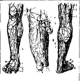
Figure 1.
Figure 2. Figure 3. Figure 1. Superficial veins of the leg: 1-patella; 2 and 3-v. saphena magna; 4-v. marginalis medialis; 5-vv. digitales communes pedis; 6-v. inter-capitularis; 7-vv. digitales pedis dorsales; 8-rete venosum dorsale pedis cutaneum. Figure 2. Superficial veins of the thigh: 1- v. femoralis profunda; 2-v. saphena magna. Figure 3. Superficial veins of the leg: 1-v. saphena parva; 2-connection with deep veins; 3-anastomosis; 4-v. saphena magna; 5-v. femoro-poplitea; 6-branch to v. profunda femoris; 7-branch to v. poplitea; 8-malleus lat.; 9-rete venosum dorsale pedis cutaneum; 10-v. marginalis lat. of the femur, it runs along the inner side of the thigh and flows into the front-inner semicircle of the femoral vein in the area of the femoral triangle (fig. 2). Branches flowing into S. v. magna from the leg (fig. 1) sometimes form v. cruris anterior and posterior. Often, branches of the anterior surface of the thigh before flowing into S. v. magna form a common trunk - v. femoralis anterior; the same is observed with branches of the inner side - v. femoralis posterior. The veins of the anterior semicircle of the thigh sometimes form a separate trunk (v. saphena accessoria), accompanying the main trunk and flowing into it at various heights. V. saphena minor- a more superficial trunk. V. epigastrica, v. circumflexa ilium, v. pudendae superf. often flow into saphena vena magna instead of their usual flow into the femoral vein. Along the entire length of the leg, S. v. magna is accompanied by the n. saphenus (see Thigh). V. saphena parva, or the posterior saphenous vein, begins behind the outer marginal vein (v. marginalis lateralis) (fig. 3). S. v. parva, receiving branches from the outer superficial and deep plantar veins, runs along the outer edge, then along the posterior surface of the Achilles tendon, and lies between the heads of the gastrocnemius muscles in the split of the fascia. In the popliteal fossa, S. v. parva always divides into two branches - one flows into the popliteal vein, the other into the deep femoral vein (fig. 3). The latter of these branches receives a vessel coming from the posterior semicircle of the thigh (v. femoro-poplitea), in which blood flows mostly from top to bottom, to a lesser extent - into the gluteal veins (remnant of embryonic circulation). S. v. parva throughout its course runs with the n. cutaneus surae medialis. The systems of S. v. magnae and S. v. parvae have rich anastomoses, forming on the foot the dorsal network (rete venosum dorsales pedis); on the leg, a plexiform network of veins from the front-inner side flows into S. v. magna, from the outer-posterior side - into S. v. parva. The frequent anastomosis in the popliteal region (fig. 3) has great practical significance. The superficial veins form numerous anastomoses with the deep veins. All these anastomoses create a powerful drainage system. The emptying of the veins here is regulated mainly by changes in the position of the trunk and extremities, since the contraction of muscles has little effect on the change in the lumen of veins located in the subcutaneous tissue. Together with other causes, a uniformly prolonged position of the lowered extremities can cause vein dilation. The significant number of valves in both S. v. magna and the femoral vein creates better conditions for drainage. The absence or insufficiency of valves is usually explained by the Trendelenburg symptom. Anomalies of the superficial veins of the lower extremities, like the entire venous system, are extremely diverse (see above). A major anomaly is the flow of the entire venae saphenae parvae into vena saphena magna. Lit.- see lit. to art. Varicose veins, Blood vessels.
Related articles
Mentioned in
Cite this page
“Saphenous Veins.” Soviet Medical Encyclopedia. English translation of Bolshaya Meditsinskaya Entsiklopediya, 1st ed. (Moscow, 1928–1936), ed. N. A. Semashko. https://sovietmedicalencyclopedia.pages.dev/article/saphenous-veins/